Article Text

Abstract
Objective We explored the hypothesis that TGR5, the bile acid (BA) G-protein-coupled receptor highly expressed in biliary epithelial cells, protects the liver against BA overload through the regulation of biliary epithelium permeability.
Design Experiments were performed under basal and TGR5 agonist treatment. In vitro transepithelial electric resistance (TER) and FITC-dextran diffusion were measured in different cell lines. In vivo FITC-dextran was injected in the gallbladder (GB) lumen and traced in plasma. Tight junction proteins and TGR5-induced signalling were investigated in vitro and in vivo (wild-type [WT] and TGR5-KO livers and GB). WT and TGR5-KO mice were submitted to bile duct ligation or alpha-naphtylisothiocyanate intoxication under vehicle or TGR5 agonist treatment, and liver injury was studied.
Results In vitro TGR5 stimulation increased TER and reduced paracellular permeability for dextran. In vivo dextran diffusion after GB injection was increased in TGR5-knock-out (KO) as compared with WT mice and decreased on TGR5 stimulation. In TGR5-KO bile ducts and GB, junctional adhesion molecule A (JAM-A) was hypophosphorylated and selectively downregulated among TJP analysed. TGR5 stimulation induced JAM-A phosphorylation and stabilisation both in vitro and in vivo, associated with protein kinase C-ζ activation. TGR5 agonist-induced TER increase as well as JAM-A protein stabilisation was dependent on JAM-A Ser285 phosphorylation. TGR5 agonist-treated mice were protected from cholestasis-induced liver injury, and this protection was significantly impaired in JAM-A-KO mice.
Conclusion The BA receptor TGR5 regulates biliary epithelial barrier function in vitro and in vivo through an impact on JAM-A expression and phosphorylation, thereby protecting liver parenchyma against bile leakage.
- bile acid
- tight junction
- biliary epithelium
- cholestaticliver diseases
Statistics from Altmetric.com
Significance of this study
What is already known on this subject?
Mechanisms by which biliary epithelial barrier function is maintained or altered during liver diseases have not received much attention, while this barrier might have crucial role in protecting liver parenchyma, especially in obstructive context.
While the biliary tract is continuously bathed with bile acids (BA), the impact of these signalling molecules on epithelial permeability has not been addressed yet.
What are the new findings?
We discovered that TGR5-dependent bile acid signalling unexpectedly strengthens biliary epithelial barrier function, both in vitro and in vivo.
We identified a yet unreported pathway in which BAs regulate expression and phosphorylation of a central tight junction protein, junctional adhesion molecule A (JAM-A).
We showed that this new pathway has critical protective effects in the setting of obstructive cholestasis in mice.
In human patients with primary biliary cholangitis and primary sclerosing cholangitis, we found JAM-A upregulation in cholangiocytes, suggesting a conserved mechanism of barrier protection in the biliary epithelium during cholestasis.
How might it impact on clinical practice in the foreseeable future?
It remains a challenge to study if this would be relevant in human biliary diseases.
Our findings may have translational impact, as the new pathway we identified might be targetable in the setting of chronic cholestatic diseases.
Introduction
After liver injury and during repair, biliary homeostasis is finely tuned in order to preserve liver parenchyma from bile-induced damage, through incompletely understood mechanisms. Besides cytokines and growth factors, bile acids (BA) and their receptors constitute a signalling network, particularly involved in mounting adaptive/protective responses during liver repair.1–5 BA signal through both nuclear (mainly farnesoid X receptor) and membrane (mainly G protein-coupled BA receptor 1 [GPBAR1] or TGR5) receptors that are widely distributed in the organism and whose activation elicits a wide array of biological responses.4 TGR5 is poorly expressed in hepatocytes but is highly enriched in the biliary tract in which it has been proposed to control chloride secretion in bile,6 cholangiocyte proliferation7 and gallbladder (GB) filling.8–10 We previously demonstrated that partial hepatectomy was followed by an immediate and massive BA overload in rats, mice and humans and that the lack of TGR5 in mice resulted in impaired liver regeneration mainly through an alteration of BA homeostasis.1 3–5 Because after bile duct ligation (BDL), the lack of TGR5 was associated with exacerbated bile infarcts as compared with wild-type (WT) mice,5 we hypothesised that TGR5 protects the liver parenchyma against bile leakage across the biliary epithelium, through the regulation of paracellular permeability.
In polarised epithelia, tight junctions (TJ) ensure a diffusion barrier function and regulate selective exchanges (ions and other small molecules) between apical and basolateral environments, through the establishment and regulation of a complex assembly of plasma membrane and cytoplasmic proteins.11 The main TJ transmembrane proteins occludin, claudins and junctional adhesion molecules constitute a core around which a number of cytoplasmic proteins (including zonula occludens proteins) associate to link the transmembrane proteins with the actin cytoskeleton.12 Their extracellular domains interact with each other between adjacent cells, contributing to seal plasma membranes. Signalling mechanisms involved in paracellular permeability regulation are in general largely unknown. Post-translational modifications, including phosphorylation of TJ transmembrane proteins like occludin, junctional adhesion molecule A (JAM-A) and claudin 4 have been in particular reported to impact paracellular permeability in different epithelial cells,13 14 but this remains completely unexplored in the biliary epithelium.
The impact of BA on paracellular permeability in pathophysiological conditions has been scarcely studied and is generally viewed as deleterious, increasing paracellular passage in intestinal15 and respiratory epithelial cells.16 However, the precise signal transduction mechanisms, in particular the BA receptors involved, were not investigated in these studies. In contrast, it has been suggested that TGR5-dependent BA signalling reinforces intestinal and endothelial barriers in mice,17 18 indicating that BA through multiple signalling pathways may elicit different effects on paracellular permeability, depending on the pathophysiological context and on the cell type involved. In the biliary epithelium, the impact of BA signalling on permeability has not been addressed yet, although the biliary tract is continuously bathed with BA.
Epithelial paracellular permeability has been very scarcely explored in the fields of biliary homeostasis19 20 and liver diseases.21–23 During clinical (primary biliary cholangitis [PBC] and primary sclerosing cholangitis [PSC]) and experimental cholestasis, alteration in the expression of TJ-associated cytoplasmic proteins in hepatocytes and cholangiocytes has been described.22 23 However, whether these alterations are primarily involved in the disease pathogenesis or that they occur secondarily in the pathological course is currently unknown.
In this study, we found that TGR5-dependent BA signalling interfered with processes regulating epithelial paracellular permeability, both in vitro and in vivo, through an impact on the expression and phosphorylation of the TJ protein JAM-A. We further provided evidence that TGR5-dependent signalling and the associated impact on JAM-A had critical protective effects in the setting of obstructive cholestasis in mice. We finally suggest that this pathway may also be relevant in human cholestatic diseases.
Materials and methods
Epithelial barrier function measurements
In vitro transepithelial electric resistance (TER) was measured in the normal rat cholangiocyte (NRC), Madin-Darby Canine Kidney (MDCK) II and MDCK II TGR5-GFP cell lines, either with the Millicell ERS-2 Volt-ohmmeter (Millipore, France) with a chopstick electrode or with the Cellzscope 2 (Nanoanalytics, Germany). Ten kilodalton FITC-dextran diffusion was measured by adding the dextran solution (2 mg/mL in serum-free culture medium) to the upper chamber and sampling the lower compartment at 1-hour interval for 5 hours. For TER and dextran transfer measurements, cells were seeded at 3–4×10⁵/cm2 on polyethylene transparent inserts (0.4 µm pore size, BD Falcon; BD Biosciences, France) and cultured until a confluent monolayer was obtained.
In vivo, to study biliary epithelial permeability, 4 kDa fluorescent dextran or a modified BA (glycocholic acid [GCA] C13 N15) were injected in the GB lumen and traced respectively by spectrofluorimetry and mass spectrometry in plasma and liver, within minutes after injection. These in vitro and in vivo methods are detailed in online supplementary figure 1 and in online supplementary materials and methods.
Supplemental material
Supplemental material
Bile flow and biliary dextran excretion
Interhepatocyte paracellular permeability was explored after 40 kDa FITC-dextran intravenous injection, through the analysis of biliary FITC-dextran excretion (blood to bile passage), as described24–26 (see online supplementary materials and methods).
Surgical and other procedures used on animals
C57BL/6J Gpbar1−/− mice (referred in this study as TGR5-KO mice) and their C57BL/6J wild type littermates were initially provided by Merck Research Laboratories (Kenilworth, USA)27 and used to fund our colonies of TGR5-KO and control animals. JAM-A-KO mice were kindly provided by Pr Dejana (IFOM, Milan, Italy).28 Bile duct ligation (BDL) performed together with cholecystectomy (CCT),1 as well as treatment with alpha-naphtylisothiocyanate (ANIT) are described in more details in online supplementary materials and methods. In series of experiments, mice were treated (oral gavage) with vehicle (gelatin/NaCl [7.5%/0.62%]) or the specific TGR5 agonist RO552723929 (10 mg/kg/day, Roche, Basel, Switzerland) for 2 days before BDL or ANIT treatment. The specificity of RO5527239 was confirmed in this study based on the lack of GB dilation on treatment in TGR5-KO mice, whereas this previously reported effect9 was constantly found in WT mice (online supplementary figure 2).
Supplemental material
GB injection with RO5527239 or vehicle
The same protocol was used as described for dextran and BA injection in the GB lumen (online supplementary materials and methods). A solution of 30 µM RO5527239 in PBS or the same volume of vehicle was injected. The GB was removed 20 min after injection and stored in a protein lysis buffer at −80°C until used for WB analysis.
Tissue fragments were removed at various times after surgery, and either frozen in nitrogen-cooled isopentane or in RNAlater (R0901, Sigma) and stored at −80 °C until use, or fixed in 4% FA and embedded in paraffin.
Patient samples
Liver biopsies came from the Biological Resource Center of Kremlin-Bicêtre Hospital (CRB PARIS SUD - UG 1203, HOPITAL BICETRE – APHP, France), as explained in more details in the online supplementary materials and methods: PBC, PSC and control normal liver samples (non-tumoural liver samples from hepatectomies). All patients signed an informed consent form.
Immunohistochemistry
Antibodies against: JAM-A, phospho (S285)-JAM-A, claudin-1, claudin-2, claudin-3 and claudin-5, occludin, cingulin, ZO-1, ZO-2, E-cadherin, the cholangiocyte marker cytokeratin-19, PKC ζ were used on ethanol acetone-fixed 10 µm liver or GB cryosections, and images were acquired in epifluorescence (Axiovert, Zeiss) and confocal (EZ-C1, Nikon) microscopy and analysed with the Image J software. Fluorescence intensity was analysed on liver sections. Details related to immunohistochemistry are provided in online supplementary materials and methods and online supplementary table 1.
Cell culture
NRC, MDCK II and F258 cell lines were cultured as previously described.21 30 Previously generated MDCK II Tet-Off cells were used as described.14 Further details are provided in online supplementary materials and methods.
Immunoblot
Proteins were analysed by sodium dodecyl sulfate (SDS)-polyacrylamide gel electrophoresis with antibodies (see online supplementary methods and online supplementary table 1 for references and dilutions) specific for JAM-A, phospho (S285)-JAM-A, claudin-3 and claudin-4, occludin, ZO-1, ZO-2, α-actin, phospho-PKC ζ-λ, GFP, GAPDH, PKC ζ, CK-19 and α-tubulin. The protein bands were detected by enhanced chemiluminescent reaction and signal intensity was semiquantified by densitometry (Fusion Fx7, Vilber Lourmat, Germany).
Biochemical assays
Alanine aminotransferase (ALT), alkaline phosphatase (ALP) and total bilirubin in the plasma were measured as detailed in online supplementary materials and methods.
Further applied methods
Additional animal treatments, cell culture, immunohistochemistry, immunoblots, biochemical assays and RT PCR experiments are further described in the online supplementary methods and online supplementary table 2.
Statistical analysis
Mann-Whitney U test and Student’s t-test were used to compare sample means with paired controls. Results are expressed as means±SEM. P values: *≤0.05; **≤0.01; ***≤0.001.
Results
TGR5 controls paracellular permeability in biliary epithelial cells in vitro and in vivo
As an in vitro test for TGR5 involvement in the regulation of paracellular permeability, rat biliary epithelial cells (NRC and F258)30 expressing TGR5 (online supplementary figure 3A) were cultured on membranous inserts and stimulated with a TGR5 specific agonist (RO552723929) or vehicle. We found that this agonist significantly increased TER and reduced 10 kDa FITC-dextran diffusion (figure 1A and online supplementary figure 3B). In line with these data, TGR5 overexpression (MDCK II TGR5-GFP cells) resulted in significantly increased TER, in both basal and TGR5 agonist stimulated conditions (figure 1B). As an in vivo test for evaluating the impact of TGR5 on basal biliary epithelial permeability, we focused on the GB, as it is the tissue most enriched in TGR527 31 32 (online supplementary figure 3C). Transepithelial diffusion of 4 kDa FITC-dextran or of the modified BA GCA C13 N15 was investigated after GB-restricted injection, as explained in details (online supplementary figure 1 and online supplementary materials and methods). As indicated by plasma FITC fluorescence and liver GCA C13 N15 concentration after GB injection, biliary transepithelial paracellular permeability was significantly increased in TGR5-KO as compared with WT mice (figure 1C). Interestingly, interhepatocyte paracellular permeability was not impaired by TGR5 loss, as shown by similar FITC-40 kDa dextran biliary excretion after intravenous injection in WT and TGR5-KO mice (figure 1D). These data are in line with no or weak TGR5 expression in hepatocytes, as opposed with strong expression in the biliary tract and GB epithelial cells33 (online supplementary figure 3C). Together, these observations identify TGR5 as a positive regulator of biliary epithelium barrier function.
Supplemental material

TGR5 impacts biliary epithelium permeability in vitro and in vivo. (A) NRC cells cultured on inserts were stimulated with RO5527239 (RO) or with the vehicle (DMSO). TER and 4 kDa FITC-dextran transfer were measured after 5 hours as explained in Materials and methods. Data are means of 3–5 experiments per condition. Student’s t-test. (B) TGR5 overexpression (in TGR5-GFP MDCK II cells) is correlated with an increase in both non-stimulated (left, histogram, n=10) and RO5527239 (RO) stimulated (right, arrows on graphs) TER, as compared with control MDCK II cells (right part, n=2–6). Student’s t-test. (C) In vivo assessment of biliary epithelial permeability, as detailed in Materials and methods, after 4 kDa FITC-dextran or modified BA (GCA C13 N15) GB injection (n=3–4 experiments, Student’s t-test). (D) In vivo assessment of interhepatocyte permeability as detailed in Materials and methods. FITC-dextran fluorescence passage in bile was analysed after intravenous injection, in WT and TGR5-KO mice. For C and D, n=6–8 mice/group. Student’s t-test. BA, bile acid; NRC, normal rat cholangiocyte; TER, transepithelial electric resistance; WT, wild-type.
TGR5 controls the expression and phosphorylation of the TJ protein JAM-A
As TJ proteins are central to the control of paracellular permeability in epithelia, we analysed their expression and localisation in WT and TGR5-KO mice liver and GB. At the mRNA level, the main TJ proteins (claudins, occludin, JAM-A and Zonula Occludens-1 (ZO-1)) were similarly expressed in TGR5-KO as compared with WT mice (online supplementary figure 4A). At the protein level, only JAM-A expression was altered in TGR5-KO intrahepatic bile ducts (IHBD) and GB epithelium (figure 2, online supplementary figure 4B-F). As shown on GB sections from WT and TGR5-KO mice, while JAM-A and pS285-JAM-A (referred as pJAM-A in this study) were expressed at the TJ in WT GB epithelial cells (colocalising with ZO-1), they were significantly less expressed (figure 2A) and TJ-localised in the TGR5-KO GB epithelium, as confirmed by confocal microscopy analysis (online supplementary figure 5). In the liver, similar observations were made in IHBD (figure 2B). JAM-A expression at hepatocyte TJ, previously reported in cell lines,34 was unaltered in TGR5-KO livers (Figure 2B, online supplementary figure 6A), in line with weak or no TGR5 expression in this cell type.33 WB on whole liver extracts did not show any significant difference in total tissue JAM-A expression between WT and TGR5-KO mice at the basal state, whereas in GB, JAM-A expression was significantly decreased in TGR5-KO as compared with WT mice (online supplementary figure 6B,C). These observations suggested that TGR5 regulates the biliary epithelium barrier function by influencing expression and possibly phosphorylation of the TJ protein JAM-A, which has been reported (Ser285 phosphorylation) to be important for the development and maintenance of functional TJs in other epithelial (intestine and kidney) cell lines.14 35 36 In contrast to the biliary tract, in the ileum, JAM-A and pJAM-A were similarly expressed in WT and TGR5-KO mice (online supplementary figure 7A).
Supplemental material
Supplemental material
Supplemental material
Supplemental material

The lack of TGR5 alters JAM-A expression in biliary epithelia. (A) Epifluorescence analysis of JAM-A (upper panel) and pJAM-A (lower panel) coimmunostaining with ZO-1 on GB cryosections from WT and TGR5-KO mice. *indicates GB lumen. Semiquantification histograms are shown. (B) Epifluorescence analysis of JAM-A (upper panel) and pJAM-A (lower panel) coimmunostaining with CK-19 (cholangiocyte marker) on liver cryosections from WT and TGR5-KO mice. Semiquantification histogram is shown. Representative images and semiquantification from n=4 mice per group (Student’s t-test). Scale bar=10 µm. n=5 (liver) to 8 (GB) mice/group. GB, gallbladder; JAM-A, junctional adhesion molecule A; WT, wild-type.
To address the molecular mechanism in more detail, we analysed the Ser285 phosphorylation of JAM-A in response to TGR5 engagement. TGR5-specific agonist treatment rapidly (minutes) resulted in strong JAM-A phosphorylation in vitro in biliary epithelial cell lines (figure 3A and online supplementary figure 8A,B). In addition, in TGR5-transfected MDCK II cells, increasing TGR5 expression correlated with rising JAM-A expression and JAM-A Ser285 phosphorylation (figure 3B). Importantly, in vivo injection in the GB lumen with RO5527239, but not with the vehicle, rapidly (20 min) triggered Ser285 JAM-A phosphorylation (figure 3C). These observations thus strongly suggested that TGR5 expression, in a previously unreported manner, regulates the TJ protein JAM-A expression and phosphorylation in the biliary tract. Longer term TGR5 agonist treatment also boosted JAM-A expression, although the pJAM-A/JAM-A ratio remained unchanged. This was observed both in vitro in NRC cells cultured with RO5527239 for up to 72 hours (figure 4A) and in vivo in WT mice after oral administration of this TGR5 agonist for 4 days as shown by western blot on GB extracts (figure 4B). These observations indicated that TGR5 stimulation triggers an acute boost in JAM-A Ser285 phosphorylation and a persistent rise in JAM-A protein levels. Interestingly, JAM-A mRNA expression was not increased on TGR5 agonist treatment (figure 4B), suggesting that TGR5-induced rise in JAM-A protein expression results from post-translational regulation. In line, JAM-A protein stability was significantly enhanced by TGR5 stimulation, as shown in cycloheximide experiments (figure 4C). Of note, other TJ or TJ-associated proteins expression (online supplementary figure 9A) and stability (figure 4C) were not affected by TGR5 stimulation. Using MDCK II cells with inducible expression of either wild type JAM-A (JAM-A/WT) or a non-phosphorylatable JAM-A mutant (JAM-A/S285A),14 we found that JAM-A protein stabilisation was dependent on its phosphorylation (figure 4C,D and online supplementary figure 9B). Furthermore, TGR5-specific stimulation did not elicit any significant TER increase in JAM-A/S285A expressing cells (figure 4D). Together, these observations highlight the TJ protein JAM-A as a main target of TGR5 signalling in biliary epithelial cells, in which this receptor modulates paracellular permeability at least in part through JAM-A phosphorylation-dependent stabilisation.
Supplemental material
Supplemental material

TGR5 controls the expression and phosphorylation of the TJ protein JAM-A. (A) Biliary epithelial cells (NRC) were stimulated with RO5527239 (RO; 30 µM) or with the vehicle, and JAM-A or pJAM-A was analysed by western blot (left panels, representative images and semiquantification, n=3 experiments) or immunofluorescence (right panel, representative image). (B) TGR5 overexpression in MDCK II cells correlates with increased JAM-A expression and phosphorylation. Representative western blots on MDCK II cells transfected with increasing amounts of the TGR5-GFP plasmid and semiquantification of JAM-A and pJAM-A. (C) In vivo JAM-A phosphorylation after GB injection with the TGR5 agonist RO5527239 was analysed in GB protein extracts, as explained in Materials and methods. One representative western blot and semiquantification. Each sample is a pool of n=3 GB in the two groups. GB, gallbladder; JAM-A, junctional adhesion molecule A; NRC, normal rat cholangiocyte.

TGR5 controls JAM-A protein stability. (A) Long-term increase in JAM-A protein expression on TGR5 agonist treatment in vitro. NRC cells cultured for 72 hours with either RO5527239 (RO; 30 µM) or with the vehicle (dimethylsulfoxide (DMSO); 0,06%). Representative western blot images and semiquantitative analysis from n=3 experiments. Right lower panel: quantitative RT-PCR measurements of JAM-A mRNAs on DMSO or RO treatment (10 µM and 30 µM) during 72 hours (n=3–4 experiments per condition). (B) Long-term increase in JAM-A protein expression on TGR5 agonist treatment in vivo. GB from WT mice treated for 4 days with RO5527239 (RO, oral gavage, 10 mg/kg/day), or with the vehicle (Veh). Three left panels: representative images and semi-quantitative evaluation of JAM-A and pJAM-A western blots. Each sample is a pool of 2–3 GB. Right panel: quantitative RT-PCR measurements of JAM-A mRNAs on Veh or RO treatment (n=4–7 experiments per condition). (C) JAM-A stability is enhanced by TGR5 agonist treatment. JAM-A/WT or a non-phosphorylatable JAM-A mutant (JAM-A/S285A) MDCK II cells were treated with cycloheximide (66 nmol/L, 24 hours) together with DMSO or RO (50 µM), and JAM-A expression was analysed by western blot. Claudin-2 (Cldn-2) stability was not impacted by TGR5 agonist treatment. Representative western blot images and semiquantitative analysis. n=3–5 cell samples/condition. ( D ) TGR5 controls paracellular permeability through JAM-A phosphorylation. RO5527239 (RO; 30 µM, 30 min) does not induce JAM-A phosphorylation in mutant S285A MDCK II cells, as revealed by western blot. Left panel and histograms are representative images and semiquantitative analysis from three experiments. Right graphs: TGR5 agonist-induced increase in TER is dependent on JAM-A phosphorylation. RO5527239 (RO) does not induce any significant increase in TER in mutant non phosphorylatable JAM-A (S285A) MDCK II cells. Data points are means of n=3–5 experiments. Mann-Whitney U test. GB, gallbladder; JAM-A, junctional adhesion molecule A; NRC, normal rat cholangiocyte; TER, transepithelial electric resistance; WT, wild-type.
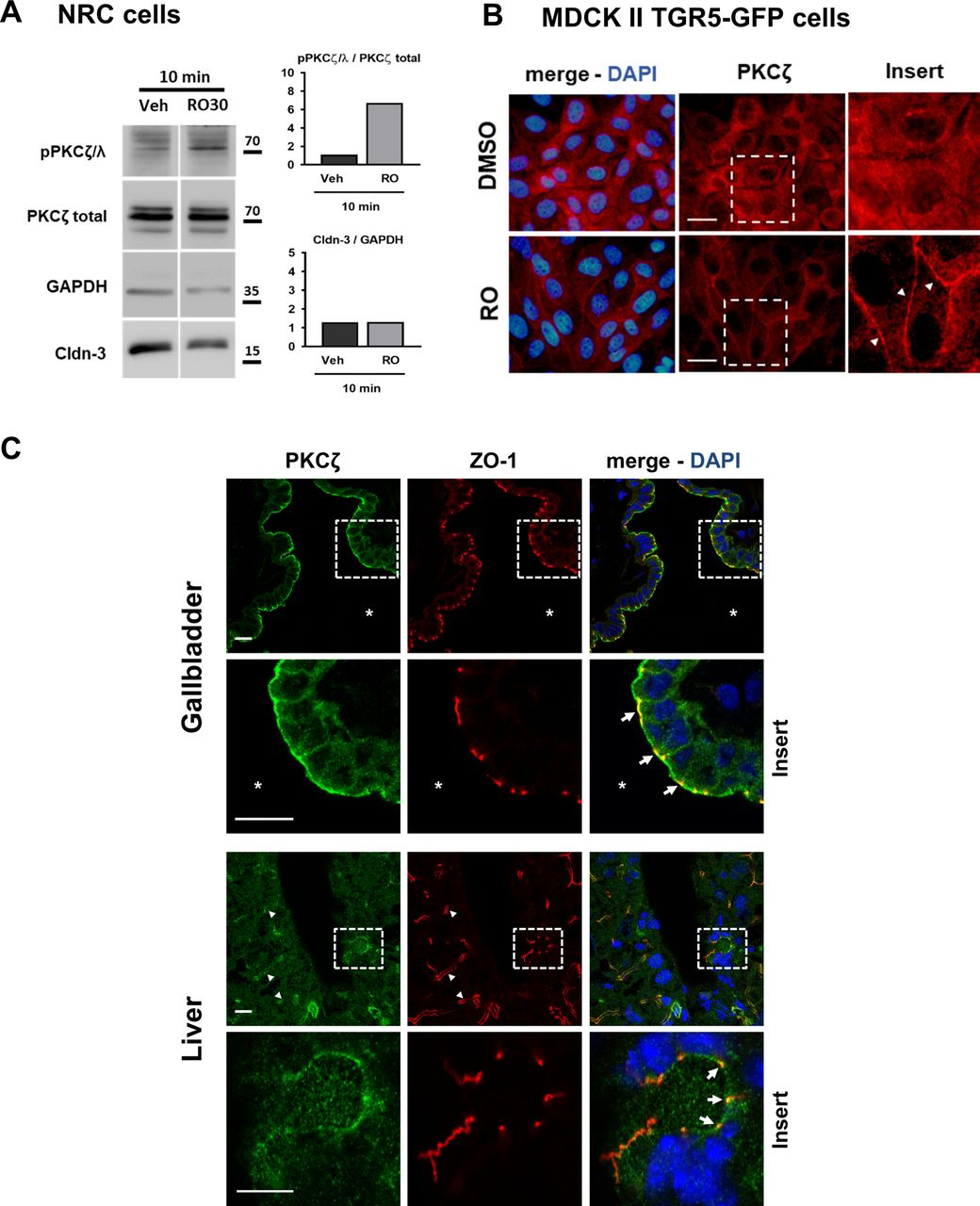
To analyse the TGR5-triggered signalling pathway upstream of JAM-A, we focused on atypical protein kinase C-ζ (PKCζ), which has previously been reported as a main contributor of JAM-A Ser285 phosphorylation at TJs in kidney epithelial cells.14 Interestingly, TGR5-specific stimulation rapidly (10 min) induced significant phosphorylation of PKCζ-λ (figure 5A). In line, PKCζ translocation from cytosolic pools to the plasma membrane was observed on TGR5 agonist treatment in MDCK II TGR5-GFP cells (figure 5B). Finally, in the GB epithelium as well as in IHBD, we found that PKCζ was localised at the apical membrane domain of cholangiocytes, including at the TJ (figure 5C). These data together suggest that TGR5-mediated BA signalling in the biliary epithelium triggers rapid PKCζ-dependent JAM-A Ser285 phosphorylation.

PKCζ activation is associated with TGR5-mediated JAM-A phosphorylation. (A) TGR5 agonist (RO5527239) induces PKCζ phosphorylation, analysed on western blots in NRC cells. One representative image and semiquantitative analysis are shown. (B) PKCζ translocation to plasma membrane on TGR5 agonist treatment (arrow heads). TGR5-GFP MDCK II cells were incubated with vehicle or RO5527239 for 30 min, and PKCζ was immunostained (red). (C) Confocal microscopy analysis of GB and liver immunostaining for PKCζ and ZO-1. Apical and TJ staining of PKCζ in the biliary epithelium (arrows in the GB insert panels). In upper liver panels, * indicates GB lumen, arrow heads indicate bile canaliculi and the insert corresponds to a bile duct. Arrows in the insert panel show TJ. Representative images from n=4 GB and livers. GB, gallbladder; JAM-A, junctional adhesion molecule A; NRC, normal rat cholangiocyte; PKCζ, protein kinase C-ζ; TJ tight junction.
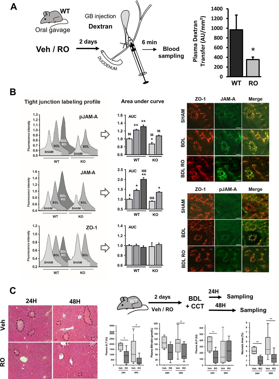
TGR5 stimulation increases biliary epithelium barrier function and provides hepatoprotection in cholestatic mice through an impact on JAM-A
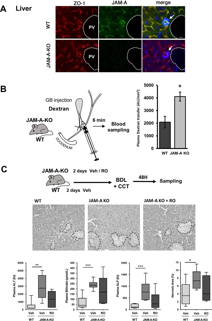
As it became clear that TGR5 had a positive impact on JAM-A expression and phosphorylation in the biliary tree resulting in paracellular permeability modulation, we focused on potential pathophysiological consequences of this previously unreported pathway in the context of obstructive BA overload. We first controlled if in vivo treatment with the TGR5 agonist had functional impact on biliary epithelial permeability. As shown in Figure 6A, 4kDa FITC-dextran transfer towards the plasma after GB-restricted injection was significantly reduced in WT mice pretreated with the TGR5 agonist as compared with vehicle-treated mice. We then looked at the potential effects of TGR5-mediated JAM-A expression and phosphorylation in the setting of experimental cholestasis, after BDL or ANIT intoxication. Interestingly, we observed that these models by themselves induced the expression and phosphorylation of JAM-A in the biliary epithelium (figure 6B and online supplementary figure 10A). This was not the case in the ileum, in which JAM-A and pJAM-A were similarly expressed before and after BDL (online supplementary figure 7B). We also observed that after ANIT intoxication, TGR5-KO mice exhibited a more severe phenotype as compared with WT (figure 6B), as previously shown in the BDL model.5 Of note, JAM-A phosphorylation was significantly more induced in WT than in TGR5-KO BDL mice (figure 6B), and JAM-A mRNA expression was not significantly induced in GB from BDL mice (online supplementary figure 8C). These data suggest once again that TGR5-dependent post-translational JAM-A processing was responsible for enhanced protein stability in the biliary epithelium in these models. Importantly, in livers from patients with PBC and PSC, JAM-A protein (but not mRNA) expression was also induced in the biliary epithelium as compared with control patients (online supplementary figure 11A), suggesting a conserved JAM-A-dependent mechanism of barrier protection in the biliary epithelium in cholestatic context. In contrast, pJAM-A expression was not enhanced in those patients: in cholangiocytes, it was surprisingly detected in the cytosol, in addition to a weak expression at the TJ; in hepatocytes it was mostly suppressed, in line with a decreased whole liver pJAM-A expression (online supplementary figure 11B,C). We then pretreated WT mice with either the vehicle or the TGR5 agonist (RO5527239) and performed BDL or ANIT intoxication. After BDL or ANIT, bile infarcts were reduced in TGR5 agonist-treated as compared with vehicle-treated mice, thus pointing to a protective effect of TGR5 agonism in these models (figure 6C and online supplementary figure 10B). Of note, TGR5 agonist treatment was not associated with any significant impact on cytokine gene induction after BDL (online supplementary figure 12). In contrast, we observed that JAM-A and pJAM-A were induced in bile duct epithelial cells from BDL or ANIT mice treated with RO5527239 as compared with vehicle-treated mice (figure 6B and online supplementary figure 10A). Our data thus suggest that although JAM-A is induced in these cholestatic models, TGR5 stimulation further boosts (stabilises) its expression. Interestingly, 4 kDa FITC-dextran GB injections demonstrated significantly enhanced transparietal passage towards the plasma in JAM-A-KO as compared with WT mice (figure 7A,B). In line, the lack of JAM-A was by itself associated with more severe bile infarcts after BDL (figure 7C), confirming the importance of this TJ protein in the control of biliary epithelial permeability. Importantly, the expression of other TJ proteins in livers from JAM-A-KO mice was not altered as compared with WT animals (online supplementary figure 13). Finally, the protective effect of TGR5 specific agonism after BDL observed in WT mice was significantly impaired in JAM-A-KO mice (figure 7C), further reinforcing the idea that TGR5 operates at least in part through an impact on JAM-A to build a protective response against bile leakage in these experimental conditions.
Supplemental material
Supplemental material
Supplemental material
Supplemental material

TGR5 agonism provides hepatoprotection in BDL mice. (A) GB-restricted injections with 4 kDa FITC-dextran (see online supplementary materials and methods), after vehicle or TGR5 agonist treatment (RO5527239 [RO]), in WT mice. Semiquantification of dextran passage in the plasma after 6 min (data are means of n=5–7 mice per group, Student’s t-test). (B) JAM-A expression and phosphorylation in the liver are increased after BDL. RO5527239 treatment in BDL mice further increases JAM-A and pJAM-A. Semiquantitative analysis of intrahepatic bile duct immunostainings (see online supplementary materials and methods). Left graphs: representative profiles of tight junctions fluorescence for each condition. Middle graphs: areas under curve (AUC) determined from representative profiles for each condition (see online supplementary materials and methods). Right images: JAM-A, pJAM-A and ZO-1 immunofluorescence on liver sections from WT and TGR5-KO mice. Scale bar=25 µm. Data are means of n=4–10 mice per group, Mann-Whitney U test. Symbols: * comparison with WT sham; $ comparison with WT BDL. (C) TGR5 agonist treatment significantly protects WT mice from BDL-induced liver injury (see the experimental design), as revealed by H&E-stained liver sections (necrosis area, surrounded by doted lines) as well as plasma alanine aminotransferase (ALT), bilirubin and alkaline phosphatase (ALP) n=4–11 mice/group (Mann-Whitney U test). Scale bar=50 µm. BDL, bile duct ligation; GB, gallbladder; JAM-A, junctional adhesion molecule A; WT, wild-type.

TGR5-dependent hepatoprotection is impaired in JAM-A KO mice. (A) JAM-A and ZO-1 immunofluorescence on liver sections from WT and JAM-A-KO mice. Arrows showing TJ-localised JAM-A in WT bile ducts, lacking in KO mice. Scale bar: 20 µm. (B) GB-restricted injections with 4 kDa FITC-dextran (see Materials and methods), in WT and JAM-A-KO mice. Semiquantification of dextran passage in the plasma after 6 min (data are means of n=5 mice per group, Student’s t-test). (C) TGR5 agonist treatment significantly protects WT but not JAM-A-KO mice from BDL-induced liver injury, as revealed by H&E-stained liver sections (necrosis area, surrounded by doted lines) as well as plasma ALT, total bilirubin and alkaline phosphatase (ALP). n=6–9 mice/group, Mann-Whitney U test. ALT, a lanine aminotransferase; BDL, bile duct ligation; JAM-A, junctional adhesion molecule A; TJ, tight junction; WT, wild-type.
Discussion
While hepatocyte TJ proteins have received attention regarding their regulatory impact on bile secretion,19 20 the pathophysiological role of these proteins in the biliary epithelium has been scarcely explored. Sparse evidence has been reported on TJ or TJ-associated proteins alteration in experimental or clinical liver diseases,22 23 but these alterations were not defined as secondary or primary to the disease process. The main underlying concept in this field is that alteration in intercholangiocyte TJs would result in leaky bile ducts, allowing bile reflux towards hepatic parenchyma, contributing to injury during the disease process. This concept is strikingly illustrated in patients with neonatal ichtyosis and sclerosing cholangitis syndrome, a primary (mutational) claudin-1 defect.21 37 More recently, mutations in the TJ-associated protein ZO-2 gene together with severe liver disease were reported.38 However, the relative impact of intercholangiocyte versus interhepatocyte TJ protein defects on the pathophysiology of these diseases is far from being delineated, in part because TJ protein repertoire is not the same in hepatocytes and cholangiocytes.26 Secondary TJ alterations in the liver also take place especially in inflammatory contexts during a number of cholestatic diseases.22 23 39 Cytokines and lipopolysaccharide have impact on both barrier and pore functions of TJ proteins in biliary epithelial cells, with in general a global effect of enhanced permeability,39 but only few reports are available on TJs in the liver diseases context. Recently, dramatic alteration in TJ protein expression and TJ structure observed in a double KO mouse model for γ-catenin and β-catenin was associated with severe cholestatic liver injury due to a complete loss of hepatocyte blood–biliary barrier, pointing out the crucial hepatoprotective role of TJ.40
In the present study, we provide both in vitro and in vivo evidence that BA signalling, through an impact on JAM-A expression and phosphorylation, strengthens the cholangiocyte blood–biliary epithelial barrier and protects the liver parenchyma against bile leakage during obstructive experimental liver diseases. Although BAs are generally viewed as factors increasing paracellular passage via receptors and signalling pathways that remain mostly elusive,15 16 41 they were however reported as reinforcing agents for endothelial18 and intestinal17 barriers, through TGR5-mediated mechanisms. Importantly, although the biliary epithelium is continuously bathed with highly concentrated BA, the BA regulatory impact on intercholangiocyte TJ proteins and functions had never been specifically investigated. JAM-A is reported as one of the first proteins to be present at cell–cell junctions when cultured cells form intercellular contacts.42 43 With the identification of JAM-A (and JAM-A Ser285 phosphorylation) as downstream effectors of TGR5-dependent BA signalling, our study demonstrates that TGR5 targets a central regulator of TJ and epithelial barrier formation.
TJ proteins are regulated both at transcriptional and post-transcriptional levels, although precise mechanisms remain poorly defined in most cases.13 44 Different studies revealed in particular that the phosphorylation state of individual TJ proteins including occludin, claudins or ZO-1 had impact on paracellular epithelial barrier function.13 44 Previous in vitro studies have reported that JAM-A expression and phosphorylation were crucial for paracellular permeability and identified the atypical PKCζ as a main kinase involved in JAM-A phosphorylation.14 We found that TGR5-specific stimulation signals at least in part through PKCζ activation and thereby may control JAM-A phosphorylation on S285. To our knowledge, our report is the first alighting an in vivo impact of JAM-A phosphorylation on epithelial permeability. Previous in vivo studies reported impact of JAM-A on gut epithelial permeability, which is compromised in JAM-A-KO mice, but the role of JAM-A phosphorylation was not analysed.45 46
In the pathophysiological course of cholestatic liver diseases (online supplementary figure 14), the following scenario may thus be proposed based on our data. TGR5-mediated signalling in the basal state as well as in the context of BA overload leads to JAM-A phosphorylation and JAM-A protein stabilisation, at least in part through PKCζ activation. This leads to decrease TJ permeability and protects the liver parenchyma from BA leakage. TGR5 thus appears as crucial for BA-mediated hepatoprotection. Our data in PBC and PSC patients suggest that mechanisms contributing to JAM-A phosphorylation may be dysregulated with unknown impact in this disease context (online supplementary figure 11). Finally, targeting cell specifically this pathway in the liver, although far from being available, remains a challenge for future therapeutic intervention.
Supplemental material
Acknowledgments
We would like to thank Aurélie Bonilla, Caroline Rousseau, Julie Sempé, Céline Dubois, Maura De Almeida and Olivier Fayol for their technical help. We would like to thank Dr Dominique Lagadic-Gossmann for providing us with F258 cells. We would like to thank Galya Vassileva and the Merck Research Laboratories (Kenilworth, USA) for providing us with the C57Bl/6J Gpbar1-/- mice, and Pr Elisabetta Dejana (Milan, Italy) for providing us with the C57Bl/6J JAM-A-/- mice. We thank Colette Rey and Chantal Housset (Inserm U938) for help with mice IBDU isolation.
References
Footnotes
NK and JU-B contributed equally.
Contributors JU-B and NK contributed equally to this work. TT: study concept and design, obtained funding, wrote the manuscript and study supervision; GM, JU-B, NK, HS, VB-J, ID, ZT, NP, JG, DC and TT: acquisition, data analysis and interpretation; GM, JU-B, DC, ZT and KE: critical revision of the manuscript for important intellectual content; CU, KE, AD-S and CG: technical and material support.
Funding This study was funded by Institut National de la Santé et de la Recherche Médicale, Université Paris-Sud and Agence Nationale de la Recherche (Grant Number:15-CE14-0007-01).
Competing interests None declared.
Patient consent This manuscript does not contain identifiable patients data.
Provenance and peer review Not commissioned; externally peer reviewed.