Article Text

Abstract
Objective The extent to which tryptophan (Trp) metabolism alterations explain or influence the outcome of inflammatory bowel diseases (IBDs) is still unclear. However, several Trp metabolism end-products are essential to intestinal homeostasis. Here, we investigated the role of metabolites from the kynurenine pathway.
Design Targeted quantitative metabolomics was performed in two large human IBD cohorts (1069 patients with IBD). Dextran sodium sulphate-induced colitis experiments in mice were used to evaluate effects of identified metabolites. In vitro, ex vivo and in vivo experiments were used to decipher mechanisms involved. Effects on energy metabolism were evaluated by different methods including Single Cell mEtabolism by profiling Translation inHibition.
Results In mice and humans, intestinal inflammation severity negatively correlates with the amount of xanthurenic (XANA) and kynurenic (KYNA) acids. Supplementation with XANA or KYNA decreases colitis severity through effects on intestinal epithelial cells and T cells, involving Aryl hydrocarbon Receptor (AhR) activation and the rewiring of cellular energy metabolism. Furthermore, direct modulation of the endogenous tryptophan metabolism, using the recombinant enzyme aminoadipate aminotransferase (AADAT), responsible for the generation of XANA and KYNA, was protective in rodent colitis models.
Conclusion Our study identified a new mechanism linking Trp metabolism to intestinal inflammation and IBD. Bringing back XANA and KYNA has protective effects involving AhR and the rewiring of the energy metabolism in intestinal epithelial cells and CD4+ T cells. This study paves the way for new therapeutic strategies aiming at pharmacologically correcting its alterations in IBD by manipulating the endogenous metabolic pathway with AADAT.
- inflammatory bowel disease
Data availability statement
Data are available on reasonable request.
This is an open access article distributed in accordance with the Creative Commons Attribution Non Commercial (CC BY-NC 4.0) license, which permits others to distribute, remix, adapt, build upon this work non-commercially, and license their derivative works on different terms, provided the original work is properly cited, appropriate credit is given, any changes made indicated, and the use is non-commercial. See: http://creativecommons.org/licenses/by-nc/4.0/.
Statistics from Altmetric.com
Disclaimer: this video summarises a scientific article published by BMJ Publishing Group Limited (BMJ). The content of this video has not been peer-reviewed and does not constitute medical advice. Any opinions expressed are solely those of the contributors. Viewers should be aware that professionals in the field may have different opinions. BMJ does not endorse any opinions expressed or recommendations discussed. Viewers should not use the content of the video as the basis for any medical treatment. BMJ disclaims all liability and responsibility arising from any reliance placed on the content.
WHAT IS ALREADY KNOWN ON THIS TOPIC
Kynurenic acid (KYNA) and xanthurenic acid (XANA) can activate aryl hydrocarbon receptor (AhR)-related genes but their agonist activity is unknown.
Some tryptophan metabolites are essential for intestinal homeostasis, but activation of IDO pathway can be deleterious.
AhR activation impacts intestinal epithelial cells proliferation and gut barrier integrity.
AhR activation impacts T cell differentiation.
WHAT THIS STUDY ADDS
XANA and KYNA abundance are negatively correlated with intestinal inflammation in mice and humans.
The kynurenine pathway is composed of protective (KYNA and XANA) and deleterious metabolites (QUIN).
XANA and KYNA are bona fide AhR ligand.
XANA and KYNA promote intestinal epithelial cells survival and proliferation through enhancement of mitochondrial respiration.
XANA and KYNA promote Th17 cells differentiation in a mechanism dependent on glycolysis induction.
The use of the recombinant aminoadipate aminotransferase (AADAT), to favor KYNA and XANA production, is protective in colitis model.
HOW THIS STUDY MIGHT AFFECT RESEARCH, PRACTICE OR POLICY
Manipulating the endogenous tryptophan metabolism with AADAT is an attractive new therapeutic strategy in inflammatory bowel disease.
Introduction
Inflammatory bowel diseases (IBDs) represent a public health issue in industrialised societies.1 The precise mechanisms leading to these diseases are linked to an inappropriate immune system response,2 genetic susceptibilities involving many genes (eg, CARD9, NOD2)3 and altered intestinal gut microbiota.4
In the broad sense, metabolism is the set of chemical reactions in a living organism required for its functioning.5 The interweaving of metabolism pathways, from amino acid catabolism to glycolysis and mitochondrial respiration, makes it an intricated network crucial for cellular homeostasis,6 activation or differentiation.7 Logically, its deregulation is involved in many pathogenesis events.8 9 Literature has recently pointed out the role of tryptophan (Trp) metabolism in the context of intestinal inflammation.3 10 Trp can be metabolised following three major pathways: the serotonin and kynurenine (KYNU) pathways (KP), while the indole pathway.11 The production of indoles by the gut microbiota, which is altered in patients with IBD, is crucial to intestinal homeostasis, notably through their effect on activating the aryl hydrocarbon receptor (AhR).3 12 The role of serotonin and KP have been highlighted in several chronic inflammatory pathological contexts, from IBD10 13 14 to metabolic syndrome.15 The KP is of particular interest as it leads to the production of several end-product metabolites, which have strong biological effects through identified receptors such as AhR (kynurenic acid (KYNA)),16 GPR35 (KYNA),17 N-methyl-D-aspartate receptor (KYNA, quinolinic acid (QUIN)).18 The KP and its host receptor targets are particularly relevant in inflammatory contexts, including IBD, as these conditions are associated with KP activation.10 Indeed, some KP metabolites impact immune cells functions notably via AhR activation19 that is involved in the differentiation of T cells,20 the production of IL-17 and IL-22,21 22 the maintenance of type 3 innate lymphoid cells (ILC3) in the gut23 or the antioxidant response.24 However, the role of many metabolites from the KP in IBD remains poorly defined.
Here, we used targeted quantitative metabolomics to characterise the trp metabolism in colitis models in mice and two large independent human cohorts. We observed a strong negative correlation between the amount of xanthurenic acid (XANA) and KYNA, and intestinal inflammation. Treatment with XANA or KYNA exhibits protective effects in colitis models by modulating the energy metabolism in intestinal epithelial cells (IEC) and T cells. Oxidative phosphorylation is boosted in IEC, leading to an improvement in proliferation and ultimately tissue repair, mediated by the AhR-IL-22 axis. Glycolysis is stimulated in T cells with positive effects regarding activation and polarisation towards TH17 phenotype. From a therapeutic perspective, we proposed a recombinant enzyme-based strategy to rewire the endogenous Trp metabolism toward the production of XANA and KYNA. We show the efficacy of this strategy in colitis models, thus paving the way for the development of a new class of drugs for IBD.
Results
XANA and KYNA abundance negatively correlates with colitis severity in mice
The KP (figure 1A) is globally considered to play a role in the inflammatory process. To gain insight into the precise role of metabolites downstream of KYNU in intestinal inflammation, we performed a dense time-course experiment in mice submitted to Dextran Sodium Sulphate (DSS)-induced colitis (figure 1B). The peak of inflammation, evaluated by histological scoring and colon length measurement, was observed on day 9. Quantitative targeted metabolomics was used to measure the metabolites of the Indoleamine 2,3-dioxygenase (IDO) pathway in serum and colon. We observed that the dynamics of the metabolites composing this pathway differed from one to another (figure 1C). In particular, the dynamics of KYNU and QUIN positively correlated with inflammation, while the opposite pattern was observed for XANA or KYNA (figure 1C). Similarly, a negative correlation was observed between weight loss and amounts of KYNA and XANA in caecum content (figure 1D), while the opposite was observed with KYNU and QUIN (online supplemental figure S1A,B). These results suggest that specific metabolites of KP, notably KYNA and XANA, could play a protective role in colitis.
Supplemental material
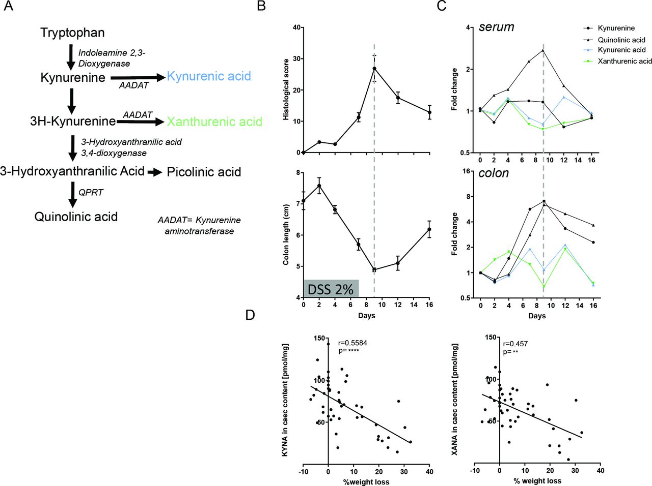
KYNA and XANA abundances are negatively correlated with inflammation during DSS-induced colitis. (A) Kynurenine pathway, (B) histological score and colon length dynamic during the course of DSS-induced colitis, (C) dynamic of kynurenine pathway metabolites in serum and colon tissue during the course of DSS-induced colitis, (D) correlation between KYNA and XANA abundance in the caecum and weight loss. Statistical analysis: n=6–8 per time point, for correlation n=47, Spearman’s rank correlation, **: p<0.01, ****: p<0.0001. AADAT, aminoadipate aminotransferase; DSS, dextran sodium sulphate; KYNA, kynurenic acid; XANA, xanthurenic acid.
XANA and KYNA abundance correlates with disease activity in humans with IBD
We then studied the relevance of these results in a human context. We analysed faecal (n=104) and serum (n=108) samples from 15 patients enrolled in a pilot clinical trial evaluating the effect of faecal microbiota transplantation in Crohn’s disease.25 Quantitative metabolomics analysis targeting Trp metabolites was performed on these samples (online supplemental figure S1C). We observed a negative correlation between several markers of disease activity, including various clinical, endoscopic and biological parameters, with KYNA and XANA (as well as the ratio of KYNA, XANA with their precursors, KYNU and 3-hydroxykynurenine, respectively), while the reverse was noted with KYNU and QUIN (online supplemental figure S1D,E). We then took advantage of a large independent cohort (Suivitheque) of 1069 patients with IBD and 98 healthy subjects (HS) for which serum and faeces (for a subpopulation) were available (online supplemental table S1, figure 2A). Trp metabolites measured in serum by targeted quantitative metabolomics approach significantly discriminated patients with IBD from HS (figure 2B). The differential analysis identified many strongly statistically significant differences between HS and patients with IBD (figure 2C) and also between patients with IBD in flare and in remission (figure 2D). Notably, XANA and KYNA (as well as the ratio of KYNA, XANA with their precursors) were decreased in patients with IBD compared with HS and in patients in flare compared with those in remission (figure 2C–E and online supplemental figure S1F–H). In this second cohort, we also observed a negative correlation between several clinical26 and biological markers of disease activity and XANA and KYNA (figure 2F). The signal was also confirmed when we correlated the serum concentration of Trp metabolites and several cytokines in a subgroup of 149 patients with IBD, taken randomly in the Suvitheque cohort (the characteristics of these 149 patients are described in online supplemental table S2). Here again, XANA and KYNA negatively correlated with inflammatory cytokines, while the opposite was observed for QUIN (online supplemental figure S1I). A similar signal, although weaker, was observed when looking at metabolites in the stools (online supplemental figure S1J,K). Finally, when considering the subgroup of patients with IBD in remission (n=333), a lower serum level of XANA and KYNA was predictive of a higher risk of occurrence of disease-related clinical events (surgery or flare with the need to initiate a new treatment) (figure 2G).
Supplemental material
Supplemental material

KYNA and XANA abundances correlate with disease activity in humans with IBD: Suivitheque cohort. (A) Design of the Suivitheque cohort. (B) PCA analysis based on trp metabolites in serum. (C) Differential analysis of the abundance of TRP metabolites in serum between patients with IBD versus HS (adjusted p value bH). (D) differential analysis of the abundance of TRP metabolites in serum between patients with IBD in flare versus remission (adjusted p value bH). (E) XANA and KYNA amount in serum (ANOVA with correction for FDR bH). (F) Correlation between the amount of tryptophan metabolites in serum and disease activity parameters. Spearman, p<0.05, q<0.1 (BH). (G) Clinical event-free survival according to XANA and KYNA levels in serum. Only patients in remission were included in this analysis. Two groups were defined according to the median level of XANA and KYNA. Statistical comparison was performed with a log-rank test. ANOVA, analysis of variance, ****: p<0.0001; CRP, C reactive protein; FDR, false discovery rate; Hb, haemoglobin; HBI, Harvey-Bradshaw Index; HS, healthy subjects; IBD, inflammatory bowel disease; IDO, indoleamine 2,3-dioxygenase; KYNA, kynurenic acid; PCA, principal component analysis; SCCAI, Simple Clinical Colitis Activity Index; XANA, xanthurenic acid.
Taken together, these results in mice and two independent human cohorts show strong negative correlations between the serum and faecal amounts of XANA and KYNA, and intestinal inflammation, and suggest a potential protective role of these metabolites.
Administration of XANA and KYNA protects from DSS-induced colitis
To explore the potential protective role of XANA and KYNA in intestinal inflammation settings, mice submitted to DSS-induced colitis were orally gavaged with XANA or KYNA (online supplemental figure S2A), leading to an elevation in the serum levels of these metabolites (online supplemental figure S2B). As demonstrated by the reduction in weight loss (figure 3A), disease activity index (figure 3B), colon length shortening (figure 3C) and histological score (figure 3D,E), treatment with XANA and KYNA induced a strong protective effect compared with the control group. Interestingly QUIN, whose abundance was positively correlated with inflammation severity, exhibited proinflammatory effects (online supplemental figure S2C–E). The protective effect of XANA and KYNA was confirmed by colonic transcriptomic response at day 12 with the downregulation of many pro-inflammatory cytokines and chemokines genes (eg, IL-6, CXCR1, figure 3F) in mice treated with these metabolites. The same protective effect was observed when XANA and KYNA were administered through intraperitoneal injection (online supplemental figure S2F–J).
Supplemental material

IDO metabolites, XANA and KYNA protect against DSS-induced colitis. (A) Body weight loss, (B) DAI, (C) colon length, (D) histological score, (E) colon histology pictures and (F) nanostring colon transcriptomic results at day 12. Each column represents a mouse. Statistical analysis: n=16–20 mice per group, two-way or one-way ANOVA, with Bonferroni post test, *: p<0.05, **: p<0.01, ***: p<0.001, ****: p<0.0001. ANOVA, analysis of variance; DAI, disease activity index; DSS, dextran sodium sulphate; IDO, indoleamine 2,3-dioxygenase; KYNA, kynurenic acid; XANA, xanthurenic acid.
Altogether, these results demonstrate a protective effect of XANA and KYNA in a colitis model in mice.
XANA and KYNA act on viability and proliferation of IECs through AhR activation and IL-22
To investigate the mechanism of the protective effects of XANA and KYNA in colitis, we started by exploring a potential effect on IEC. As shown by the increased staining for Ki67 in the recovery phase at day 12 in colitis experiment, XANA and KYNA stimulated the proliferation of IEC and intestinal healing compared with control mice (figure 4A). In addition, a wound healing assay, carried out with human IEC, showed that XANA and KYNA stimulate cell proliferation and, therefore, faster wound repair (figure 4B,C). KYNA has been shown to activate GPR3517 and the AhR pathway.16 While the protective effects of XANA and KYNA were maintained in GPR35-/- mice (online supplemental figure S3A,B), it was lost in AhR-/- mice (figure 4D, online supplemental figure S4A,B). Indeed the activation of AhR by XANA and KYNA was seen in vivo, as demonstrated by the induction of the expression of Cyp1a1, a major AhR target gene, in the colon of mice treated with XANA or KYNA for 12 days (figure 4E). FICZ (6-formylindolo(3,2b)carbazole), an AhR agonist, recapitulated the effect of KYNA and XANA (figure 4B,C). Moreover, the healing effect of XANA and KYNA in wound healing assay was lost in the presence of the AhR inhibitor CH223191 (figure 4F,G), suggesting that AhR, which is known to stimulate IEC proliferation,27 28 is at least partly involved in the underlying mechanisms. Experiments with a reporter cell line confirmed that both XANA and KYNA activate the AhR pathway (figure 4H). Similar results were obtained with human IEC cell lines in which the two metabolites induced the nuclear translocation of AhR (online supplemental figure S4C) and the expression of the AhR target gene Cyp1a1 (online supplemental figure S4D). Finally, the radioligand displacement assay showed that XANA and KYNA were effective in displacing radiolabeled TCDD (2,3,7,8-tetrachlorodibenzo-p-dioxin), a high-affinity AhR ligand, with a stronger effect for XANA (figure 4I). The radiodisplacement of a ligand with high affinity for AhR (TCDD), provides evidence that XANA and KYNA are true ligands for AhR.
Supplemental material

KYNA and XANA promote intestinal epithelial cells viability and proliferation through AhR. (A) Ki67 and DAPI staining in mouse colon at day 12 of a DSS model and quantification. (B, C) Scratch test on HT-29 cells treated with KYNA, XANA or FICZ: representative pictures and quantification of empty space. (D) Colitis protection by KYNA and XANA is abrogated in AhR-/- mice. (E) Oral gavage of KYNA and XANA for 12 days activate the expression of the AhR target gene, CYP1A1 in the colon. (F, G) Scratch test on HT-29 cells treated with KYNA, XANA, FICZ, with the AhR antagonist CH223191. (H) AhR reporter cell line activation and (I) radio displacement of TCDD induced by KYNA and XANA. (J–L) colitis protection by KYNA and XANA are partly conserved in AhRΔIEC mice. Data represent one out of two independent experiments. Statistical analysis: for scratch test 100 measures per slide, in vitro experiment n=3 and for in vivo and ex vivo experiment n=5–10 mice per group, two-way or one-way ANOVA, with Bonferroni post test, *: p<0.05, **: p<0.01, ***: p<0.001, ****: p<0.0001. AhR, aryl hydrocarbon receptor; ANOVA, analysis of variance;DSS, dextran sodium sulphate; KYNA, kynurenic acid; XANA, xanthurenic acid.
Interestingly, XANA and KYNA still exhibited some level of protection in AhRΔIEC mice that are deficient for AhR only in IECs (figure 4J–L), suggesting that other cell types are also involved in the mechanisms of action. In the gut, besides the direct activation of epithelial cells, AhR agonists also stimulate lymphoid cells to produce IL-22, which has a protective effect in colitis settings, notably by inducing the production of antimicrobial peptides, such as RegIIIγ and RegIIIδ, by IEC.11 Interestingly, the expression of Il-22, RegIIIγ and RegIIIδ was upregulated (statistically significant for IL-22 only) in the colon of mice treated by XANA and KYNA (online supplemental figure S4E). The protective effect of XANA and KYNA was decreased in IL-22-/- mice (online supplemental figure S4F,G), suggesting that the induction of IL-22 is involved in the mechanism underlying the effects of XANA and KYNA.
Taken together, these results demonstrate that XANA and KYNA are bona fide AhR ligands. Their protective effects are at least partly mediated by direct activation of the AhR pathway, as well as indirect effect through AhR-IL-22 axis activation. IEC are involved in the mechanisms of action of KYNA and XANA, together with other cell types.
XANA and KYNA act on mitochondrial metabolism in epithelial cells
IEC functions rely on energy and particularly on mitochondrial activity. In the colon, the dominant source of energy for epithelial cells is the short-chain fatty acid (SCFA) butyrate, which is produced from the digestion of fibres by the gut microbiota. Butyrate enters epithelial cells through specific transporters and diffuses into the mitochondria, where it undergoes β-oxidation, leading to the production of acetyl-CoA, NADH and FADH2 that feed the TCA cycle and the electron transport chain to produce ATP. The luminal content of mice treated by XANA or KYNA for 12 days showed a drop in the concentration of SCFA compared with untreated controls (figure 5A). The analysis of the gut microbiota composition by next-generation sequencing did not indicate any significant effect of KYNA and XANA after 12 days of gavage (online supplemental figure S5A–D). This suggests that the decreased SCFA concentration may be due to an increase in consumption by host cells rather than a decrease in production by the gut microbiota. The increased expression of two SCFA transporters, Smct and Mct-1, in the colon of mice treated with XANA or KYNA (figure 5B), as well as the increased consumption of butyrate by IEC treated with XANA or KYNA (online supplemental figure S5E), support this hypothesis. We then explored the effect of XANA and KYNA on the energy metabolism of human IEC. The proportion of cells with dysfunctional mitochondria (in connection with an altered energy metabolism), defined by a positive Mitotracker Green staining with a negative Mitotracker Red staining, was decreased after treatment with XANA or KYNA (figure 5C). Interestingly, even mitochondria classified as dysfunctional were in better shape in XANA or KYNA-treated cells, as shown by a higher Mitotracker Red MFI than in control cells (figure 5D). Beyond a smaller proportion of dysfunctional mitochondria, XANA and KYNA-treated cells exhibited a higher respiratory activity (figure 5E) and greater maximal respiration (figure 5F,G), while the glycolysis was not impacted (figure 5H,I). The same effect was recapitulated on colon epithelial cells isolated from mice gavaged with KYNA and XANA for 12 days (figure 5J), and in colon organoids treated with KYNA and XANA (figure 5K). The positive effect of XANA and KYNA on tissue repair disappeared when mitochondrial respiration was inhibited with oligomycin (Oligo) (figure 5L,M). Furthermore, cell proliferation increased in mouse colon organoids treated with KYNA or XANA (figure 5N). This effect was abrogated by oligomycin. Therefore, by promoting mitochondrial respiration, the effects of XANA and KYNA on epithelial cells’ energy metabolism contribute to tissue integrity and could, at least partly, explain their protective effects in colitis settings.
Supplemental material

KYNA and XANA improved mitochondrial metabolism in epithelial intestinal cells. (A) SCFA dosages in cecum content and (B) relative SMCT and MCT-1 expression in murine colon at steady state in mouse after 12 days of KYNA and XANA gavage. (C) Dysfunctional mitochondria (MitoRed- and MitoGreen+) in HT-29 cells after 72 hours of KYNA or XANA stimulation (100 µM). (D) MitoRed MFI in dysfunctional mitochondria (MitoRed- MitoGreen+) from HT-29 cells. (E) MitoRed MFI in MitoRed+ HT-29 population. (F) Mitochondrial metabolisms with ocr measurements and (G) maximal respiration in Seahorse assay on HT-29 cells after 72 hours of KYNA or XANA stimulation. (H) Glycolysis metabolism, with ECAR measurements and (I) compensatory glycolysis in Seahorse assay on HT-29 cells after 72 hours of KYNA or XANA stimulation. (J) MitoRed MFI of CD45-EpCAM+ cells from the colon of mice gavaged for 12 days with KYNA or XANA. (K) MitoRed MFI of CD45-EpCAM+ cells from mice colon organoids 72 hours after KYNA, XANA and oligomycin treatment (5 µM). (L, M) Scratch test on HT-29 cells treated with KYNA, XANA in presence of oligomycin (5 µM): representative pictures and quantification of empty space. (N) Proliferation assay with EdU on mouse colon organoids 72 hours after KYNA, XANA and oligomycin treatment (5 µM). Data represent one out of two independent experiments. Statistical analysis: for in vitro experiment n=3–6 and for in vivo experiment n=5–7 mice per group, two-way or one-way ANOVA, with Bonferroni post test, *: p<0.05, **: p<0.01, ***: p<0.001, ****: p<0.0001. ANOVA, analysis of variance; EdU, 5-éthynyl-2’-déoxyuridine; KYNA, kynurenic acid; MFI, mean fluorescence intensity; SCFA, short-chain fatty acid; XANA, xanthurenic acid.
XANA and KYNA effects are dependent on adaptative immunity and particularly TH17 cells
As the above-described results suggest that IEC are not the only cell types involved in the mechanisms of action of XANA and KYNA, we explored the potential role of other cells.
The expression of several chemokines (ie, Ccl22, Ccl20) involved in T cells recruitment and T-cell-related transcription factor (ie, Nfatc2, Nfactc3) were induced in the colon of XANA or KYNA-treated mice (figure 6A). We reasoned that T cells, which are crucial actors in colitis and IBD, might be involved in the therapeutic effects of XANA or KYNA. In Rag2-/- mice, which have no functional T and B cells, the effect of XANA and KYNA was lost (figure 6B,C, online supplemental figure S6A–H), showing that adaptive immunity is involved in the protection induced by the two metabolites. In vivo, after 7 days of DSS exposure, XANA and KYNA-treated wild-type (WT) mice exhibited an increased abundance of TH17 and Rorγt+ T cells, but not TH1 cells (online supplemental figure S6I,J). In vitro, XANA and KYNA promoted T cell differentiation towards TH17 (figure 6D and online supplemental figure S6K).
Supplemental material

Adaptative immunity is involved in KYNA and XANA protective effects. (A) Lymphocytes-related genes colonic expression after 12 days of oral gavage with KYNA or XANA. (B) Body weight loss, (C) DAI in Rag2-/- after XANA administration. (D) Action of KYNA and XANA on TH17 cell differentiation in vitro. (E) Mitotracker red MFI in Jurkat cells and (F) mouse CD4+ T cells after a stimulation with KYNA and XANA. (G) Mitochondrial metabolisms with OCR measurements, basal and maximal respiration in Seahorse assay on Jurkat cells after KYNA or XANA stimulation for 72 hours. (H) Mitotracker red MFI in AhR-/- mouse CD4+ T cells. (I) Glycolysis metabolism, with ECAR measurements, basal and compensatory glycolysis. (J) Mitochondrial dependence and glycolytic capacity of mouse MLN CD4+T cells assessed by SCENITH at day 9 of DSS-induced colitis. (K) 2DG action on TH17 differentiation in the presence of KYNA and XANA. Data represent one out of two independent experiments. Statistical analysis: for in vitro experiment n=6–9, ex vivo experiments n=10–14 and for in vivo experiment n=7 mice per group, t-test or two-way or one-way ANOVA, with Bonferroni post-test, *: p<0.05, **: p<0.01, ***: p<0.001, ****: p<0.0001. ANOVA, analysis of variance; DSS, dextran sodium sulphate; KYNA, kynurenic acid; MFI, mean fluorescence intensity; SCENITH, Single Cell mEtabolism by profiling Translation inHibition; XANA, xanthurenic acid.
Taken together, these results show that adaptive immunity is involved in the protective effects of XANA and KYNA, with a particular role for TH17 cells.
XANA and KYNA stimulate glycolysis in lymphocytes through AhR
Energy metabolism has a strong impact on T cells functions.29 As XANA and KYNA impact energy metabolism in IEC, we hypothesised it could also be the case with T cells. In Jurkat human T cell line and mouse primary CD4+ T cells, XANA and KYNA induced a reduction in mitochondrial metabolism as demonstrated by the decreased Mitotracker Red fluorescence (figure 6E,F). This result was confirmed by Seahorse assay that showed a reduction of maximal respiration (figure 6G). However, the proportion of dysfunctional mitochondria was unchanged (online supplemental figure S6L). This effect was AhR-dependent as it was recapitulated by AhR agonist (figure 6F) and lost in AhR deficient CD4+ T cells (figure 6H). Seahorse assay also showed that treatment with XANA or KYNA stimulates glycolysis in T cells, as demonstrated by the increased extracellular acidification rate (ECAR) at basal state (basal glycolysis) and after blocking mitochondrial respiration with rotenone and antimycin A (compensatory glycolysis) (figure 6I). To evaluate in vivo the effects of XANA and KYNA on T cells energy metabolism, we used a newly developed assay, SCENITH (Single Cell mEtabolism by profiling Translation inHibition).30 This method is based on quantifying metabolism-dependent translation rates through puromycin incorporation into nascent proteins. The use of specific inhibitors allows the estimation of glucose dependence, mitochondrial dependence, glycolytic capacity, fatty acid (FAO) and amino acid oxidation (AAO) capacity. Metabolic profile of T cells from the mesenteric lymph nodes of KYNA-treated and XANA-treated mice during DSS-induced colitis confirmed a decrease in mitochondria dependence and an increase in glycolytic capacity, with no change in FAO and AAO capacity (figure 6J and online supplemental figure S6M,N). In addition, blocking glycolysis with 2DG inhibited the XANA and KYNA-induced TH17 differentiation (figure 6K and online supplemental figure S6K). These results demonstrate that the TH17 polarisation induced by XANA and KYNA is mediated by the modulation of cellular energy metabolism towards glycolysis. This effect is AhR-dependant and contributes to the protective effect of XANA and KYNA.
Modulation of endogenous TRP metabolism towards XANA and KYNA using recombinant aminoadipate aminotransferase protects from DSS-induced colitis
From a therapeutic perspective, we speculated that diverting the endogenous Trp metabolism by favouring the production of anti-inflammatory metabolites while decreasing the production of potential proinflammatory ones might be an attractive strategy. Kynurenine aminotransferase 2, also called AADAT (aminoadipate aminotransferase), is the common enzyme that catalyses the transformation of kynurenine and 3H-kynurenine into KYNA and XANA, respectively (figure 1A). Moreover, its abundance is dramatically reduced in the serum of human patients with IBD (figure 7A and online supplemental figure S7A). In order to evaluate the therapeutic potential of AADAT in intestinal inflammation, we first expressed murine AADAT in Escherichia coli and purified it using affinity chromatography. The functionality of the newly generated enzyme was confirmed in vitro by showing the consumption of kynurenine and 3H-kynurenine and the production of XANA and KYNA (online supplemental figure S7B–H). Three different doses of AADAT were then administered intraperitoneally to mice submitted to DSS-induced colitis. AADAT administration induced a strong dose-response protective effect, as demonstrated by the improved weight loss, DAI, colon length and histological score (figure 7B–G). The KYNA/KYN and XANA/3OH-KYN ratio significantly increased in serum on day 9, 1 hour after AADAT injection, clearly showing the functional effect on the targeted metabolism pathway (figure 7H).
Supplemental material

AADAT administration protects from DSS-induced colitis. (A) AADAT quantified in serum of healthy subjects (HS, n=39) and patients with IBD in remission (R, n=238) or in flare (F, n=248), (B) body weight loss, (C) DAI, (D) colon length, (E) relative genes expression of IL-1β, IFNγ, RORc and LCN2, (F) colon histology pictures and (G) histological score. (H) Serum metabolite levels in mice at day 9 of DSS-induced colitis, 1, 6 and 12 hours after AADAT injection. Statistical analysis: n=8–10 mice per group, two-way or one-way ANOVA with Bonferroni post-test, *: p<0.05, **: p<0.01, ***: p<0.001, ****: p<0.0001. AADAT, aminoadipate aminotransferase; ANOVA, analysis of variance; DAI, disease activity index; DSS, dextran sodium sulphate; IBD, inflammatory bowel disease; ns, not significant.
In summary, these last results show that recombinant AADAT delivery has an attractive therapeutic potential in IBD, through the modulation of the endogenous Trp metabolism.
Discussion
The incidence of IBD is growing everywhere in the world, representing a global pandemic. To better understand the disease and develop new therapeutic options, several actors involved in the pathogenesis are actively investigated, including the immune system and the gut microbiota. Metabolism emerged as a new player in immune functions and inflammation with the advent of the new field of immunometabolism.29 However, the role of specific metabolites and their mode of action remain mostly elusive. Here, we deliberately focused on Trp metabolism, which has been recognised as a crucial actor in intestinal homeostasis through the generation of active end-products both by host cells and the gut microbiota.11 Using targeted quantitative metabolomics on samples from a dense time-course experiment in mice and two large human datasets, we identified XANA and KYNA as candidate anti-inflammatory metabolites. We showed that XANA and KYNA exhibit anti-inflammatory effects through action on IEC and T cells. This protective effect was at least partly AhR-dependent and mediated by the modulation of cellular energy metabolism. By administrating the recombinant AADAT enzyme, we were able to hijack the endogenous Trp metabolism toward the production of XANA and KYNA and protect mice from colitis.
Although Trp metabolism was previously explored in IBD,10 11 the current study, encompassing the analysis of samples from a randomised controlled trial evaluating FMT in CD, and from a large cohort of 1069 patients with IBD, is the greatest effort with this aim to date. In order to generate FAIR data, we used a sensitive, easily reproducible and validated technology for the absolute quantification of all tryptophan metabolites by LC-MS/MS (Liquid Chromatography coupled to tandem Mass Spectrometry), which can be used for the exploration of a large number of samples.31 We identified XANA and KYNA as negatively correlated with several clinical, biological and endoscopic markers of disease activity, as well as with several proinflammatory cytokines. Moreover, lower levels of XANA and KYNA were predictive of relapse in patients with IBD in remission, supporting a potentially active role in the inflammatory process.
In colitis models in mice, we confirmed the anti-inflammatory effects of XANA and KYNA and demonstrated their effects on IEC and T cells. XANA and KYNA promoted IEC proliferation, supporting tissue integrity and healing processes. The two protective metabolites impacted T cells phenotype, notably by promoting TH17 differentiation. Depending on the context, stimulation of the TH17 axis can have detrimental or protective effects.32 Indeed, IL-17A promotes the integrity of the epithelial barrier by inducing claudin expression and thus reinforcing tight junctions in IEC.33 It is also involved in mounting the appropriate response towards bacteria and fungi, a crucial function at the host–microbiota interface in the gut.21 34 This protective effect of IL-17 has been pointed out in humans, as blocking IL-17 in patients with Crohn’s disease exacerbates intestinal inflammation,35 and it can promote intestinal inflammation in patients with psoriasis or spondyloarthritis. The precise contributions of IEC and T cells in the therapeutic effects of XANA and KYNA could not be determined. However, T cells seem to predominate as effect of XANA and KYNA was completely lost in Rag2-/- mice while it was only partially lost in AhRΔIEC mice.
KYNA is a known agonist for GPR35, an IBD susceptibility gene.36 However, its protective effect and the one of XANA were not dominantly mediated by this receptor as it was still observed in GPR35-/- mice. On the other hand, AhR activation was largely involved. KYNA was previously shown to be able to activate AhR.37 We confirmed this result and demonstrated that XANA, together with KYNA, are bona fide AhR agonists. The protective effect on IEC and T cells was at least partly linked to a modulation of cellular energy metabolism. In IEC, mitochondrial activity, which is the dominant energy source in colonocytes,29 38 was boosted and promoted cellular proliferation and intestinal barrier restoration. In T cells, XANA and KYNA induced an energy switch towards glycolysis, the favourite energy source of activated effector T cells.29 39 These effects on energy metabolisms were involved in the protective effects of XANA and KYNA. Interestingly, AADAT, the enzyme producing both XANA and KYNA, is a mitochondrial protein. Along with the transformation of KYNU and 3-H kynurenine into KYNA and XANA, AADAT transform 2-oxoglutarate, a TCA cycle intermediate, into glutamate according to the following reactions: KYNU+2-oxoglutarate<=>KYNA+ glutamate+ H2O and 3-H kynurenine+2-oxoglutarate<=>XANA+glutamate+ H2O. This is consistent with the impact of manipulating this metabolic pathway on cellular energy metabolism.
In IBD, the current treatment options are based on immunosuppressants that can induce significant side effects, including an increased risk of neoplasias and infections. In this setting, the concepts and therapeutic targets are relatively archaic. As a final step, we evaluated a new therapeutic strategy aiming at engineering the endogenous Trp metabolism to promote the production of XANA and KYNA. The exogenous administration of a recombinant version of AADAT was successfully protecting mice from colitis. In contrast to most of the therapeutic strategies that aim at blocking a proinflammatory agent or pathway, this approach aims at restoring the normal production of anti-inflammatory metabolites while decreasing at the same time the production of proinflammatory ones, such as quinolinic acid.
In summary, our study identified a new mechanism linking Trp metabolism to intestinal inflammation and IBD. The skewed metabolism in the overactivated kynurenine pathway is associated with a relative deficiency in the anti-inflammatory metabolites XANA and KYNA. Bringing back these metabolites has protective effects involving AhR and the rewiring of the energy metabolism in IEC and CD4+T cells. In addition to providing key evidence of the importance of Trp metabolism in the maintenance of intestinal homeostasis, this study paves the way for new therapeutic strategies aiming at pharmacologically correcting its alterations in IBD by manipulating the endogenous metabolic pathway with AADAT.
Material and methods
see online supplemental material and methods section.
Supplemental material
Mouse model
All gene-deficient and WT mice were on the C57BL/6 background. All mice used were female, between 7 and 10 weeks old. Animal experiments were performed according to local ethical panel and the Ministère de l’Education Nationale, de l’Enseignement Supérieur et de la Recherche, France under agreement Apafis 19 750-2019041014309428.
IBD cohorts
The population from the IMPACT clinical trial that evaluated the effect of FMT in CD (NCT02097797) was described elsewhere.25
KP metabolites
KYNA and XANA were administered daily by oral gavage (400 mg/kg and 300 mg/kg, respectively, Sigma) or by intraperitoneal injection (5 mg/kg and 4 mg/kg, respectively).40 For in vitro experiments, KYNA and XANA were used at a concentration of 10–1000µM.
Seahorse
HT-29 (10 000 cells per well, plated 72 hours before the experiment) or Jurkat cells (200 000 cells per well, plated in poly-L-lysine precoated plate immediately before the experiment) were incubated with KYNA (100 µM), XANA (100 µM) or FICZ (0.2 ng/mL) for 72 hours. The experiments were performed according to manufacturer instructions.
DSS-induced colitis model
To induce colitis, mice were administered drinking water supplemented with dextran sulfate sodium (DSS; MP Biomedicals, LLC, Aurora, Ohio, USA) for 7 days and were then allowed to recover by drinking unsupplemented water for the next 5 days. Different percentages of DSS were used in the experiments, depending on the product batch and the mice genotype, to avoid a too high mortality rate.
Nanostring
RNA was extracted from the colon with RNeasy Mini Kit (Qiagen) and controlled with a bioanalyser (Agilent 2100 Bioanalyzer System) with RNA Nano protocol’s instructions. XT_PGX_MmV1 Immunology kit was used following manufacturer’s instructions.
Mitotracker assay
MitoTracker Green FM (1/1500) and MitoTracker Deep Red (1/1000) were added in FACS buffer for 15 min at RT (ThermoFisher Scientific) before analysis.
Single Cell mEtabolism by profiling translation inHibition
SCENITH assay was performed as previously described.30
Scratch test
IBIDI technology was used to realise perfect wounds on HT-29 cells’ layers. Stimulation was performed with KYNA (100 µM), XANA (100 µM) and FICZ (0.2 pg/µL) with or without CH223191 (Sigma, 10 µM), for 72 hours.
Lamina propria immune cells preparation
As previously described, colonic lamina propria cells preparation was performed.41
qPCR analysis
According to the manufacturer’s instructions, total RNA was isolated from colon samples or cell suspensions using an RNeasy Mini Kit (Qiagen) Quantitative RT-PCR was performed using QuantiTect Reverse Transcription Kit (Qiagen) and then a Takyon SYBR Green PCR kit (Eurogentec) or Luna Universal One-Step RT-qPCR Kit (New England Biolabs) in a StepOnePlus apparatus (Applied Biosystems) with specific mouse oligonucleotides.
Histology
The histological score, on H&E staining slides, was described previously.42 Immunofluorescence staining was performed according to standard staining methods on a Leica BOND RX.
SCFA dosage
SCFA dosage was performed as previously described.41
Targeted quantitative metabolomics
The method has been described previously.43
AADAT in human samples
AADAT was quantified by ELISA following manufacturer’s instruction (XpressBio, XPEH1430).
Radio-ligand binding assay
Ligand binding to the cytosolic proteins was determined by the hydroxyapatite binding protocol and scintillation counting as described elsewhere.44
Supplemental material
Data availability statement
Data are available on reasonable request.
Ethics statements
Patient consent for publication
Ethics approval
This study involves human participants and was approved by Comité de Protection des Personnes Ile-de-France IV, IRB 00003835 Suivitheque study; registration number 2012/05NICB. Participants gave informed consent to participate in the study before taking part.
Acknowledgments
Nanostring—The authors thank the UMR 8199 LIGAN-MP Genomics platform (Lille, France) which belongs to the ‘Federation de Recherche’ 3508 Labex EGID (European Genomics Institute for Diabetes; ANR-10-LABX-46). Histology and Bioanalyser- This work has benefited from the facilities and expertise of @BRIDGe (Université Paris-Saclay, INRAE, AgroParisTech, GABI, 78350 Jouy-en-Josas, France). Mice-The authors thanks Tatiana Ledent and IERP team, for breeding.
References
Supplementary materials
Supplementary Data
This web only file has been produced by the BMJ Publishing Group from an electronic file supplied by the author(s) and has not been edited for content.
Supplementary Data
This web only file has been produced by the BMJ Publishing Group from an electronic file supplied by the author(s) and has not been edited for content.
Footnotes
Twitter @ChloeMichaudel, @J_Kirchgesner, @h_sokol
Contributors CM performed the majority of the experiments. CD, AM, AAu, AAg, MS, YW, AoL, CG, GDC, MP, AnL, JP, CO, LC, LGB-H and PL contributed and/or supported us for some experiments. AL and PE performed/contributed to trp metabolites dosages. JK, NB, AB, IN-L, CL, PS and LB provided/analyzed human material/samples. RRA provided SCENITH assay. CM and HS designed the study and wrote the manuscript. PN, PI and ZD performed ligand-binding assay. CM, HS, ZD, LGB-H, PL, DM, SM, MLR, PE, NR and M-LM read the manuscript and contributed to the finalised version. All authors approved the final version of the manuscript. HS was responsible for the overall content as the guarantor.
Funding HS received funding from the European Research Council (ERC) under the European Union’s Horizon 2020 Research and Innovation Programme (ERC-2016-StG-71577) and from Agence Nationale de Recherche (ANR-20-CE14-0005-1). HS and NR received funding from Association Francois Aupetit (AFA). ZD received financial support grant from Czech Science Foundation, No 20-00449S.
Competing interests HS report lecture fee, board membership, or consultancy from Carenity, AbbVie, Astellas, Danone, Ferring, Mayoly Spindler, MSD, Novartis, Roche, Tillots, Enterome, BiomX, Biose, Novartis,Takeda, Biocodex and is cofounder of Exeliom Biosciences.
Patient and public involvement Patients and/or the public were not involved in the design, or conduct, or reporting, or dissemination plans of this research.
Provenance and peer review Not commissioned; externally peer reviewed.
Supplemental material This content has been supplied by the author(s). It has not been vetted by BMJ Publishing Group Limited (BMJ) and may not have been peer-reviewed. Any opinions or recommendations discussed are solely those of the author(s) and are not endorsed by BMJ. BMJ disclaims all liability and responsibility arising from any reliance placed on the content. Where the content includes any translated material, BMJ does not warrant the accuracy and reliability of the translations (including but not limited to local regulations, clinical guidelines, terminology, drug names and drug dosages), and is not responsible for any error and/or omissions arising from translation and adaptation or otherwise.