Article Text

Statistics from Altmetric.com
ELASTIC SCATTERING SPECTROSCOPY (ESS): A NOVEL WAY OF DETECTING DYSPLASIA IN BARRETT’S OESOPHAGUS AND REDUCING BIOPSY NUMBERS
Current guidelines suggest adequate surveillance requires 20 biopsies, most of which are normal. A technique that allows restriction of biopsies to abnormal areas would be attractive. The ESS probe passed through an endoscope, pulses light at the tissue and examines the spectrum of reflected light which is sensitive to the density of intracellular organelles. These are increased in dysplasia so ESS can distinguish adenocarcinoma and high grade dysplasia from non-dysplastic tissue (see figure). In this study, 234 matched optical and histological sites were collected from 81 patients. The sensitivity of the ESS was 92% and specificity 60%. The implications of this technique are that the number of biopsies needed per patient would drop from an average of 17 to 8. Furthermore, a negative surveillance performed with the assistance of ESS would have a negative predictive value of >99.5%. The data look impressive but the study needs repeating to see whether other centres can replicate these excellent results.
See p 1078
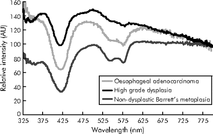
Representative spectra obtained with ESS. AU, arbitrary units.
MOLECULAR DIFFERENCES BETWEEN ENTEROCYTES ON THE CRYPT AND VILLUS
Four lineages of small intestinal epithelial cells arise from stem cells near the base of the crypts. They comprise enterocytes, paneth cells, goblet cells and enteroendocrine cells. All except paneth cells migrate towards the villi, dividing as they do so. Enterocytes secrete fluid while in the crypts but develop the capacity to absorb nutrients and water once on the villi. Little is known about the genetic changes that underlie this switch in function. Gassler and colleagues investigate this by dissecting enterocytes from human crypts and villi by laser capture microdissection and comparing gene expression using Affymatrix X3P arrays. They find that cells on villi express more genes associated with nutrient transport and metabolism than those from crypts. Villus enterocytes also express more cell cycle inhibitors consistent with their inability to divide. On the other hand, enterocytes from the crypt express more genes associated with transcription and translation.
See p 1084

The crypt/villus axis of the small intestine. LMD, laser microdissected areas.
PROBIOTIC TREATMENT PREVENTS STRESS INDUCED HYPERSENSITIVITY TO DISTENSION
Patients with irritable bowel syndrome (IBS) often report that stress induces symptoms. A key feature of IBS is hypersensitivity to rectal distension. This study assessed such sensitivity in an animal model using partial restraint stress, which in previous studies has been shown to increase gut permeability. The authors administered a lactobacillus probiotic, which releases nitric oxide (NO) in the colonic lumen and is known to exert anti-inflammatory effects. The probiotic reduced both visceral hypersensitivity and the increased gut permeability induced by stress (see figure), an effect which was abolished by the NO scavenger haemoglobin. The probiotic also reduced the stress induced increased phosphorylation of myosin light chain (MLC). Phosphorylation of MLC causes cytoskeleton contraction and hence increase in colonic permeability. This study suggests new ways in which probiotics may benefit IBS, although whether these changes will be observed in humans remains to be proved.
See p 1090

Effect of Lactobacillus farciminis on partial restraint stress (PRS) induced increase in gut permeability. Adding haemoglobin to scavenger NO blocked the benefit of the probiotic.
DOES A NEGATIVE SCREENING COLONOSCOPY EVER NEED TO BE REPEATED?
Colonoscopy is the current gold standard for screening colorectal cancer. However, colonoscopy is expensive, time consuming, uncomfortable for the patient and dangerous if performed by an unskilled endoscopist. Therefore, the fewer colonoscopies that need be performed, the better. Brenner and colleagues investigated the risk of developing colorectal cancer in patients whose initial colonoscopy is negative. They found that those screened with a negative colonoscopy had a 74% lower risk of colorectal cancer, even if the colonoscopy was performed 20 years before. Risks were particularly low for cancer of the rectum and sigmoid colon. They propose that screening colonoscopy need only be repeated after 20 years if the initial colonoscopy is negative. If this recommendation is supported by other studies the savings in terms of patient comfort and cost will be substantial.
See p 1145
IMPORTANCE OF ENDOSCOPIC RESECTION OF SPORADIC ADENOMA IN ULCERATIVE COLITIS
Endoscopic surveillance looking for dysplasia in ulcerative colitis may sometimes reveal an adenoma. This series describes 148 patients with chronic ulcerative colitis and an adenoma. In 87 of the patients, the adenoma was removed completely endoscopically and of these only 4 developed low grade intra epithelial neoplasia at a later stage. Sixty of the patients, however, did not undergo endoscopic removal. Twenty nine of these developed a neoplasm in the same segment of the colon previously containing the adenoma. As can be seen in the figure, the risk of developing intra-epithelial and neoplasia was substantially increased in those not undergoing polypectomy. The message is clear that detecting an adenoma in such patients mandates endoscopic removal and regular further surveillance to detect and treat subsequently developing neoplasia in adequate time.
See p 1151

Kaplan-Meier survival curve showing reduced risk of developing colitis associated intraepithelial neoplasia after polypectomy compared with medical treatment alone.
INHIBITION OF COX-2 REDUCES INFLAMMATION AND MACROPHAGE INFILTRATION
Chronic pancreatitis is characterised by inflammation and necrosis leading to fibrosis, stone formation and ultimately the destruction of the pancreas, causing diabetes, malabsorption and cancer. The role of COX-2 in the pathogenesis of chronic pancreatitis is not well understood. The authors investigate this using the well known WBN/Kob mouse model of chronic pancreatitis and inhibited COX-2 by feeding the mice rofecoxib. They found that rofecoxib causes a significant reduction and delay in inflammation and fibrosis. In separate experiments they demonstrated that rofecoxib reduces fMLP-stimulated migration of cultured macrophages. These experiments strongly suggest that the efficacy of COX-2 inhibitors in chronic pancreatitis should be tested by randomised controlled clinical trial.
See p 1165
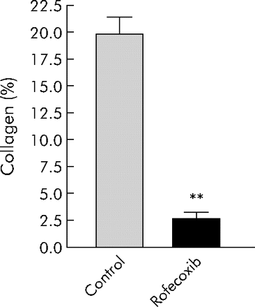
Rofecoxib reduces collagen formation in a mouse model of chronic pancreatitis.
Risk reduction of proximal colorectal cancer according to location and recorded completeness of colonoscopy
Linked Articles
- Barrett's oesophagus
- Small intestine
- Neurogastroenterology
- Colorectal cancer
- Colorectal cancer
- Pancreatitis