Article Text

Statistics from Altmetric.com
Luminal GI
Bowel cancer screening in England: the first million
In April 2006 the Department of Health in England agreed funding for a national programme of biennial gFOBt screening of 60–69-year-olds. Roll out of the programme started in July 2006 and was complete by January 2010. In this issue of Gut, Logan et al report the uptake and early outcomes of the first million people screened. Almost 2.1 million were invited to participate, and as expected, women had a higher return of tests than men (54.4% vs 49.6%). Of the 1.08 million returning tests 2.5% of men and 1.5% of women had an abnormal test. Seventeen thousand five hundred and eighteen (10 608 M, 6910 F) underwent investigation, with 98% having a colonoscopy as their first investigation. Cancer (n=1772) and higher risk adenomas (n=6543) were found in 11.6% and 43% of men and 7.8% and 29% of women investigated, respectively. Seventy one percent of cancers were ‘early’ (10% polyp cancer, 32% Dukes A, 30% Dukes B; see table 1) and 77% were left-sided (29% rectal, 45% sigmoid) with only 14% being right-sided compared with expected figures of 67% and 24% for left and right side from UK cancer registration. These early data are encouraging and suggest that the intended target of 16% reduction in overall bowel cancer mortality is achievable (see page 1439 ).
Duke's stage of bowel cancers detected after first investigation of first million people screened in England
The promise of microRNA's as biomarkers for colorectal cancer
MicroRNAs (miRNA) are short (approximately 20 basepairs in length) non-coding RNA's that affect the expression of genes. They have considerable potential as prognostic biomarkers and therapeutic targets in cancer, but that potential still remains to be realised. Yanlei and colleagues have profiled the miRNAs in human colorectal cancers and found that miR-150 is a promising tissue-based predictive and prognostic marker (see figure 1). They carried out a series of carefully done studies that validated their results. This study opens the possibility that miR-150 will be a molecular marker that will enhance our ability to precisely treat patients with colorectal cancer (see page 1447 ).
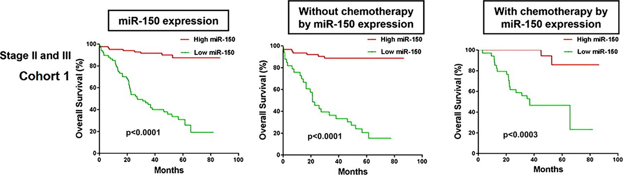
Association of miR-150 expression levels with chemotherapy outcome in the CRC patients with TNM stage II or III in the cohort 1 and cohort 2. A. For the 195 CRC patients with stage II or III, low miR-150 expression was associated with poor survival for those who received chemotherapy in the cohort 1.
Identifying driver genes in colorectal cancer
Colorectal cancer arises as the consequence of mutations in colon epithelial cells that deregulate signalling pathways in these cells. One of the most common signalling pathways that is activated in colorectal cancer is the Wnt signalling pathway. This pathway induces the expression of a variety of genes that appear to be important promoters of tumour cell growth, including CYCLIND1, CMYC, CDK4, etc. Expression of one of these genes, ASCL2, is present in almost 80% of colorectal tumours suggesting it helps drive tumour formation in the colon. Reed et al have now created a sophisticated mouse model to demonstrate that Ascl2 is not of likely importance in mediating the effects of increased Wnt signalling, which provides valuable information regarding the search for ‘druggable’ targets for novel therapies for colorectal cancer (see figure 2). Studies such as these are critical for sorting out the relevant effects of gene mutations on the biology of cancers (see page 1435 ).

Ascl2-TG mice were interbred with ApcMin mice to generate the required ApcMin and ApcMinAscl2-TG cohorts. (A) Kaplan-Meier survival curve of aged cohorts of ApcMin (n=21) and ApcMinAscl2-TG (n=18) mice, demonstrating that no significant differences in survival of the two cohorts.
Hepatology
Does the role of insulin resistance change in HCV genotype 1 patients?
Insulin resistance is a negative predictive factor for sustained viral response (SVR) to treatment with interferon and ribavirin in patients with chronic hepatitic C virus infection. Direct antiviral agents may improve insulin sensitivity together with a decrease of viral load as recently reported in this journal (Moucari R, et al. Gut 2010;59:1694–8).The interesting paper by Serfaty et al (page xxxx) investigates the predictive role of metabolic factors in patients receiving a triple therapy with telaprevir in addition to interferon and ribavirin. This subanalysis of an international phase II trial found that insulin resistance was not a predictor of SVR and moreover, that at 24 weeks follow-up insulin resistance was significantly lower in SVR patients (figure 3). Thus, telaprevir may overcome insulin resistance and an as yet important predictive factor of response seems irrelevant for treatment with triple therapy. These results were obtained in young patients without advanced liver disease and thus need to be confirmed in further trials (see page 1473).

Association between log HOMA-IR and virological response during telaprevir-based treatment. HOMA-IR, homoeostatic model assessment-insulin resistance; LN, natural logarithm; SVR, sustained virological response; T0, baseline; W, week; FU24, follow-up week 24.
The role of immunohistochemical markers for the diagnosis of small hepatocellular carcinoma
The early diagnosis of hepatocellular carcinoma (HCC) is crucial for the success of treatment and for patient survival. Unfortunately, imaging often fails to detect very small HCC in a cirrhotic liver and therefore biopsy is considered the gold standard for nodules smaller than 2 cm. Histology can be flawed by false negative results. Therefore, some immunohistochemical markers have been proposed to improve the accuracy of histology. Several studies investigated the value of such markers in tissues known to be HCC but prospective analysis as demanded in this journal (Lamerz R. Gut 2011;60:881–2) has not been performed so far. This important study from Bruix et al for the first time prospectively evaluated a panel of three recently proposed immunohistochemical markers of HCC. They found a sensitivity of 60% and a specificity of 100% when at least two markers were positive (table 2). However, since the expert pathologists involved in this study performed extremely well with only 7.5% false negative results the additional value of the marker panel was poor. The conclusion may be that immunohistochemical markers could be valuable particularly for scarce biopsy material, difficult cases and less experienced pathologists (see page 1481 ).
Final diagnosis of the 60 nodules according to size at baseline ultrasound (≤10 mm; 11–15 mm and 16–20 mm)
Diagnostic accuracy for detection of hepatocellular carcinoma using one, two or three of the markers under study
Linked Articles
- Original articles
- Original articles
- Original articles
- Colon
- Original articles