Article Text

Statistics from Altmetric.com
Luminal gastroenterology
What is the incidence of oesophageal adenocarcinoma in non-dysplastic Barrett's?
The most widely cited annual incidence of oesophageal adenocarcinoma (OAC) in Barrett's oesophagus (BO) is 0.5% but this estimate did not account for the presence of baseline dysplasia. In this issue of Gut, Desai et al report an updated meta-analysis of observational studies and provide an accurate estimate of the risk of OAC in patients with BO who were free of dysplasia at baseline. The 57 included studies comprised 11 434 patients and 58 547 patient-years of follow-up. The pooled annual incidence of OAC was 0.33% (95% CI 0.28% to 0.38%). During surveillance, patients with non-dysplastic BO may be at least 10 times more likely to die from an unrelated cause than to develop OAC. Among 16 studies that provided information on patients with short-segment BO, the annual incidence of OAC was only 0.19%. The incidence of OAC in non-dysplastic BO is around 1 per 300 patients per year while that in short-segment BO is under 1 per 500 patients per year. These estimates are much lower than previously thought and suggest that surveillance strategies for patients with non-dysplastic BO, particularly those with short segments, may need to be reconsidered (see page 970).
Microbial fingerprinting and IBS subtypes
Irritable bowel syndrome (IBS) onset may follow enteric infections. The gut microbiota is emerging as a key determinant of colonic health and disease. Various changes in the microbiota have been described in IBS but their primacy has not been defined. In this issue of Gut, Jeffery et al performed a detailed analysis of the faecal microbiota in IBS and control subjects and correlated the findings with key clinical and physiological parameters. The authors report that a detailed assessment of the faecal microbiota in IBS does not reveal a uniform change in the microbiota. However, analysis of microbial populations in IBS reveals distinct clusters, of which some overlap with normal controls while others are quite different. An increased Firmicutes:Bacteroidetes ratio best characterises those IBS subjects who differ from normal populations (figure 1). Furthermore, the microbial signature is related to the clinical phenotype in a subset of IBS patients. This work suggests that microbial fingerprinting may help to identify IBS subpopulations with differing prognosis and varied therapeutic responses (see page 997).

Visualisation of Taxonomic levels. Pie charts showing proportion of reads in each phylum (top) and genus (bottom) for the controls, normal-like IBS and the IBS clusters.
Figuring out ways to make colonoscopy better
Colonoscopy is central to colorectal cancer screening and its effectiveness in colon cancer prevention and early detection depends on colonoscopy quality. Using the NHS Bowel Cancer Screening Programme (BCSP), which offers biennial faecal occult blood testing and colonoscopy to those who are faecal occult blood testing positive, Lee and colleagues have examined measures to assess and maintain the quality of colonoscopy, which are critical to optimise the use of this resource (figure 2). They found that the NHS BCSP provides high quality colonoscopy as demonstrated by high caecal intubation rates, high adenoma detection rates, high comfort scores and low adverse event rates. The quality of colonoscopy appeared to be achieved by ensuring BCSP colonoscopists meet a high standard prior to commencing screening and through ongoing quality assurance assessment, particularly of adenoma detection measures. These findings provide the basis for standards to which colonoscopy will be held in the future (see page 1050).
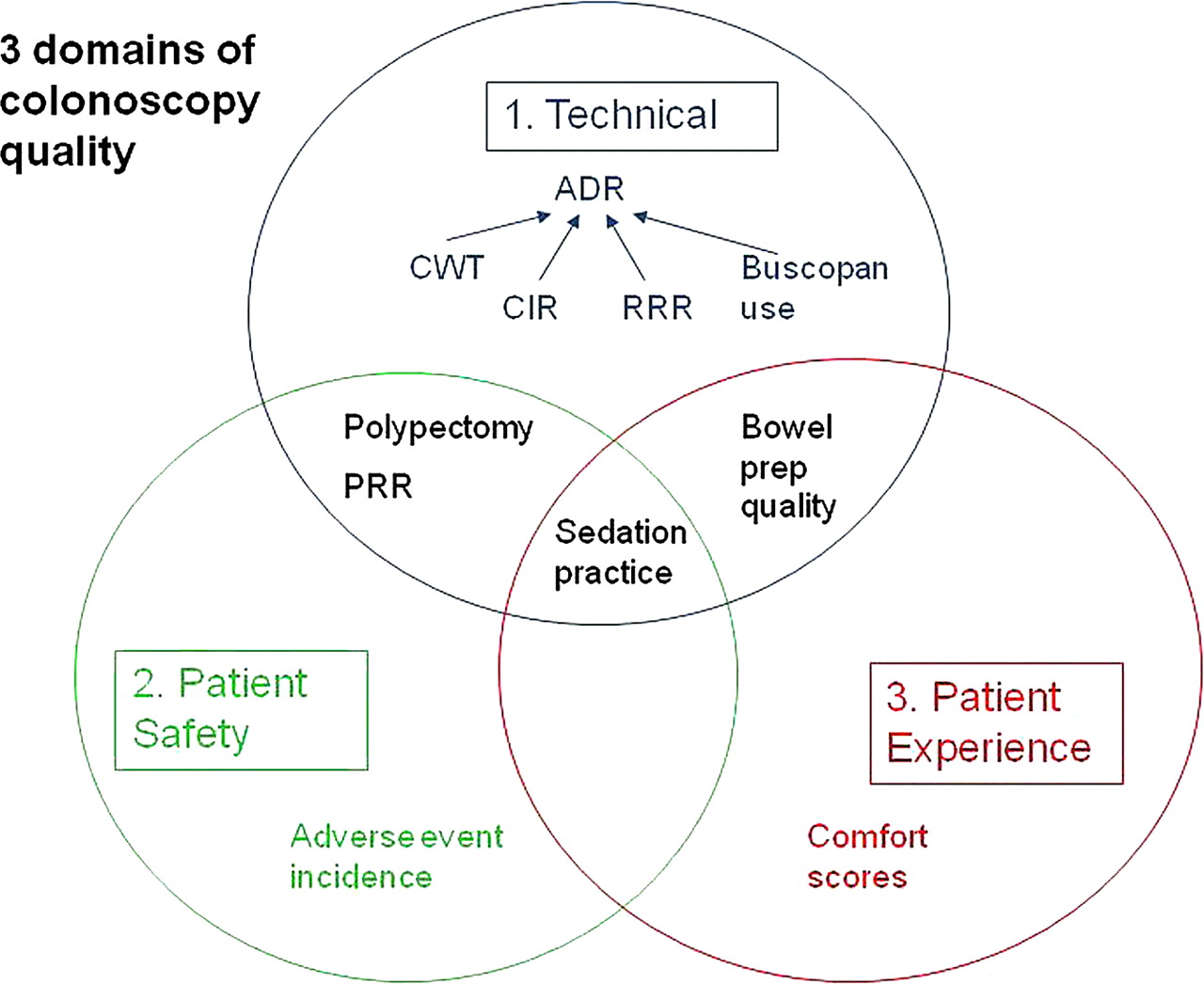
Three domains of colonoscopy quality assessment.
Why does red meat associate with an increased risk of colorectal cancer?
Colon cancer is a leading cause of cancer deaths in Western countries and is associated with diets high in red meat. Haeme, the iron-porphyrin pigment of red meat, induces cytotoxicity of gut contents and damages colon surface epithelium. The resulting compensatory hyperproliferation leads to epithelial hyperplasia, which has been proposed to increase colon cancer risk. Despite this level of understanding regarding how haeme may affect colon cancer risk, it is not known what specific genes and signalling pathways are actually driving these effects on proliferation. Ijssennagger and colleagues have now identified many of the potential mechanisms driving this process. They have found that the cells at the surface of the colon mucosa are the colon cells that are damaged by haeme and that these cells appear to drive the proliferation of the crypt cells by activating signalling pathways that drive proliferation in the crypt cells. Their results suggest that these genes could be used as biomarkers for diet-related colon cancer risk (see page 1041).
Hepatology
T cell impairment in patients after successful treatment of chronic viral hepatitis C
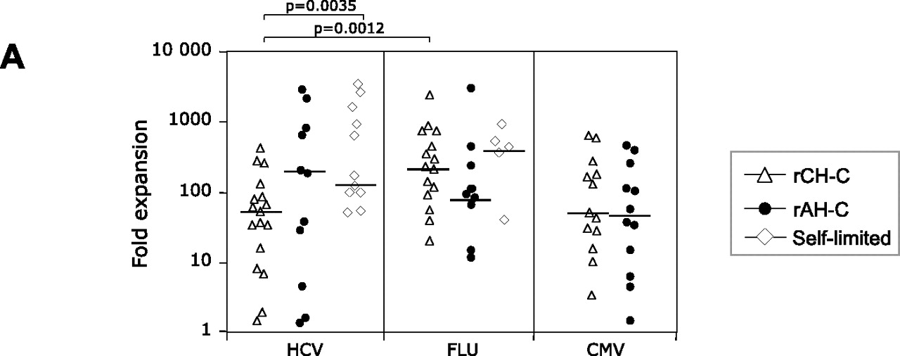
It is well known that function of CD8 T cells is reduced in patients with hepatitis C virus infection. This interesting study from Carlo Ferrari's group (see page 1076) investigates the restoration of T cell function following sustained viral response to treatment. The authors found that T cell function was restored to a lesser degree in patients with chronic as compared to patients with acute infections and did not fully recover in either group (figure 3). These findings have important implications for immunomodulatory strategies in these patients.

Expansion capacity and cytotoxicity of HCV-specific CD8-cells are efficient in rAH-C but not in rCH-C patients. In vitro expansion of HCV-, Flu- and CMV-specific CD8-cells after 10 days of peptide stimulation (panel A).
A novel prognostic score for acute liver failure
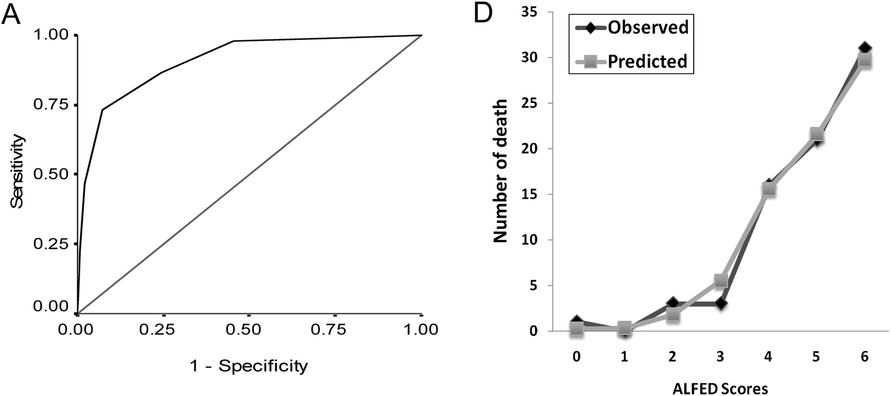
Prognosis of mortality is crucial for the decision to list a patient for urgent liver transplantation. Several scores have been developed to predict whether a patient will need transplantation. These scores with the Kings College criteria being the most widely used have rather good positive predictive values (‘this patient will die without transplant’), but poor negative predictive values (‘this patient will survive without transplant’). This intriguing study from India (see page 1068) proposes a novel score based on the change of four parameters during 3 days: INR, serum bilirubin, arterial ammonia and hepatic encephalopathy. This score had an excellent accuracy also in a validation cohort (figure 4A and B). The authors report a positive predictive value of their score of 85% compared to 68% obtained with the Kings College criteria and a negative predictive value of 87% which also exceeded the Kings score performance. Two aspects deserve special attention: patients did not have the option of liver transplant which supports the strength of the predictive values. Moreover, acute liver failure was due to viral hepatitis E in many patients. This is a rare cause of liver failure in Western countries and thus the validity of the score needs to be confirmed in other cohorts.
Receiver operating characteristic (ROC) curves showing mortality and the ALFED model scores in the derivation cohort (A). The observed and predicted number of death in ALF patients for all risk strata of ALFED model in the validation cohort (B).
Linked Articles
- Colon
- Colon
- Hepatology
- Hepatology
- Stomach
- Oesophagus
- Irritable bowel syndrome