Article Text

Abstract
Introduction There is an urgent need to create tools to quantify collagen in liver fibrosis to facilitate stratification of disease and development of anti-fibrotic agents. Multiphoton microscopy enables imaging of unstained biopsies using endogenous sources of non-linear signals such as Two-Photon Excitation Fluorescence (TPEF) and Second Harmonic Generation (SHG). SHG allows specific detection of non-centrosymmetric structures such as fibrillar collagen, mainly type I. The SHG score is a measure of relative collagen area and is obtained by post-acquisition SHG/TPEF image processing. We have assessed the ability of our method to quantify collagen in advanced fibrosis due to CHC, with respect to Ishak stage (IS).
Methods Biopsies from patients with advanced fibrosis (IS ≥3) were selected from 1 centre in the Trent Study of Patients with Hepatitis C Virus, a prospective cohort study formed in 1991. Index biopsies prior to 2008 were selected and notes reviewed for subsequent liver related outcomes (LRO). LRO was defined as variceal bleed, ascites, encephalopathy, HCC or liver related death. SHG was measured on 4μm FFPE sections. A mask of the biopsy area was created with TPEF. Image processing was performed by two independent researchers, blinded to Ishak stage, using in-house macros and each using different software (Image J & Matlab). PASW 17.0 was used for statistical analysis.
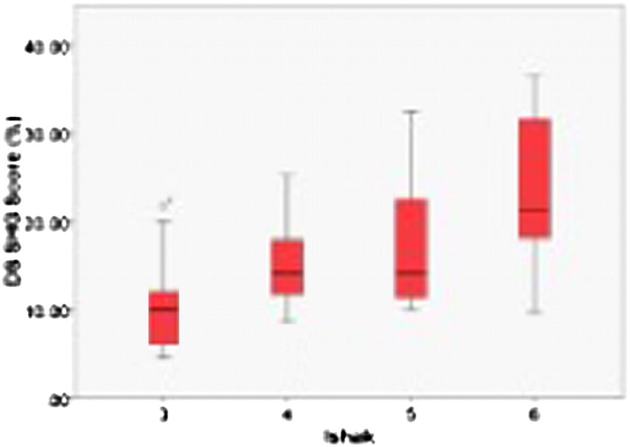
Results The SHG score was acquired in 58 of 83 biopsies (66%). 25 were excluded due to signal artefact from paraffin, obscuring SHG signal from collagen. There was no significant difference in scoring by two researchers (p<0.001). The median SHG score was 15.96% (IQR 11.3–21.3%). Abstract PMO-150 figure 1 shows the median SHG score for each IS. SHG signal increased with disease severity (IS3:10.1%; IS4:14.1%; IS5:14.1%; IS6:21.2%). LRO occurred in 15 patients after a median of 57 months post-biopsy. The mean SHG score at index biopsy was 19.1% in those with, and 16.6% in those without subsequent LRO (non-significant difference, p>0.05).

Conclusion SHG has proved to be a valuable method of quantifying collagen in liver fibrosis and does not require standard histochemical stains. Further development of this quantitative measure may result in a tool to assess response to anti-fibrotic therapy and progression to clinical endpoints.
Competing interests None declared.
References 1. Gailhouste L, et al. Fibrillar collagen scoring by second harmonic microscopy: A new tool in the assessment of liver fibrosis. J Hep 2010;52:398–406.
2. Guilbert T, et al. A robust collagen scoring method for human liver fibrosis by second harmonic microscopy. Opt Expr 2010;18:25794–807.