Article Text

Abstract
Introduction Directly-acting antiviral (DAA) therapy achieves sustained virological response (SVR) in over 90% of those with chronic hepatitis C (CHC), but even in those with SVR, the risk for liver related morbidity and mortality persists albeit at a lower level. It is hypothesised that the residual risk of liver related outcome in those with SVR is related to the progression or non-regression of structural and haemodynamic changes of advanced liver disease at the time of HCV therapy. Here, we investigate the effect of DAAs on the hepatic architecture and splanchnic haemodynamics in patients with decompensated cirrhosis, using quantitative MRI techniques.
Method We prospectively recruited patients receiving DAAs for CHC related decompensated cirrhosis from the East Midlands hub of the Early Access Programme commissioned by NHS England. MRI was performed before and at the end of a 12-week treatment with Daclatasvir, Sofosbuvir and Ribavarin. A respiratory-triggered inversion recovery scheme was used to measure the longitudinal (T1) relaxation time of the liver employing a fat suppressed Echo Planar Imaging-acquisition. Phase-contrast (PC)-MRI was used to assess the velocity, area and bulk flow in the splanchnic circulation without any intravenous contrast agents.
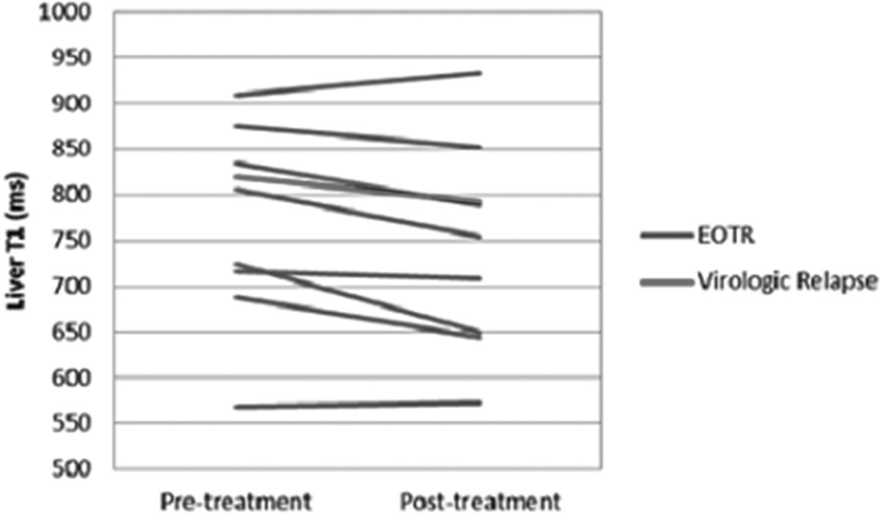
Results 9 patients have undergone baseline and post-treatment MR scans and end-of-treatment response (EOTR) was achieved in all 9 patients, but one patient has had virologic relapse. There was a significant reduction in the T1 relaxation time in patients with SVR (baseline T1: 765 ± 112 ms; post-treatment 737 ± 118 ms; p = 0.043) (Figure 1). There were no significant changes in the hepatic artery, portal vein, superior mesenteric artery and splenic artery flow. The changes in the Model for End-Stage Liver Disease (MELD) and United Kingdom Model for End-Stage Liver Disease (UKELD) score are shown in Table 1.

Conclusion Treatment of decompensated CHC related cirrhosis with DAA is associated with early improvement in the MR markers of liver architecture. These early changes are likely to reflect the reduction in the inflammation associated with EOTR and is evident before any improvement in conventional liver function tests. This novel quantitative MR methodology will allow us to non-invasively monitor HCV related liver disease.
Disclosure of interest None Declared.