Article Text

Abstract
Introduction Detection of early neoplasia in Barrett’s oesophagus (BO) by white-light endoscopy is challenging due to the inconspicuous nature of dysplasia. Molecular imaging using fluorescently labelled wheat-germ agglutinin (WGA) is a promising tool as this topically applied imaging agent shows lower binding to dysplastic versus non-dysplastic BO.1 However in an endoscopy setting, the detection of fluorescence in the blue/green range is limited by high-levels of tissue autofluorescence. This limitation can be overcome by using near infra-red (NIR) imaging. We aimed to assess in an ex-vivo model the feasibility of WGA-based NIR imaging for detection of dysplasia in BO.
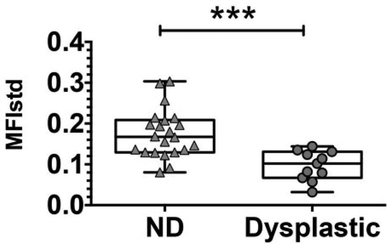
Methods we recruited patients with early BO-related neoplasia undergoing endoscopic mucosal resection (EMR). Freshly collected EMR specimens were sprayed with WGA-IR800CW and then imaged with a high-sensitivity NIR camera. Fluorescence images were captured and up to two punch biopsies were collected from each EMR under fluorescence guidance. The EMRs were paraffin embedded, cut every 2 mm and processed for histopathological assessment. Each section was scored by an expert GI pathologist every 1 mm to construct a pathology grid, which was manually co-registered with the fluorescence image. The mean fluorescence intensity (MFI) of cells in dysplastic and non-dysplastic areas was compared by the Wilcoxon matched-pairs signed rank test. Only EMR specimens with at least one dysplastic gland were included in the analysis. In addition, the MFI of punch biopsies taken from dysplastic and non-dysplastic areas was also compared by Mann-Whitney test.
Results Ten patients were recruited at a single centre. A total of 18 EMR specimens and 33 punch biopsies were collected, of which 10 were dysplastic. In the whole EMR analysis, we found a significantly lower MFI for dysplastic versus non-dysplastic areas (P = 0.0012), in accordance with the reported reduced binding of WGA to neoplastic BO epithelium. Similarly, we found a nearly 2 fold reduction in the MFI of punch biopsies taken from dysplastic as compared to non-dysplastic (ND) areas (P = 0.0002) (Figure 1).

Conclusion WGA-based NIR imaging is an effective method for differentiating dysplastic from non-dysplastic BO mucosa ex vivo, which reduces the effects of tissue autofluorescence. In-vivo studies are now required to test the efficacy of this method for detecting dysplasia during endoscopic surveillance.
Reference 1 Bird-Lieberman, et al. Nat Med 2012.
Disclosure of Interest None Declared