Article Text

Abstract
Aim: A subset of functional dyspepsia patients respond to acid suppressive therapy, but the prevalence of non-erosive reflux disease in functional dyspepsia and its relevance to symptoms have never been established. The aim of the present study was to study 24 hour pH monitoring in consecutive functional dyspepsia patients.
Methods: A total of 247 patients with dyspeptic symptoms (166 women, mean age 44 (SEM 1) year), with a negative upper gastrointestinal endoscopy and without dominant symptoms of heartburn participated in the study. In all patients, the severity of dyspeptic symptoms and the presence of heartburn was assessed by a questionnaire and a 24 hour oesophageal pH monitoring study was performed. All patients underwent a gastric emptying breath test and in 113 a gastric barostat study was performed.
Results: Abnormal pH monitoring (acid exposure >5% of time) was found in 58 patients (23%). Of 21 patients with a positive heartburn questionnaire, 76% had pathological pH monitoring, while this was the case in only 18.5% of patients with a negative heartburn questionnaire. Demographic characteristics and the prevalence of other pathophysiological mechanisms did not differ between heartburn negative patients with normal or abnormal acid exposure. Pathological acid exposure in heartburn negative patients was associated with the presence of epigastric pain (65 v 84%, p<0.005) and of moderate or severe pain (48 v 69%, p = 0.005).
Conclusion: Pathological oesophageal acid exposure is only present in a subset of heartburn negative functional dyspepsia patients, which are characterised by a higher prevalence of epigastric pain.
- GORD, gastro-oesophageal reflux disease
- MDP, minimal distending pressure
- functional dyspepsia
- gastro-oesophageal reflux disease
- heartburn
- pH monitoring
Statistics from Altmetric.com
Functional dyspepsia and gastro-oesophageal reflux disease (GORD) are among the most prevalent upper gastrointestinal disorders.1,2 According to the Rome consensus, functional dyspepsia is defined as persistent or recurrent pain or discomfort centred in the upper abdomen, with no evidence of organic disease that is likely to explain the symptoms.3 It is suggested that these symptoms differentiate dyspepsia from GORD, in which heartburn is the predominant symptom.3 Identifying patients with GORD is important, as effective management strategies, based on acid suppressive therapy, are available for these patients.4
Studies have reported that a subgroup of functional dyspepsia patients also respond to acid suppressive therapy,5,6 but elimination of GORD patients from these studies was not always adequate, as they also included patients with “reflux-like dyspepsia”. Separating GORD from functional dyspepsia is hampered by a number of confounding factors. As the majority of patients with GORD will not have erosive oesophagitis, endoscopy is an insensitive test for reflux disease.7 It has been proposed that symptom evaluation is the most effective method to recognise reflux induced symptoms, based on recognition of heartburn as the most typical symptom.5 However, studies have revealed that both patients and physicians have difficulties in recognising heartburn,6 and it has been suggested that a descriptive questionnaire may help to improve the identification of functional dyspepsia patients with underlying heartburn.8 Indeed, a structured questionnaire to recognise heartburn was able to identify putative functional dyspepsia patients that were likely to respond to acid suppressive therapy.6,9
It has also been suggested that functional dyspepsia symptoms, in the absence of heartburn, may actually be an atypical manifestation of GORD.10,11 In these patients, a heartburn questionnaire is unlikely to differentiate GORD from functional dyspepsia. Oesophageal pH monitoring may be the method of choice to demonstrate reflux in patients with atypical manifestations.12 Few studies reported on pH monitoring in functional dyspepsia patients, and they all failed to adequately exclude patients with heartburn.13,14 Hence, the true prevalence of non-erosive reflux disease in typical functional dyspepsia, after exclusion of heartburn, and its relevance to symptoms have not been established.
The aim of the present study therefore was to study the prevalence of abnormal 24 hour oesophageal pH monitoring and its relevance to the symptom pattern in presumed functional dyspepsia patients. We also examined whether the use of a structured heartburn questionnaire is able to predict abnormal pH monitoring in presumed functional dyspepsia patients.
MATERIALS AND METHODS
Study subjects
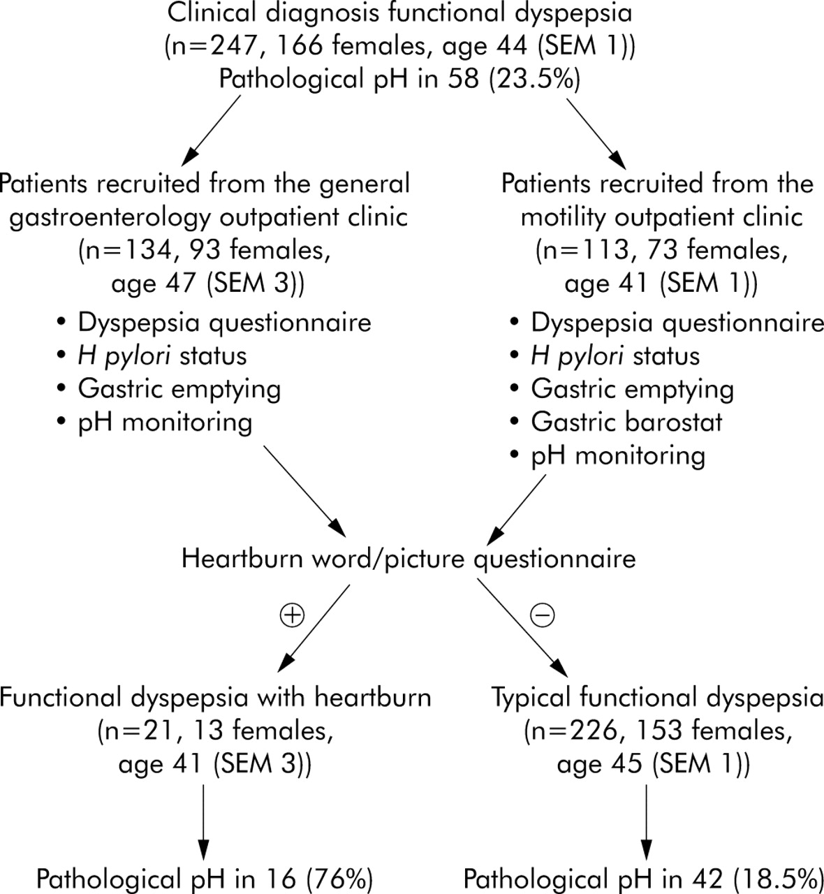
Consecutive patients with a new clinical diagnosis of functional dyspepsia according to the Rome II criteria were recruited. At the time of the study, acid suppressive drugs were only reimbursable in Belgium for peptic ulcer disease or for erosive oesophagitis. Patients presented to the general gastroenterology outpatient clinic and to the motility outpatient clinic because of pain or discomfort centred in the upper abdomen, and all underwent careful history taking and clinical examination, upper gastrointestinal endoscopy, routine biochemistry, and upper abdominal ultrasound (fig 1). Inclusion criteria were the presence of dyspeptic symptoms for at least three months, in the absence of organic, systemic, or metabolic disease. Exclusion criteria were the presence of oesophagitis, gastric atrophy or erosive gastroduodenal lesions on endoscopy, heartburn as a predominant symptom (that is, when retrosternal burning sensation was the most bothersome symptom), a history of peptic ulcer, major abdominal surgery, underlying psychiatric illness, and the use of non-steroidal anti-inflammatory drugs, steroids, or drugs affecting gastric acid secretion. During upper gastrointestinal endoscopy, biopsies were taken from the antrum and the corpus to stain with cresyl violet for the presence of Helicobacter pylori. A psychiatrist ruled out anorexia nervosa in patients with weight loss in excess of 5% of the initial body weight. All drugs potentially affecting gastrointestinal motility were discontinued at least one week before the questionnaires and tests of gastric and oesophageal function. Informed consent was obtained from each participant. The Ethics Committee of the University Hospital had previously approved the protocol.

Flow chart depicting the selection of functional dyspepsia patients, their separation according to the heartburn word-picture questionnaire and the result of 24 hour oesophageal pH monitoring.
Symptom questionnaire
Before the 24 hour pH studies, each patient completed a functional dyspepsia questionnaire as reported and validated previously.15–17 The patient was asked to grade the intensity (0–3; 0 = absent, 1 = mild, 2 = moderate, and 3 = severe, interfering with daily activities) of epigastric pain and of seven discomfort symptoms (bloating, postprandial fullness, early satiety, nausea, vomiting, belching, and epigastric burning).3 Also, the amount of weight lost since the onset of the symptoms was noted.
In addition, all patients completed a four item heartburn questionnaire which consisted of the following questions:
-
Do you frequently experience a rising, spreading uncomfortable feeling behind your breastbone?
-
Is this feeling often combined with a burning sensation in your chest?
-
Do antacids relieve your symptoms?
-
Have you had your symptoms during four or more days in the last week?
It has been shown that a positive answer to all four items is associated with an 85% probability of oesophagitis or pathological pH monitoring.9 Furthermore, items 1–3 were shown to be associated with a favourable response to a proton pump inhibitor in reflux disease and in dyspepsia.6,9
Ambulatory pH monitoring
Ambulatory oesophageal pH monitoring was performed using an antimony pH electrode with a separate skin reference electrode (Synectics Medical, Stockholm, Sweden). The data were stored on a portable digital recorder (Digitrapper MkIII, Synectics Medical, Stockholm, Sweden). Before each study, the pH probe was calibrated in buffer solutions of pH 7 and 1. An episode of acid reflux was defined as a decrease in oesophageal pH to less than 4 during more than 10 seconds.18
The pH probe was introduced via a nasal orifice into the oesophagus 5 cm proximal to the lower oesophageal sphincter, defined by previous stationary oesophageal manometry. The probe was then attached with adhesive tape to the subject’s nose and cheek. In addition, appropriate positioning in the oesophagus was confirmed by brief fluoroscopy. A data collection device was connected to the probe and worn in a belt on the patient’s waist. Ambulatory monitoring lasted for approximately 22 hours after which the probe was removed and the data transferred to a personal computer for analysis. Patients recorded the time of food or fluid consumption and posture changes on a diary card. They were instructed to stay upright during the daytime. Patients were asked not to eat between meals and to preferably drink water during the recording. As dyspeptic symptoms do not have acute onset and disappearance, the symptom marker was not used during pH monitoring.
Gastric emptying studies
Gastric emptying for solids was measured in all patients, using the previously validated 13C octanoic acid breath test.19 Briefly, all studies were carried out in the morning after an overnight fast. The test meal consisted of 60 g of white bread, one egg, the yolk of which was doped with 74kBq of 13C-octanoic acid sodium salt, and 300 ml of water. Breath samples were taken before the meal and at 15 minute intervals for a period of 240 minutes postprandially.
Sensitivity to gastric distention and gastric accommodation
Sensitivity to gastric distention and gastric accommodation to a meal were studied using a gastric barostat. Following an overnight fast of at least 12 hours, a double lumen polyvinyl tube (Salem sump tube 14Ch, Sherwood Medical, Petit Rechain, Belgium) with an adherent plastic bag (1200 ml capacity; 17 cm maximal diameter), finely folded, was introduced through the mouth and secured to the subject’s chin with adhesive tape. The position of the bag in the gastric fundus was checked fluoroscopically.
The polyvinyl tube was then connected to a programmable barostat device (Synectics Visceral Stimulator, Stockholm, Sweden). To unfold the bag, it was inflated with a fixed volume of 300 ml of air for two minutes with the study subject in a recumbent position, and again deflated completely. The subjects were then positioned in a comfortable sitting position with the knees bent (80°) and the trunk upright in a specifically designed bed.
After a 30 minute adaptation period, minimal distending pressure (MDP) was first determined by increasing intrabag pressure by 1 mm Hg every three minutes until a volume of 30 ml or more was reached. This pressure level equilibrates the intra-abdominal pressure. Subsequently, isobaric distensions were performed in stepwise increments of 2 mm Hg starting from MDP, each lasting for two minutes, while the corresponding intragastric volume was recorded. Subjects were instructed to score their perception of upper abdominal sensations at the end of every distending step, using a graphic rating scale that combined verbal descriptors on a scale graded 0–6.15,16,20,21 The end point of each sequence of distensions was established at an intrabag volume of 1000 ml, or when the subjects reported discomfort or pain (score 5 or 6). We previously established that more complex distending protocols yield similar results.21
After a 30 minute adaptation period with the bag completely deflated, the pressure level was set at MDP+2 mm Hg during at least 90 minutes. After 30 minutes, a liquid meal (200 ml, 300 kcal, 13% proteins, 48% carbohydrates, 39% lipids, Nutridrink, Nutricia, Bornem, Belgium) was administered. Gastric tone measurement was continued for 60 minutes after the meal.
Data analysis
The data were analysed with the aid of commercially available software (Gastrosoft Inc Synectics Medical, Irvine, TX, USA). The following acid reflux variables were obtained from computerised analysis: number of reflux episodes, number of reflux episodes lasting longer than five minutes, fraction of time of oesophageal acid exposure. The recording was divided in upright and supine periods. In keeping with the normal values established at our centre, pathological acid exposure was defined as oesophageal acid exposure more than 5% of time.18 Studies in asymptomatic controls demonstrated that the 95th percentile for oesophageal acid exposure in asymptomatic controls is 4.7% for total time and 5.1% for upright reflux.18 In addition, similar to the study of Small et al, we also considered the presence of borderline pathological acid exposure (% of time acid exposure between 4% and 6%).13
Gastric half emptying time (t½) was calculated from the 13CO2 content of breath samples as previously described.19 Delayed emptying was defined as t½ above the 95% confidence interval in healthy volunteers.22
In the gastric sensitivity studies, for each two minute distending period, the intragastric volume was calculated by averaging the recording. Discomfort threshold was defined as the first level of pressure and the corresponding volume that provoked a score of 5 or more. Pressure thresholds were expressed as pressures relative to MDP.15 Hypersensitivity to gastric distention was defined as a distending pressure inducing discomfort below the mean –2 SD in healthy volunteers.15
Gastric tone before and after administration of the meal was measured by calculation of the mean balloon volume for consecutive five minute intervals. The meal induced gastric relaxation was quantified as the difference between the average volumes during 30 minutes before and 60 minutes after the administration of the meal.16 Impaired accommodation to a meal was defined as a meal induced relaxation below the mean –2 SD in healthy volunteers.16
Statistical analysis
Patients were subdivided in patients with normal oesophageal acid exposure, borderline pathological oesophageal acid exposure, and pathological acid exposure. In agreement with the literature, patients who responded “yes” to all four questions were considered as patients with a positive heartburn questionnaire. However, we also investigated the use of other cut offs (one, two, or three “yes” responses). The relation between the heartburn questionnaire (using different cut offs) and the outcome of pH monitoring was analysed using χ2 testing.
Demographic variables, gastric half emptying time, perception threshold and discomfort threshold to gastric distention, and the meal induced accommodations in different patient groups were compared using Student’s t test. The prevalence of dyspeptic symptoms, and of putative pathophysiological abnormalities in different patient groups were compared by χ2 testing.
Stepwise multiple logistic regression analysis was used to identify the association between the risk of pathological pH monitoring, the presence of heartburn dyspeptic symptoms and their severity, other pathophysiological mechanisms and demographic features. p values of 0.05 and 0.1 were chosen as cut off points to respectively enter and exit the stepwise procedure. Odds ratio with 95% confidence interval (CI) were computed. Differences were considered to be significant at the 5% level. All data are given as mean (SEM). Statistical evaluations were performed using specialised software (SAS, SAS Institute, Cary, NC, USA).
RESULTS
A total of 253 consecutive patients were recruited for the study. In three patients, pH data were not available as a result of technical failure and three patients did not tolerate pH probe insertion. Data from the remaining 247 patients (166 women; mean age 44 (SEM 1) years) were the basis for the present analysis.
Characteristics of functional dyspepsia patients
Table 1 summarises the grading of dyspeptic symptoms in the patient group. Postprandial fullness and bloating were the most prevalent symptoms. Helicobacter pylori was demonstrated on gastric biopsies in 27 patients (11%). Sixty four patients (26%) had delayed solid gastric emptying. Gastric sensitivity and accommodation testing was performed in all patients recruited from the motility clinic (n = 113) (fig 1). Of these, 29 (26%) had hypersensitivity to gastric distention and 38 (34%) had impaired accommodation.
Frequency of severity grading for each of eight dyspepsia symptoms in 247 patients with functional dyspepsia
Results of 24 hour pH monitoring and relation to dyspepsia mechanisms
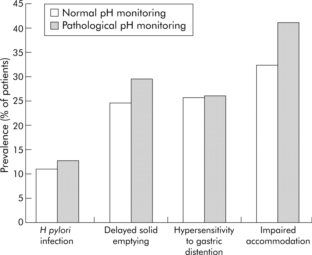
Oesophageal pH monitoring was normal (<5% of time pH<4) in 189 patients (77%), whereas 58 patients (23%) had a pathological pH monitoring. Daytime pH monitoring was pathological in 52 patients (21%) and night-time acid exposure was pathological in 46 patients (19%). Demographic features did not differ between patients with or without pathological pH monitoring (130/189 v 36/58 females, mean age 44.1 (SEM 1.0) v 45.4 (SEM 2.0) years, BMI 22.7 (SEM 0.3) v 23.8 (SEM 0.7) kg/m2; all non-significant). The prevalence of H pylori infection, of delayed gastric emptying, of hypersensitivity to gastric distention, and of impaired accommodation, did not differ between patients with or without pathological pH monitoring (fig 2).

Putative pathophysiological mechanisms in functional dyspepsia with (n = 58) or without (n = 189) pathological oesophageal pH monitoring. The figure depicts for each group the prevalence of Helicobacter pylori infection, of delayed solid gastric emptying, of hypersensitivity to gastric distention, and of impaired accommodation to a meal. No significant differences between groups occurred.
Oesophageal pH monitoring was borderline pathological (4–6% of time pH<4) in 20 patients (8%), below 4% in 180 patients (73%), and above 6% in 47 patients (19%). Again, demographic features did not differ between the three groups. Borderline pathological pH monitoring was not associated with a different symptom profile or with specific pathophysiological mechanisms (data not shown).
Results of heartburn questionnaire and relation with 24 hour pH monitoring
The vast majority of patients (175, 71%) responded “no” to all four questions. Eighteen patients (7%) gave a positive response to one question, 20 (8%) to two questions, and 13 (5%) to three questions. The heartburn questionnaire was considered positive in the case of four positive answers, which occurred in 21 patients (9%). Demographic features did not differ between patients with or without positive heartburn questionnaire (153/226 v 13/21 females, mean age 44.7 (SEM 1.0) v 40.6 (SEM 3.1) years, BMI 23.0 (SEM 0.3) v 22.4 (SEM 0.9) kg/m2; all non-significant).
The relation between the number of positive items on the heartburn questionnaire and 24 hour pH monitoring is summarised in table 2. In the 21 patients with positive responses to all four questions, pH monitoring was pathological in 16 (76%) and borderline pathological in none (fig 3). Daytime pH monitoring and night-time acid exposure were each pathological in approximately half of the patients (table 2). Hence, in this population of functional dyspepsia patients, the heartburn questionnaire had a specificity of 76% in predicting pathological acid exposure, but a sensitivity of only 28%.
Relation between heartburn questionnaire and pH monitoring in 247 patients with functional dyspepsia
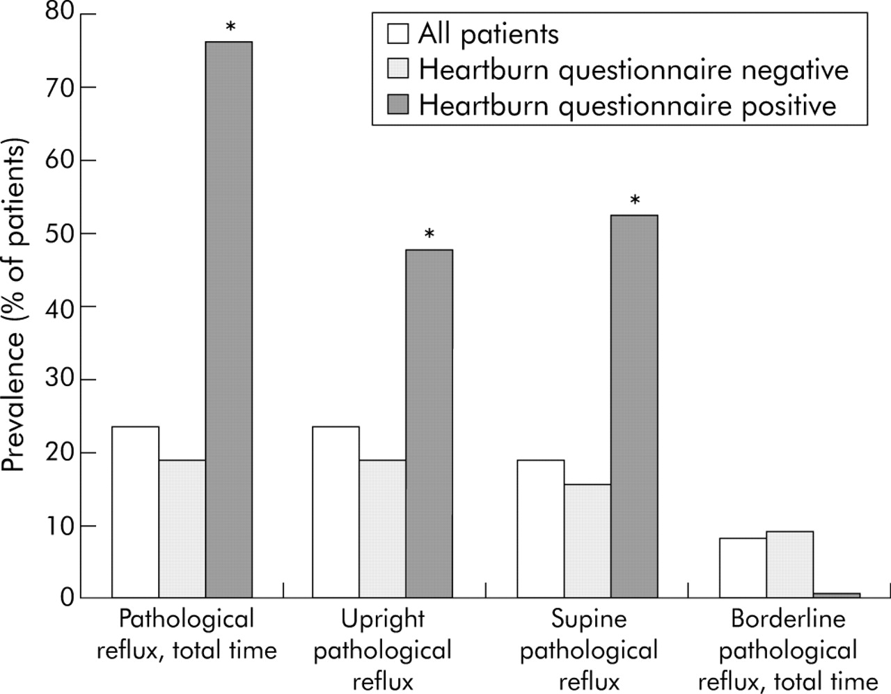
Prevalence of pathological oesophageal pH monitoring in all functional dyspepsia patients (n = 247), and in functional dyspepsia patients with a negative (n = 226) or a positive (n = 21) heartburn questionnaire. *p<0.05 compared to patients with a negative heartburn questionnaire.
Of the 226 patients with a negative heartburn questionnaire (<4 positive responses), pH monitoring was pathological in only 42 patients (18.5%) (fig 3). The prevalence of pathological pH monitoring was significantly higher in patients with a positive heartburn questionnaire compared to those with a negative heartburn questionnaire (p<0.0001). However, when patients with a positive heartburn questionnaire were eliminated, the prevalence of pathological pH monitoring was not significantly lower. Similar findings were obtained when considering a cut off of three positive responses (n = 34, pathological pH monitoring in 59% compared with 18% in those with less than three positive responses, p<0.0001).
In the 189 patients who had normal acid exposure, the heartburn questionnaire was positive in five (2.5%). Of the 58 patients with pathological acid exposure, the heartburn questionnaire was positive in 16 (27.5%). None of the 20 patients with a borderline pathological pH monitoring had a positive heartburn questionnaire. The specificity and the sensitivity of the heartburn questionnaire in predicting pathological acid exposure were 82.8% and 27.5% respectively.
Relation between 24 hour pH monitoring and dyspepsia symptoms
The association between symptom severity scores and pH monitoring was investigated without and with elimination of patients with a positive heartburn questionnaire. When all patients were included, the presence of moderate or severe (score ⩾2) symptoms of epigastric pain (91/189 v 40/58, p = 0.005) was significantly higher in patients with pathological pH monitoring, bloating tended to be more prevalent (130/189 v 47/58, p = 0.07) and early satiety tended to be less prevalent (93/189 v 21/58, p = 0.08) (fig 4). Similar results were obtained when the presence of the symptoms (score ⩾1) was considered: epigastric pain (123/189 v 49/58, p<0.005) and bloating (155/189 v 54/58, p<0.05) were significantly more prevalent in the subgroup of patients with pathological pH monitoring. The prevalence of other symptoms did not differ significantly between both groups. Similar results were obtained with daytime or night-time pH monitoring. The sensitivity and specificity for moderate or severe epigastric pain in predicting abnormal oesophageal acid exposure were 69% and 31% respectively in the total functional dyspepsia patient group. When patients with a positive heartburn questionnaire were eliminated, sensitivity and specificity for moderate or severe epigastric pain in predicting abnormal oesophageal acid exposure were 67% and 24% respectively.

Prevalence of dyspepsia symptoms in patients with (n = 58) or without (n = 189) pathological 24 hour oesophageal pH monitoring. The figure depicts the percentage of patients grading individual symptoms as moderate or severe (score ⩾2) in each group. Moderate or severe epigastric pain was significantly more prevalent in patients with pathological pH monitoring (*p<0.05), bloating tended to be more prevalent, and early satiety tended to be less prevalent (†0.06<p<0.09).
Stepwise multiple logistic regression analysis was used to identify the association between the risk of pathological pH monitoring and different patient variables and symptoms. The likelihood of pathological pH monitoring increased with the number of positive answers to the heartburn questionnaire (table 3). Furthermore, the presence of pain (OR = 2.478, 95% CI 1.130 to 5.434, p = 0.02), the presence of moderate or severe fullness (OR = 2.562, 95% CI 1.027 to 6.390, p = 0.04), the presence of moderate or severe bloating (OR = 2.598, 95% CI 1.058 to 6.377, p = 0.04), the absence of moderate or severe early satiety (OR = 0.458, 95% CI 0.220 to 0.952, p = 0.04), and the presence of severe epigastric burning (OR = 2.992, 95% CI 1.243 to 7.200, p = 0.01) were independently associated with the risk of pathological pH monitoring.
Likelihood of pathological pH monitoring according to number of positive responses on the four item heartburn questionnaire in 247 patients with functional dyspepsia
After elimination of the patients with a positive heartburn questionnaire, the prevalence of pain (119/184 v 15/42, p = 0.02) and the prevalence of moderate or severe pain (89/184 v 28/42, p = 0.03) remained significantly increased in those with pathological pH monitoring compared to those with normal pH monitoring.
Stepwise multiple logistic regression analysis after elimination of patients with a positive heartburn questionnaire demonstrated that the presence of pathological pH monitoring was associated with the presence of pain (OR = 3.330, 95% CI 1.228 to 9.027, p = 0.02), the presence of moderate or severe pain (OR = 2.344, 95% CI 1.046 to 5.255, p = 0.04), and the presence of severe epigastric burning (OR = 2.746, 95% CI 1.095 to 6.887, p = 0.03).
DISCUSSION
Both functional dyspepsia and GORD are extremely common disorders of the gastrointestinal tract which may both cause chronic or recurrent upper gastrointestinal symptoms.1–3 The pathophysiology of GORD is reasonably well understood and successful treatment modalities are available.4,23,24 The pathophysiology of functional dyspepsia is poorly understood and treatment options for this condition are limited.25 However, in clinical practice, distinguishing GORD from functional dyspepsia has proven difficult. Studies have established that clinicians often fail to recognise typical reflux symptoms. It has been suggested that a systematic questionnaire aimed to better recognise heartburn might be useful under these circumstances.6,9 Furthermore, GORD may present with atypical symptoms, that may mimic other conditions including functional dyspepsia.7,10,13 In atypical presentations, although a true gold standard is lacking, oesophageal pH monitoring is probably the method of choice to demonstrate reflux disease.
In the present study, we selected consecutive patients with a clinical diagnosis of functional dyspepsia after appropriate testing. All patients had negative upper gastrointestinal endoscopies in the absence of acid suppressive therapy, which adequately excluded erosive oesophagitis. None of the patients were judged by the treating gastroenterologist to have predominant reflux disease. All patients subsequently underwent oesophageal pH monitoring, which was normal in the majority of them. This finding confirms that functional dyspepsia is a separate entity from endoscopy negative reflux disease. It has been suggested that endoscopy negative GORD with atypical symptomatic presentation might be an important and underestimated cause of functional dyspepsia symptoms.7,10 However, oesophageal pH monitoring was normal in over 75% of these functional dyspepsia patients.
Nevertheless, the 24 hour pH monitoring revealed that almost one quarter of these patients had pathological acid exposure of the distal oesophagus. Demographic features and the presence of putative pathophysiological mechanisms such as delayed gastric emptying, hypersensitivity to gastric distention, or impaired accommodation did not distinguish patients with or without pathological pH monitoring. However, symptoms of epigastric pain were more prevalent in dyspeptic patients with pathological pH monitoring. This is in keeping with findings from controlled studies that dyspeptic patients with predominant pain are more likely to respond to proton pump inhibitor treatment.5 Similarly, Carlsson et al found that epigastric pain was a prominent feature of dyspeptic patients that were likely to respond to acid suppressive therapy.6
The heartburn questionnaire identified a subgroup of patients with a particularly high prevalence of pathological pH monitoring, which is reflected in a specificity of 76%. This high specificity is in agreement with observations in a subset of patients that participated in a controlled trial of proton pump inhibitor therapy in functional dyspepsia.14 These patients may have had both functional dyspepsia and GORD, and the high prevalence of both disorders would certainly allow overlap.1,2 However, the heartburn questionnaire had a low sensitivity of only 28%. Thus, it failed to identify the majority of patients with pathological pH monitoring, and it failed to significantly decrease the prevalence of pathological pH monitoring after elimination of patients with a positive questionnaire.
Symptoms of epigastric pain were more prevalent in dyspeptic patients with pathological pH monitoring. Previously, we also demonstrated that epigastric pain was associated with hypersensitivity to gastric distention in functional dyspepsia,15 but in the present study, pH monitoring and sensitivity to gastric distention were not related. The sensitivity of the presence of moderate or severe pain to predict pathological acid exposure was 69%, but the specificity was only 31%. Hence, the presence of epigastric pain is not a very accurate marker for endoscopy negative reflux disease in dyspeptic patients without heartburn.
The present study does have some implications for clinical practice. In dyspeptic patients, the use of a descriptive questionnaire helps to identify a population with heartburn. As the majority of these will have evidence of reflux disease on oesophageal pH monitoring, this group of patients might be expected to benefit from acid suppressive therapy. Almost 20% of functional dyspepsia patients with a negative heartburn questionnaire will also have pathological oesophageal acid exposure. Most of these will report moderate or severe epigastric pain. One could consider to also treat these patients with acid suppressive therapy. However, as other mechanisms like visceral hypersensitivity are also associated with increased prevalence of epigastric pain, and as the specificity of epigastric pain in predicting pathological acid exposure was low, it seems unlikely that the benefit from acid suppressive therapy will be very high in this group. Nevertheless, a prospective study evaluating the use of epigastric pain as a predictor of protein pump inhibitor responsiveness in functional dyspepsia seems warranted based on our data.
The present study has a number of limitations that should be taken into account when generalising our findings. Firstly, the setting of the study was a large hospital with both secondary care (general gastroenterology clinic, referrals from general practitioners) and tertiary care (specialised motility clinic, referrals from specialists) functions. The findings are not necessarily relevant to primary care dyspeptic patients. Secondly, the heartburn questionnaire was developed and validated for prediction of responsiveness to acid suppression in Scandinavia, in a different linguistic and cultural setting. Moreover, since this study was started, a number of new GORD questionnaires have been developed which may allow better recognition of reflux symptoms.26 On the other hand, the systematic study of a large group of consecutive well characterised patients with pH monitoring, symptom assessment, heartburn evaluation, and additional pathophysiological studies represents a strength of the present study compared with previous efforts.
In summary, we have shown that the majority of patients with a clinical diagnosis of functional dyspepsia seen at a tertiary care centre have normal 24 hour oesophageal pH monitoring, and less than one quarter of the patients have abnormal acid exposure.
Compared with patients with normal pH monitoring, patients with pathological acid exposure have more prevalent symptoms of epigastric pain. Both groups do not differ in the prevalence of H pylori infection, of delayed gastric emptying, of hypersensitivity to gastric distention, or of impaired accommodation. A four item heartburn questionnaire identified a subset with a high prevalence of pathological pH monitoring, but failed to identify the majority of patients with pathological acid exposure.
REFERENCES
Footnotes
-
Competing interest: none declared.
-
Published online first 21 June 2005