Article Text

Abstract
BACKGROUND Propensity to colonic neoplasia differs between the right and left colon.
AIMS To examine whether this difference may be related to regional differences in epithelial apoptosis, in expression of a proapoptotic regulatory protein, Bak, and in proliferation.
PATIENTS Individuals with no history of colorectal neoplasia.
METHODS Archival blocks of colorectal tissues were immunostained for proliferating cells (antibody to Ki-67 antigen), and Bak expression (polyclonal antiserum). Cells containing DNA strand breaks, a marker of apoptosis, were identified by terminal deoxyuridine nucleotidyl nick end labelling (TUNEL).
RESULTS There were fewer TUNEL positive epithelial cells in the right colon (mean 1.2 (SE 0.1)% of all epithelial cells) than the left colon (2.2 (0.1)%, p<0.0001) or rectum (2.2 (0.3)%, p<0.05). Bak expression was less common in the right colon (mean 46 (2.3)% of epithelial cells immunoreactive) than the left colon (66 (2.7)%, p<0.0001), or rectum (67 (2.3)%, p<0.001). Bak expression and TUNEL positivity were highly positively correlated (p<0.0001). In contrast to apoptosis, mean whole crypt proliferation labelling index was similar throughout the colorectum (right colon: 15.6 (3.2)%; left colon: 13.5 (1.2)%; rectum: 13.3 (2.3)%).
CONCLUSION The percentage of proliferating colonic epithelial cells is constant throughout the colon, but fewer epithelial cells undergo Bak mediated apoptosis in the right than in the left colon or rectum. This suggests that colonocytes may be lost by methods other than apoptosis in the right colon.
- programmed cell death
- Bcl-2
- cell cycle
- cell proliferation
- colon carcinogenesis
Abbreviations used in this paper
- TUNEL
- terminal deoxyuridine nucleotidyl nick end labelling
Statistics from Altmetric.com
The human colon develops embryologically from the primitive midgut and hindgut. The midgut gives rise to the ascending and proximal transverse colon whereas the distal transverse, descending, and sigmoid colon, and the rectum are derivatives of the hindgut. The junction between the midgut and the hindgut is proximal to the splenic flexure, approximately one third of the way along the transverse colon, and the right colon is conventionally defined as the area proximal to the splenic flexure.1
Cancers of the right and left colon differ in their epidemiology, molecular genetics, and behaviour.2 For example, populations at high risk of colon cancer (defined as more than 25 cases per 100 000 annually) have a predominance of left sided cancers, whereas low risk populations (defined as less than 10 per 100 000 annually) present with a predominance of right sided cancers. Of note, in recent years there has been a shift to the right in high risk nations, for unknown reasons.3 ,4 Genetic differences between right and left colon cancers include the findings that left sided lesions are more likely to have 18q deletions than are cancers of the right colon,5 and right sided cancers are relatively more common in hereditary non-polyposis colon cancer than in sporadic cancer.6
These differences between right and left colon cancers may reflect differences in the physiology and function of the normal colonocytes from which they arise. For example, the embryonic origins, blood supply, and the normal expression of differentiation markers such as blood group and carbohydrate antigens, mucins, and lectin binding capacities differ between the right and left human colon.7-9 There may also be differences in the proliferation and migration of normal colonocytes located at different sites within the human colon. Three studies have been performed in patients who did not have cancer at the time of biopsy. In these studies proliferation was measured ex vivo, either with tritiated thymidine,10 ,11 or by a stathmokinetic method to measure the crypt cell production rate.12 One of these studies found increased cell proliferation rates in the caecum compared with other regions,12 another found significantly less proliferation in the caecum than in other regions,10 and a third reported no differences in proliferation labelling indexes between biopsy specimens taken from different sites.11Using the stathmokinetic method to measure crypt cell production rate, three small studies in rats have been similarly inconclusive.12-14 Significantly higher crypt cell production rate in the caecum compared with other regions of the colon was reported in one of these studies, but the other two did not find any significant differences between the proximal and distal colon.
Recently there has been increasing interest in the possibility that the growth of cancers is linked to an imbalance between proliferation and apoptosis. Apoptosis normally occurs principally in colonocytes at the luminal surface, following migration and terminal differentiation from the base of the crypt,15 and it has been suggested that a deficiency of physiological apoptosis may contribute to the growth of colonic neoplasms.16 ,17 In the colon, as in other tissues, it is necessary for apoptosis to be highly regulated and coupled to rates of cell proliferation to ensure tissue homoeostasis.18 Among the genes regulating apoptosis are the Bcl-2 family of proteins. These proteins interact with themselves as well as an expanding repertoire of Bcl-2 associated proteins, to govern a checkpoint regulating the common downstream effector pathway of programmed cell death.19 In the colon, Bak has been implicated as the Bcl-2 family member principally involved in the promotion of apoptosis. Bak expression colocalises with apoptotic epithelial cells in the colonic epithelium and Bak expression increases during the apoptosis of colonocytes induced by a number of different stimuli, without consistent changes in other Bcl-2 family members.20 Whether there are regional differences in apoptosis or in the expression of the apoptosis related protein Bak in the colon has not been examined. Therefore, in this study we evaluated the extent of epithelial cell apoptosis and expression of Bak in the normal human right and left colons. In addition the numbers of cycling epithelial cells in serial sections of the same paraffin wax blocks were measured, in order to determine whether physiological differences in apoptosis or in the ratio of apoptotic to proliferating cells may explain the different predispositions of the right and left colon to cancer.
Methods
TISSUE COLLECTION
Archival paraffin wax blocks of well orientated normal colorectal tissue were studied. The specimens consisted of biopsy samples from macroscopically normal mucosa taken from patients undergoing colonoscopy and in whom no abnormalities were detected, and from the normal resection margins of colonic tissues removed at laparotomy for ischaemia. Patients with any colonic neoplasia or inflammation were specifically excluded. In total, 38 cases were examined, from patients aged 53–72 years, 15 of whom were men. The tissue was designated as right colon (proximal to the splenic flexure, n=11), left colon (n=17), or rectum (n=9).
TUNEL AND IMMUNOHISTOCHEMISTRY
All tissues had been fixed in formalin for diagnostic clinical pathology. Sections of 4 μm thickness were cut from the paraffin wax blocks onto polylysine treated slides. Epithelial cells with DNA strand breaks, a surrogate marker of apoptosis, were identified after staining by terminal deoxyuridine nucleotidyl nick end labelling (TUNEL), using a method modified from Gavrieli et al,21 as previously described.15Serial sections were then immunostained for Bak, using polyclonal antisera raised to a synthetic peptide corresponding to amino acids 14–36 of human Bak, as described previously,22 and proliferating cells were detected with the Mib-1 antibody to the Ki-67 antigen using established methods.23
QUANTIFICATION AND STATISTICAL ANALYSIS
In all cases, the quantification of positively stained cells was determined by a single observer who was unaware of the site of the tissue. For Bak and Mib-1 immunohistochemistry, and after staining by TUNEL, positively stained cells were counted in a mean of 20 colonic crypts, and the number of positive cells expressed per 100 total cells counted as a whole crypt labelling index (mean (SEM)%), as previously described.15 The Mann-Whitney U test was used to compare differences between right and left colon and rectum for each parameter, and any correlation between numbers of apoptotic and Bak staining cells was determined by Spearman’s rank correlation, a non-parametric method. All statistical tests were two sided.
Results
TUNEL histochemistry showed the presence of epithelial cells carrying fragmented DNA. These were located predominantly at the luminal aspect of colonic crypts (fig 1). The mean labelling index of TUNEL positive epithelial cells in crypts from the right colon was 1.2 (0.1)%, and was lower than in the left colon and the rectum. The mean labelling index in the left colon was 2.2 (0.1)%, and in the rectum was 2.2 (0.3)%. The differences between the right and the left colon and between the right colon and rectum were both statistically significant (p<0.0001 and p<0.05 respectively; fig2).
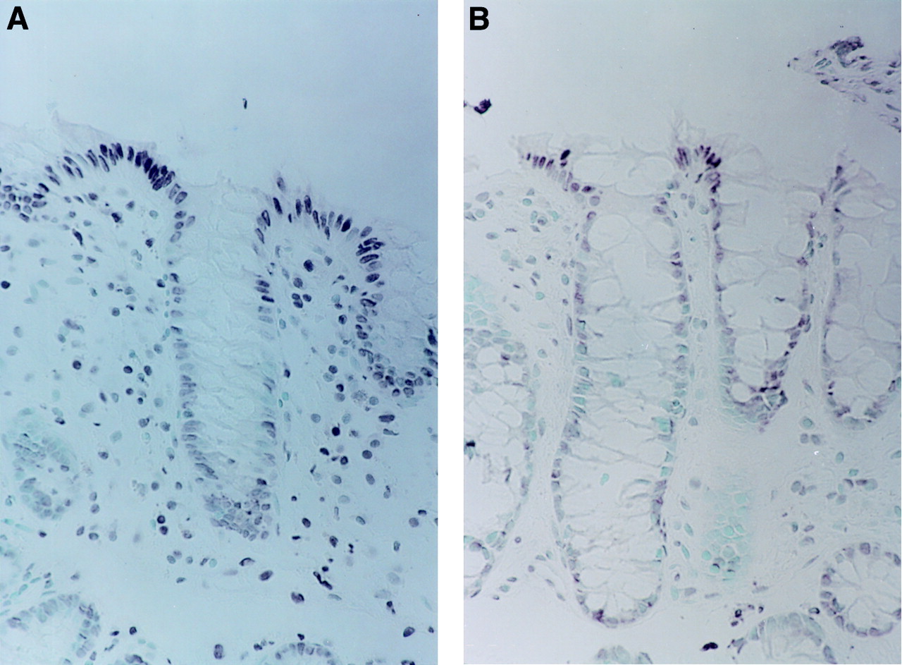
TUNEL staining showing dark, positively stained, nuclei of cells carrying DNA strand breaks at the surface of colonic crypts, more frequent in the left colon (A) than in the right colon (B). Some TUNEL positive cells are also evident in the lamina propria, especially in the left colon. Original magnification ×200.

Differences in the regional distribution of TUNEL positive epithelial cells within the colorectum. Individual data points are shown with means and standard errors represented by the bars.
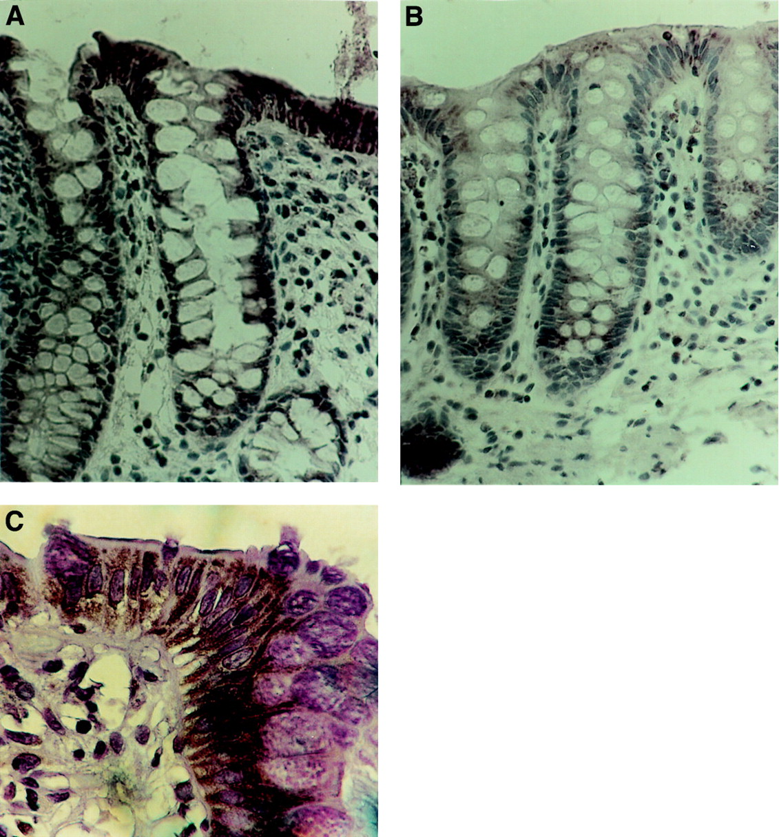
Bak expression in epithelial cells was mainly confined to cells located towards the luminal portion of the crypt, although, as for TUNEL, occasional labelled cells were observed in the proliferative zone at the crypt base. Many more cells were labelled by Bak immunohistochemistry than were positive by TUNEL, although the geographical distribution of maximal staining at the luminal end of the crypt was similar for both Bak and for TUNEL. Bak immunostaining was evident both above and below the nucleus, in a granular pattern, consistent with a location within intracellular organelles. Crypts from the the left colon and rectum (fig 3A,C) contained more epithelial cells labelled with the Bak antibody, than did crypts from right colon (fig 3B). In the right colon, a mean of 46 (2.3)% of colonic epithelial cells expressed Bak immunoreactivity compared with 66 (2.7)% in the left colon (p<0.0001), and 67 (2.3)% in the rectum (p<0.001 compared with the right colon, which was not statistically significantly different compared with the left colon) (fig 4). There was a strong positive correlation between the expression of Bak and the percentage of TUNEL positive cells (p<0.0001) (fig 5).

Expression of Bak in colonic crypts from left colon (A) and right colon (B). Bak is expressed in the cytoplasm of superficial colonocytes throughout the colon but the immunointensity and percentage of cells staining with Bak antibody is greater in the left compared with the right colon. At higher magnification (C), Bak immunostaining is evident in the supranuclear and infranuclear regions of surface colonocytes. Original magnification ×200 (A,B); ×400 (C).

Percentage of Bak immunoreactive epithelial cells in different regions of the normal human colorectum. Individual data points are shown with mean and standard errors shown as bars.
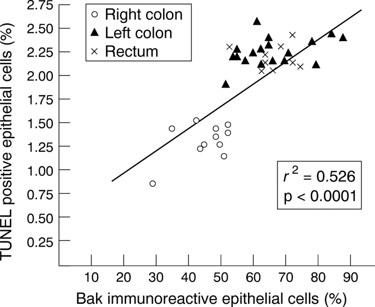
Correlation between Bak immunoreactivity and TUNEL positive cells in normal human colonic epithelium.
The Mib-1 antibody localised to the nuclei of proliferating cells, located in the base of the crypt column (fig 6). The mean whole crypt proliferation labelling indexes for the right colon, left colon, and the rectum were 15.6 (3.2)%, 13.5 (1.2)%, and 13.3 (2.3)%, respectively. There were no significant differences in the proliferation labelling indexes between different regions of the colorectum.

Immunostaining with the Mib-1 antibody to Ki-67 shows the nuclei of proliferating cells in the base of normal colonic crypts from right colon. Original magnification ×200.
Discussion
Our results show that compared with the proximal colon, the distal colon and rectum contain a greater percentage of cells expressing the pro-apoptotic protein Bak, and containing fragmented DNA, a marker of cells undergoing apoptosis. No significant differences in proliferation were found between different regions of the colon.
Why should cells in the left colon be more sensitive to apoptotic cell death than those located in the right colon and can these differences help in understanding the different propensity of these colonic regions to neoplasia? One possible explanation for our findings is that the luminal contents of the left colon may be more pro-apoptotic. Several components of the colonic luminal contents are candidate inducers of apoptosis. These include butyrate,24 ,25 protein kinase C agonists such as diacylglycerol,26 ,27 and luminal bile salts.26 Evidence for bile salt induced colonocyte apoptosis has been obtained from human studies of biopsy specimens incubated with deoxycholate ex vivo28 and with malignant colonocytes studied in vitro.26 A recent study has reported that the normal appearing colonocytes from patients with a history of previous colorectal cancer are relatively resistant to bile salt induced apoptosis,28 a finding supported by a similar phenomenon in the azoxymethane rodent model of colon cancer29 and in support of the concept that apoptosis resistance contributes to the development of colorectal neoplasia.16 ,17 However, contrary to these purported explanations, the concentrations of bile salts,30 and butyrate31 are actually higher in the right colon than the left, so that these known inducers of colonocyte apoptosis are at higher concentration where apoptosis is lower. Recent findings may help understand this apparent paradox. Of note, although butyrate induces apoptosis in transformed colonic epithelial cells,24-26it inhibits the apoptosis of the normal colonic epithelium in vivo32 and may also do so in malignant cells in vitro, at least under conditions of glucose depletion.33 Thus, increased butyrate concentations in the right colon may contribute to the higher incidence of “physiological” apoptosis here.
Differences in the sensitivity of epithelial cells of the small and large intestines to apoptosis induced by chemotherapeutic drugs or irradiation have been observed in rodents.34 Whether colonocytes from the right and left colons also differ in their sensitivity to these agents in these models is not known. Experiments involving colonic transposition in animals or xenografts of human colonocytes into immunodeficient mice may be of value in elucidating the relative effects of luminal factors and site on the induction of colonic epithelial cell apoptosis.
Our results show that in parallel with more apoptosis in the left colon, Bak is expressed in more colonocytes in crypts from the left than from the right colon. Bak expression and cells containing DNA fragmentation (TUNEL positive) are rarely seen in the base of colonic crypts where proliferation occurs, but Bak expression and apoptotic cells are more frequent towards the luminal surface of colonic crypts where physiological apoptosis occurs.15 Furthermore, the expression of Bak and the number of apoptotic cells was highly correlated, which supports the concept that Bak is an important promotor of apoptosis in the normal colonic epithelium.20
Reservations have been expressed concerning the applicability of the TUNEL method to evaluate apoptosis in the gastrointestinal tract.35 It is conceivable that the differences we have described reflect an in vitro artefact due to a differential sensitivity to TUNEL staining between the right and left colon. However, in a previous study in which we compared the sensitivity of TUNEL with morphological criteria of apoptosis by haematoxylin and eosin staining,17 we have subsequently reviewed our scores and have found no difference in TUNEL staining sensitivity between right and left colonic specimens.
In the current study we have also found that the percentage of proliferating cells, as indicated by labelling with the Mib-1 anti-Ki-67 antibody, is similar throughout the normal human colon. This is in agreement with the previous study by Terpstraet al,11 although others have found evidence for both decreased10 and increased12 numbers of proliferating cells in the human caecum compared with other sites. The reasons for discrepancies between these studies are not immediately apparent. None included patients with a known history of colorectal neoplasia, as it is known that these patients have a relatively high proliferation labelling index.10 ,11 However, unlike the other studies,10-12 we did not incubate biopsy specimens in vitro but instead assessed proliferation with an endogenous marker (Ki-67), thus reducing artefacts related to in vitro manipulation. In our hands, measuring Ki-67 in colonic specimens provides a more reproducible assessment of cycling cells than the alternative endogenous marker, PCNA.23 In the rat, the more exacting and rigorous stathmokinetic techniques have been used to assess the crypt cell production rate after metaphase arrest induced by vincristine, and although one small study reported increased proliferation in the caecum,12 two other larger studies did not.13 ,14 Overall, therefore, our results are supported by most of the published literature, suggesting that the percentage of proliferating crypt epithelial cells is constant throughout the large intestine.
In conclusion, our study suggests that although the proliferation rate is constant throughout the colon, cell loss by apoptosis associated with the expression of Bak is higher in the left than the right colon. Whether in the right colon, cell loss occurs by processes other than apoptosis is not apparent from this study. However, it is conceivable that a difference in apoptotic cell loss between right and left colons may be related to the different epidemiology, genetics, and behaviour of cancers arising from these sites, so that examining regional differences in colonic epithelial cell loss is worthy of further investigation.
Acknowledgments
The authors are grateful to Shaobai Wang for technical assistance, Stephen F Ryan for help in the selection of cases from the Department of Pathology and Laboratory Medicine at St Luke’s-Roosevelt Hospital Centre, and to Stanislas Krajewski and John C Reed for providing antibodies to Bak. This work was presented at Digestive Disease Week, Washington DC, May 1997 and published in abstract form (Gastroenterology1997;112:A605).
Abbreviations used in this paper
- TUNEL
- terminal deoxyuridine nucleotidyl nick end labelling