Article Text

Abstract
BACKGROUND T cell responses to normal intestinal bacteria or their products may be important in the immunopathogenesis of chronic enterocolitis.
AIMS To investigate the T cell specificity and cross reactivity towards intestinal bacteria.
PATIENTS/METHODS T cell clones were isolated with phytohaemagglutinin from peripheral blood and biopsy specimens of inflamed and non-inflamed colon from five patients with inflammatory bowel disease (IBD) and two controls. T cell clones were restimulated with anaerobicBacteroides andBifidobacteria species, enterobacteria, and direct isolates of aerobic intestinal flora. T cell phenotype was analysed by single-cell immunocyte assay.
RESULTS Analysis of 96 T cell clones isolated from peripheral blood and biopsy specimens from two patients with IBD showed that bothBifidobacterium andBacteroides species specifically stimulate proliferation of CD4+TCRαβ+ T cell clones from both sites and that cross reactivity exists between these anaerobic bacteria and different enterobacteria. Analysis of 210 T cell clones isolated from three patients with IBD and two controls showed that indigenous aerobic flora specifically stimulate T cell clones from peripheral blood and biopsy specimens from a foreign subject. Some of these flora specific T cell clones were cross reactive with defined enterobacteria. In addition, T cell clones stimulated by their own indigenous aerobic flora were identified in patients with IBD.
CONCLUSION Immune responses to antigens from the intestinal microflora involve a complex network of T cell specificities.
- inflammatory bowel disease
- T cells
- intestinal flora
- mucosal immunity
Abbreviations
- IBD
- inflammatory bowel disease
- PCR
- polymerase chain reaction
- BsA
- bacteria from autologous intestine—that is, sonicates and mononuclear cells from the same subject
- BsH
- bacteria from heterologous intestine—that is, sonicates and mononuclear cells from different subjects
- TCR
- T cell receptor
Statistics from Altmetric.com
Inflammatory bowel disease (IBD) comprises two forms of chronic intestinal inflammation, Crohn’s disease and ulcerative colitis. The cause of IBD is still not known, but there are numerous indications that T cells are important in IBD pathogenesis.1-4 The antigens that drive T cell activation2 and clonal expansion4 in IBD still need to be defined but both clinical and experimental evidence strongly incriminate normal luminal bacteria as a source of antigen.5 Normal luminal bacteria comprise an estimated number of 400 different bacterial species and reach their highest concentrations in the terminal ileum and colon.6 Anaerobes outnumber aerobes by a factor of 103 in the lumen, but are represented in almost equal numbers at the bowel wall.7 A large variety of observations support the hypothesis that IBD is due to T cell hyperresponsiveness towards otherwise harmless components of the normal intestinal flora. As a clinical observation, IBD manifests itself most frequently in the terminal ileum and colon—that is, those sites with the highest bacterial concentrations in the intestine. It has been found that Crohn’s disease relapses are prevented by diversion of the faecal stream,8 increases in gut permeability precede acute flares in Crohn’s disease,9 and antibiotic treatment is effective in both Crohn’s disease and ulcerative colitis.10-13 In addition, intestinal lesions of patients with IBD are characterised by increased T cell reactivity towards bacterial antigens,14 an increase in enterobacteria specific T cells,15 and a loss of tolerance towards indigenous flora.16 A role for bacteria in the pathogenesis of chronic intestinal inflammation has also been well established in different inducible animal models of intestinal inflammation.17-20 Furthermore, in interleukin-2 knockout mice,21 interleukin-10 knockout mice,22 and HLA-B27 transgenic rats,23 intestinal inflammation improves under germ free or specific pathogen free conditions, whereas germ free HLA-B27 transgenic rats develop chronic intestinal inflammation when colonised with normal luminal bacteria, especiallyBacteroides vulgatus.24 As the critical bacteria in most animal models are still not known, it may be of note that B vulgatus has also been shown to play an essential role in the pathogenesis of carrageenan induced colitis in guinea pigs.25 A variety of changes implicating luminal bacteria in disease pathogenesis were observed in rodent models of colitis induced by the hapten reagent trinitrobenzenesulphonic acid.26-29 Colitis induced by this reagent in mice is associated with a TH1 cytokine profile30 and can be transferred to naive recipient animals with purified CD4 T cells from the colon of trinitrobenzenesulphonic acid treated animals.31 In addition, administration of broad spectrum antibiotics prevents chronic colitis.32 Thus a relation between T cells, luminal bacteria, and chronic enterocolitis is well established in both animal models of chronic intestinal inflammation and patients with IBD. Identification of the critical bacterial antigens or products, either by reconstitution of germ free animals manipulated to develop chronic intestinal inflammation when exposed to bacteria or by investigation of the immune response against luminal bacteria in patients with IBD, may be crucial for the better understanding of IBD pathogenesis and for new treatment strategies to be devised. In order to understand the specificity and cross reactivity of the T cell response towards luminal bacteria more precisely, we have here chosen the latter approach and determined clonal T cell responses towards defined anaerobic intestinal bacteria and aerobic intestinal flora.
Materials and methods
PATIENTS
Peripheral blood mononuclear cells and lamina propria mononuclear cells were obtained from five patients with IBD (two with Crohn’s disease, three with ulcerative colitis; three women, two men; mean age 39.8 years with a range of 25–52 years) and two controls (a 37 year old woman with irritable bowel syndrome and a 56 year old man with bile acid induced diarrhoea). All patients attended our outpatient IBD clinic and were diagnosed as having Crohn’s disease or ulcerative colitis using established clinical, radiological, and pathological criteria. In patients with Crohn’s disease, disease activity according to criteria established by de Dombal et al 33 was classified as moderate (n = 1) or severe (n = 1). Patients were treated with mesalazine or corticosteroids. In patients with ulcerative colitis, disease activity according to criteria established by Truelove and Witts34 was moderate (n = 2) or severe (n = 1). Patients were treated with mesalazine (n = 2) or corticosteroids (n = 1). None of the patients or controls was treated with antibiotics. Biopsy specimens from inflamed (B+) and non-inflamed (B−) intestine were taken during the same endoscopic procedure and blood was withdrawn on the same day. Inflamed or non-inflamed status of all intestinal biopsy samples was macroscopically assessed during endoscopy and histologically confirmed by examination of biopsy specimens from closely adjacent areas.The study was reviewed by the local ethics committee and all patients gave informed consent conforming with the Declaration of Helsinki ethical guidelines.
PREPARATION OF MONONUCLEAR CELLS
Peripheral blood mononuclear cells were isolated from heparinised peripheral blood by Ficoll-Hypaque (Seromed, Berlin, Germany) density gradient centrifugation. Lamina propria mononuclear cells were isolated from intestinal biopsy specimens by following an established protocol using collagenase treatment.15 For cryopreservation, mononuclear cells and T cell clones were stored in 90% fetal calf serum and 10% dimethyl sulphoxide in liquid nitrogen. For generation of monocyte- and B-cell-enriched mononuclear cells (E−), T cells were removed by rosetting with neuraminidase treated sheep erythrocytes as described.15
GENERATION OF T CELL CLONES
Lamina propria T cells and matched peripheral blood T cells were cloned using phytohaemagglutinin, as reported previously.15 In brief, T cells separated by E-rosetting were incubated in Terasaki plates at 0.3 cells/well together with 12 000 cells/well of allogeneic irradiated (40 Gy) feeder cells from buffy coats in RPMI medium containing phytohaemagglutinin (1 μg/ml; Murex, Hannover, Germany), human AB-Serum (10%) and interleukin-2 (10 U/ml; Boehringer, Mannheim, Germany). Clonal T cell populations were identified by eye at day 10 and expanded together with allogeneic irradiated (40 Gy) feeder cells. All T cell clones reported in this study are independent. Clonality of T cell clones was confirmed on a randomly selected T cell clone population (n = 10) by polymerase chain reaction (PCR) using primers specific for 20 different T cell receptor (TCR)-Vβ families as described.35 Phenotyping of T cell clones was performed using anti-CD4 (Dianova, Hamburg, Germany), anti-CD8 (Dianova), and anti-TCRαβ (ATCC HB 9283) monoclonal antibodies in a single-cell immunocyte assay.
ANTIGENS
Bacteroides thetaiotaomicron (ATCC 12290, isolated from purulent material from an incision during appendicectomy), Bifidobacterium bifidum(ATCC 35914, isolated from human faeces), andHelicobacter pylori (NCTC 11637) were obtained from ATCC. Enterobacteria were obtained from the Institute of Medical Microbiology, University of Mainz.Escherichia coli was isolated from a routine agar culture; Salmonella typhimurium andYersinia enterocolitica 03 were cultured from the stools of patients with enterogenic reactive arthritis.36 Bacteria from the indigenous intestinal flora were obtained from biopsy material and grown on blood agar plates as reported previously.16 After harvesting, all micro-organisms were washed, resuspended in phosphate buffered saline, and sonicated. Sonicates were stored at −80°C and tested for sterility by incubation on agar plates. Depending on the mononuclear cells with which they were incubated, sonicates from bacteria of the intestinal flora were used as bacteria from autologous intestine (BsA; sonicates and mononuclear cells from the same subject) or as bacteria from heterologous intestine (BsH; sonicates and mononuclear cells from different subjects). Protein concentrations were determined by the Lowry method (Bio-Rad protein assay; Bio-Rad, Munich, Germany). Tetanus toxoid was a gift from Behring-Werke, Mannheim, Germany.
PROLIFERATION ASSAYS
Proliferative responses were determined in triplicate in 200 μl culture medium (RPMI/10% AB-Serum) containing 5 × 10⁴peripheral blood mononuclear cells or 1 × 10⁴ cloned responder T cells and 2 × 10⁴ autologous irradiated (40 Gy) E− antigen presenting cells in 96-well microtitre plates (Nunc, Roskilde, Denmark). Bacterial sonicates (E coli, Y enterocolitica 03, S typhimurium, H pylori, B thetaiotaomicron, B bifidum, BsA, BsH) were added at 50 μg/ml. Medium, tetanus toxoid (10 μg/ml), and phytohaemagglutinin (1 μg/ml) served as controls. Cultures with peripheral blood mononuclear cells were pulsed with [3H]thymidine (0.5 μCi/well) at day 5, and cultures with T cell clones were pulsed with [3H]thymidine (0.5 μCi/well) after 24 h. All cells were harvested 18 hours after being pulsed. [3H]Thymidine incorporation measured in triplicate was expressed as (mean (SD)) cpm.
Results
CLONAL T CELL RESPONSES TOWARDS ANAEROBICBACTEROIDES ANDBIFIDOBACTERIUM
We have previously shown that CD4 T cells specifically recognise different enterobacteria and that enterobacteria specific T cell clones are increased in intestinal IBD lesions.15 Using new T cell clones we now determined the T cell response to major constituents of the anaerobic intestinal flora in humans—that is,Bacteriodes andBifidobacterium—and assessed whether T cell cross reactivity between anaerobes and enterobacteria occurs. T cell clones were isolated from peripheral blood (n = 26) and lamina propria T cell populations (n = 70) from two patients with IBD (both ulcerative colitis) as described above, and subsequently restimulated with sonicated preparations from enterobacteria, H pylori, B thetaiotaomicron, andB bifidum. Confirming previous data,16 T cell clones responding to enterobacteria were obtained (fig 1). In addition, [3H]thymidine incorporation identified a total of seven out of 96 T cell clones from peripheral blood and intestine with reactivity towardsB thetaiotaomicron and B bifidum. Of these seven T cell clones, two were reactive towardsB thetaiotaomicron only (RP3 and BB−22), and T cell clone RB+16 was cross reactive between B thetaiotaomicron and B bifidum. Cross reactivity, however, was not restricted to anaerobic bacteria as T cell clones reactive with B bifidum andY enterocolitica (RB−9 and BB+6) orB bifidum, B thetaiotaomicron, and Y enterocolitica (RB+2 and BB−17) were also identified. All bacteria specific T cell clones were CD4+TCRαβ+ as determined by single-cell immunocyte assay.
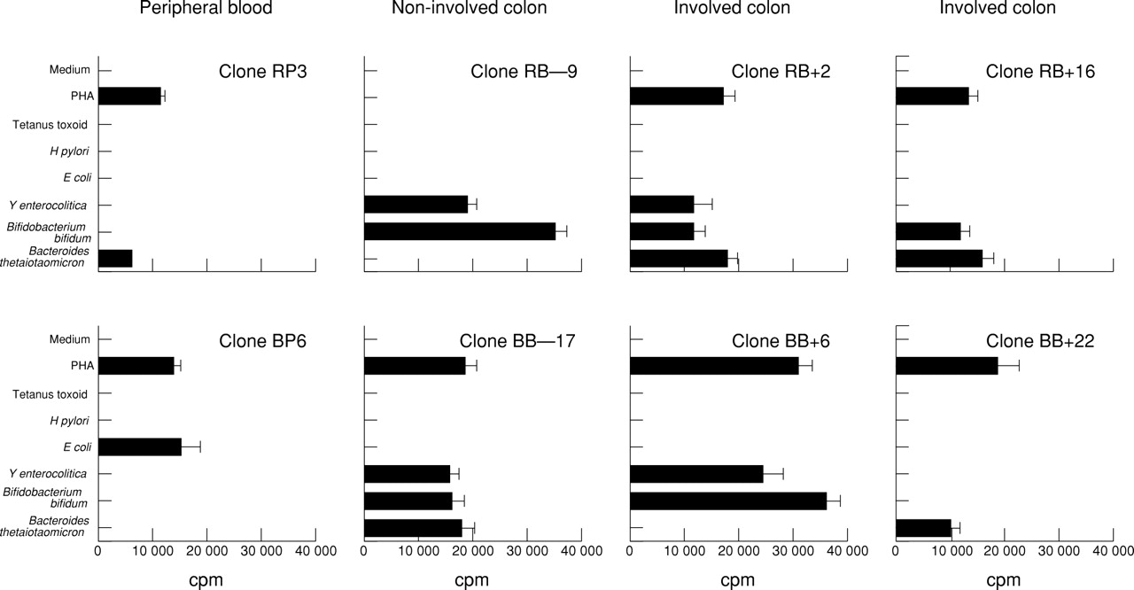
Clonal T cell responses towards Bacteroides and Bifidobacterium. T cell clones were isolated, using phytohaemagglutinin (PHA), from two patients with ulcerative colitis (patient R: peripheral blood n = 14, non-involved colon n = 12, involved colon n = 18; patient B: peripheral blood n = 12, non-involved colon n = 17, involved colon n = 23) and restimulated with the indicated antigens for 42 hours. Mean proliferation of T cell clones from peripheral blood (RP3), non-involved colon (RB−9 and BB−17), and involved colon (RB+2, RB+16, BB+6, and BB+22) stimulated by Bacteriodes thetaiotaomicron (ATCC 12290) or Bifidobacterium bifidum (ATCC 35914) is shown as measured by [3H]thymidine incorporation in triplicate cultures. Clone BP6 is representative of other T cell clones reactive with enterobacteria only. All T cell clones were CD4+TCRαβ+ by single-cell immunocyte assay.
CLONAL T CELL RESPONSES TO CRUDE SONICATES FROM AEROBIC INTESTINAL FLORA
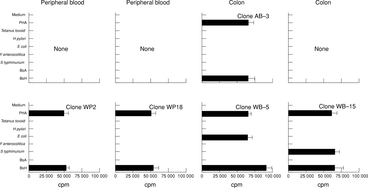
We have previously shown that crude sonicate preparations from indigenous intestinal flora stimulate proliferation in mononuclear cells from peripheral blood and intestinal lamina propria of a foreign subject but not those of the same subject.16 To determine T cell reactivity against these sonicates on a clonal level, we now isolated T cell clones from peripheral blood and lamina propria from two controls (n = 77) and three patients with IBD (n = 133) by cloning with phytohaemagglutinin. These clones were then restimulated with crude sonicates of bacteria from autologous intestine (BsA) or heterologous intestine (BsH). To assess T cell clone cross reactivity, T cell clones were also restimulated with sonicate preparations from different enterobacteria and H pylori, previously shown to selectively stimulate bacteria specific T cell clones.15 As shown in fig 2 and table 1, [3H]thymidine incorporation identified a total of five out of 77 T cell clones from controls and 21 out of 133 T cell clones from patients with IBD with reactivity towards BsH. In controls, three of five BsH reactive T cell clones were stimulated by BsH only and two of five T cell clones (WB−5 and WB−15) cross reacted with eitherE coli or S typhimurium. In patients with IBD, 15 out of 21 T cell clones were stimulated by BsH only, two out of 21 T cell clones (DB−8 and SP24) showed weak cross reactivity between BsH andH pylori or BsH, H pylori, and E coli, and four out of 21 T cell clones (DP15, DP18, DB−5 and FB+2) were cross reactive with BsA and BsH. The fraction of T cell clones recognising autologous bacteria was 0/77 in controls and 8/133 in patients with IBD. Four of eight T cell clones (DP15, DP18, DB−5, and FB+2) were cross reactive with BsA and BsH, and four of eight T cell clones (DB+5, DB+8, FP15, and FB+11) reacted to BsA only.
Clonal T cell responses to aerobic intestinal flora. T cell clones were isolated with phytohaemagglutinin (PHA) from two controls (patient A, bile acid induced diarrhoea: peripheral blood n = 23, colon n = 15; patient W, irritable bowel syndrome: peripheral blood n = 20, colon n = 19) and restimulated with the indicated antigens for 42 hours. Aerobic intestinal flora was obtained from intestinal biopsy specimens touched to blood agar plates and grown under aerobic conditions. Sonicates from outgrowing bacteria were used as BsA (sonicates and mononuclear cells from the same person) or BsH (sonicates and mononuclear cells from different people). Mean proliferation of T cell clones from peripheral blood (WP2 and WP18) and colon (AB−3, WB−5, and WB−15) stimulated by BsA or BsH is shown as measured by [3H]thymidine incorporation of triplicate cultures. All T cell clones were CD4+TCRαβ+ by single-cell immunocyte assay. Incubation of antigen presenting cells with the indicated stimuli without T cell clones resulted in <500 cpm.
Clonal T cell responses towards aerobic intestinal flora
REACTIVITY OF BsH SPECIFIC T CELL CLONE FB−10
Antigens from the indigenous intestinal flora stimulating T cell clones may be expressed by one or different bacterial species or bacterial strains. To investigate this question, we determined the proliferative response of the BsH reactive T cell clone FB−10 to sonicates from 100 different bacterial colonies, all isolated from the heterogeneous mixture of aerobic intestinal bacteria used for the generation of the original BsH sonicate (fig 3). Using these sonicates, 10 out of 100 bacterial colonies were found to stimulate T cell clone FB−10. By microbiological analysis, nine stimulatory colonies and 11 colonies without stimulatory capacity for T cell clone FB−10 were each found to contain a mixture of between one and three different enterobacteria. Thus enterobacteria constituted most of the direct aerobic intestinal isolates used in this study. In addition, the same bacterial species were found in both stimulating and non-stimulating groups of enterobacterial colonies. In all, this indicated that antigen(s) recognised by BsH reactive T cell clone FB−10 were shared by different enterobacteria but not expressed by all bacteria of a given genus.
Figure 3 Reactivity of BsH specific T cell clone FB−10. From the heterogeneous mixture of aerobic intestinal bacteria used for the generation of the original BsH sonicate, 100 colonies were isolated, expanded in Luria-Bertani medium, and sonicated. Sonicates were then incubated with T cell clone FB−10 for 48 hours at optimal concentration. Data show mean proliferation of triplicate cultures measured by [3H]thymidine incorporation. Bacterial species in nine out of 10 stimulating and 11 out of 90 non-stimulating colonies were determined and are indicated in the table underneath.
Discussion
The intestinal flora is increasingly incriminated as a source of antigens inducing or perpetuating intestinal inflammation in IBD. Therefore bacteria specific or antigen specific immunomodulation of antibacterial immune responses may represent an option for IBD treatment. The feasibility of this approach, however, will depend on the complexity of the cellular immune response to intestinal bacteria, the nature of the relevant antigens, and whether some bacterial species are more relevant to the inflammatory process than others.24
To address these questions, we determined the complexity of the CD4+TCRαβ+ immune response to antigens from the intestinal flora and show that (a) both anaerobic intestinal bacteria and enterobacteria express discrete and shared antigens, (b) T cell clones cross react with anaerobic intestinal bacteria and enterobacteria, (c) T cell clones recognising foreign intestinal flora can cross react with enterobacteria, (d) T cell clones recognising indigenous aerobic intestinal flora of the same subject can be isolated in patients with IBD. These data from peripheral blood and lamina propria T cells therefore support the concept that the CD4+TCRαβ+ T cell response to antigens from the resident intestinal flora in controls and patients with IBD is specific but also comprises the potential for broad cross reactivity between different bacterial species.
In IBD, and here especially in Crohn’s disease, abnormalities in the composition of the intestinal flora have been shown for a variety of aerobic and anaerobic bacteria.37-42 Providing a link to the pathological immune response associated with Crohn’s disease inflammation and tissue injury, these descriptive studies have been complemented by investigations of the humoral or cellular antibacterial immune response. Thus increased T cell reactivity towardsE coli and other microbial recall antigens14 and an increase in enterobacteria specific T cells15 in intestinal lesions of patients with IBD have implicated T cell responses to enterobacterial antigens in IBD. With regard to anaerobic bacteria, luminal concentrations ofPeptostreptococci, Eubacteria, andCoprococcus 40 and serum antibodies to these bacteria43 were found to be selectively increased in patients with Crohn’s disease. Furthermore, purified peptidoglycan−polysaccharide polymers fromPeptostreptococcus,Eubacteria, Salmonella faecum, and E coli were shown to induce chronic inflammation in susceptible rat strains.5
As we had previously determined the response of human CD4+TCRαβ+ T cell clones to different enterobacteria and H pylori, we now used Bacteroidesspecies, Bifidobacterium species, and direct isolates from indigenous intestinal flora to investigate the potential complexity of the immune response to different components of the intestinal flora. Importantly, the bacterial antigens used in this study differ from each other in various ways. Firstly,Bacteroides andBifidobacterium are predominant anaerobic isolates from normal faecal—that is, luminal colonic bacteria—whereas direct isolates from indigenous intestinal flora were generated using a technique favouring the growth of aerobic intestinal bacteria from the bowel wall. The bacterial flora at the bowel wall differs considerably from the faecal flora, and in the distal ileum and colon contains a much higher percentage of aerobic bacteria.7 As it is not known whether luminal bacteria or bacteria at the bowel wall are more relevant to the mucosal immune response in IBD, it may be important to study the two groups of bacteria separately. Secondly,B thetaiotomicron had been isolated from purulent material from an incision during appendicectomy, whereasB bifidum had been isolated from human faeces. Thirdly, Bacteroides andBifidobacterium were established isolates from foreign donors, whereas direct isolates from indigenous intestinal flora allowed us to test the immune response of a subject towards antigens from his/her own flora.
Using these antigens for stimulation of T cell clones, our results provide evidence that not only enterobacteria and H pylori 15 ,44 ,45 but also members of the anaerobic intestinal flora express dominant discrete protein antigens recognised by specific T cells. Cross reactive T cell clones in our study show that anaerobic bacterial species share antigens with one another andY enterocolitica. Thus, complementing T cell specificity,15 ,44 ,45 cross-reactivity, as shown between different enterobacteria,15 different intestinal anaerobic species as well as enterobacteria and intestinal anaerobic species, is a distinct feature of CD4+TCRαβ+ T cell responses to intestinal bacteria. These findings suggest that CD4+TCRαβ+ T cell mediated immune responses to different bacteria within the intestinal flora, relating to either protection or pathogenicity, are regulated by a complex network of T cell specificities. The mechanisms regulating these immune responses are not understood but certainly contrast with the non-specific antibacterial immune response of TCRγδ+ T cells.46 In this context, it may be interesting that antigenic cross reactivity between autochthonousBacteroides species and intestinal tissue of neonatal mice has been suggested as a mechanism for causing selective unresponsiveness to Bacteroides species in adult mice.47 B vulgatus in contrast has recently been identified as a critical bacterium mediating chronic colitis, gastritis, and arthritis in HLA-B27/human β2-microglobulin transgenic rats.24 In addition, the same bacterium was shown to play an essential role in the pathogenesis of carrageenan induced colitis in guinea pigs.25 The reason for the increased capacity of B vulgatus to induce intestinal inflammation in these animal models is not known and may include immunological and non-immunological causes.48 ,49 In contrast,Bifidobacterium, for which there was cross reactivity with B thetaiotamicron in this study, has been associated with qualities that are beneficial to the host. Thus it has been suggested that cell association with enterocytes and epithelial invasion by pathogenic enterobacteria are inhibited byBifidobacteria.50Furthermore, Bifidobacteria have been shown to excrete antimicrobial substances51 and to reduce the incidence of acute diarrhoea and rotavirus shedding in infants.52
As mentioned above, the intestinal flora is a complex ecosystem. It reaches stable expression shortly after birth and its individual composition is influenced by the genetic characteristics of the host.6 ,40 ,41 Therefore instead of using established or non-indigenous bacterial isolates, it may be more reasonable to investigate the immune response to bacteria isolated from an individual’s own intestinal flora. Using this approach, we have recently shown using mononuclear cell bulk cultures that controls are specifically tolerant to their own flora and that this tolerance is lost in intestinal lesions of patients with IBD.16Investigating the response of clonal T cells, we here show that CD4+TCRαβ+ T cells from peripheral blood and intestinal mucosa specifically recognise antigens from foreign flora and, at least in patients with IBD, also their own intestinal flora. The results showing that clonal T cells cross react with their own and foreign intestinal flora as well as enterobacteria were expected. They reflect the fact that different people share a pool of indigenous bacteria and that our technique for direct isolation of indigenous intestinal bacteria favoured growth of aerobic bacteria from the bowel wall. This was also shown by the fact that enterobacteria were exclusively found in an isolate stimulating BsH specific T cell clone FB−10. Adding a further level of complexity, a more detailed analysis of the bacteria recognised by BsH specific T cell clone FB−10 indicated that the relevant antigens were expressed by different enterobacteria and that antigen expression by these enterobacteria was not constitutive.
T cell clones with specificity for their own intestinal flora in IBD lesions may be the cellular equivalent of the loss of tolerance to BsA observed in mucosal mononuclear bulk cultures. However, BsA reactive T cell clones were also present in peripheral blood mononuclear cells and uninvolved mucosa of these patients, which were tolerant in bulk culture. This indicates that, in these compartments, suppression or anergy rather than deletion may normally be responsible for tolerance to BsA.
T cell cross reactivity between different intestinal bacteria suggests that targeting of single bacterial species may not significantly reduce intestinal inflammation in IBD. This does not, however, exclude the possibility that T cell reactivity in IBD is directed against a limited number of dominant bacterial antigens, especially if there is no strict correlation between expression of stimulating antigens and bacterial genus or species. Further studies using clonal T cells to define in detail the relevant antigens and their expression among different intestinal bacteria will be required to gain a better understanding of antibacterial immunity, IBD pathogenesis, and whether antigen specific immunomodulation of antibacterial immune responses is a viable option for IBD treatment.
Acknowledgments
The authors thank Professor Gattermann, Institute of Microbiology, University of Mainz, for identification of bacterial species from intestinal flora, and Iris Kaiser for expert technical assistance. This work was supported by grants from the Deutsche Forschungsgemeinschaft to R D (Du 193/2-3), M H (SFB 311,Teilprojekt C9), and M N (Ne 490/2-1).
Abbreviations
- IBD
- inflammatory bowel disease
- PCR
- polymerase chain reaction
- BsA
- bacteria from autologous intestine—that is, sonicates and mononuclear cells from the same subject
- BsH
- bacteria from heterologous intestine—that is, sonicates and mononuclear cells from different subjects
- TCR
- T cell receptor