Article Text

Abstract
INTRODUCTION As large scale genetic analysis becomes increasingly efficient, attention is turning to problems arising from inaccurate measurement of the phenotype. We have investigated the underlying basis of variation in disease severity in the large intestine of familial adenomatous polyposis (FAP) patients. The development of objective and reproducible measures may have future use in genetic studies, such as analysis of modifier genes.
METHODS We examined the ratio of adenomas to crypts from microscopic slides taken from all parts of the colon of 44 resected FAP specimens. These findings were compared with a carefully reported macroscopic polyp count. Age dependency of adenoma counts (in the period around colectomy) was also analysed.
RESULTS The adenoma:crypt ratio strongly correlated with reported macroscopic polyp count (r=0.82, p<0.001) with no significant residual variation. Polyp density measured using the adenoma: crypt ratio did not vary significantly within an individual colon. Apparent visible variation in polyp density within any colon was not found at the microscopic level. There was no detectable age related increase in macroscopic adenoma count between siblings over the age range at which colectomies were performed.
DISCUSSION The severity of colonic polyposis in FAP can be determined accurately by counting the adenoma:crypt ratio in sections derived from stored tissue blocks. Variation between patients—dependent on APC genotype and, probably, modifier genes—is manifest at both the microscopic and macroscopic levels. Thus variation in disease severity is more likely to result from different rates of tumour initiation than from differences in progression of microadenomas to macroscopic tumours. The absence of a detectable relationship between adenoma number and age (over the range studied) suggests that most tumours may be initiated relatively early in the patient's life, perhaps at a time of particular susceptibility.
- familial adenomatous polyposis
- adenoma
- microadenoma
- APC
- modifier genes
Abbreviations used in this paper
- FAP
- familial adenomatous polyposis
Statistics from Altmetric.com
Although rapid, high throughput genetic analysis is now commonplace, many studies rely on measures of disease phenotype which are based on retrospective and/or potentially inaccurate clinical measurements. Familial adenomatous polyposis (FAP) is a case in point. FAP is inherited in an autosomal dominant fashion and usually results in hundreds to a few thousand polyps developing in the colon. Variation in the FAP phenotype may be marked. Different germlineAPC mutations produce varying FAP phenotypes (such as numbers of colonic polyps) although family members who share the same mutation may also have different severities of disease.1-3 Large differences in polyp counts, from four or five polyps to more than a thousand in the same patient, have also been described depending on whether or not dye spraying has been used at the time of colonoscopy4; this may have led to a number of cases being reported incorrectly as having attenuated disease. Often, operations are performed on different individuals within the same family, or having the same mutation, at different ages, so that reported differences in polyp count might simply reflect different patient ages, instead of fundamental differences in disease pathogenesis.
Colonic polyposis in FAP has traditionally been quantitated macroscopically to assess severity although some pathologists do not perform a count, and those that do may use different methods (such as the naked eye or hand lens assessment). When counts are made, representative areas from an excised colon are sampled on a regional basis—the ascending, transverse, and descending colon—then counted by eye and an estimate calculated for the total number of polyps in the colon, after correction for total colonic mucosal area. Severity of macroscopic colonic polyposis assessed in this way correlates with colorectal cancer risk for FAP5 but it is not known to what extent macroscopic data correlate with underlying microscopic adenomatosis. Furthermore, determining the relationship between microscopic and macroscopic appearances may give insight into more fundamental mechanisms of adenoma development in FAP.
Initiation of polyp formation has been postulated to be a spontaneous event both in FAP colons and also for sporadic polyps.6The proportion of GC:AT transitions, by spontaneous deamination, is similar at APC in the germline and soma, which suggests that both are mostly spontaneous events and that the environment does not make a major contribution to the initiation of adenomas. Most new colonic crypts develop in the postnatal period and then slowly increase in number with age. It is therefore possible that adenomas are mostly initiated at this very early stage as the colonic crypt population expands. This concept is further supported by the observation of an elevated crypt fission index and an absence of increased mitotic counts in apparently normal epithelium from adult FAP patients.7 Theoretically, one would expect that if progression of adenomas is a purely random event (unaffected by environmental factors or inherited factors), then the macroscopic count should be proportional to the microscopic count. Alternatively, it is possible that macroscopic disease may depend both on microscopic disease (initiation) and on variability in the rate of progression.
To permit objective and reproducible ways of describing disease severity in the large intestine of FAP patients and to investigate the basis of variation among individuals, we have compared the severity of carefully assessed macroscopic polyposis with microscopic colonic polyposis by examining pathological data and specimens from FAP colons operated on at St Mark's Hospital over a 30 year period. We have determined the degree to which macroscopic disease correlates with microscopic disease, the variation in microscopic disease within the colorectum, the relationship between polyposis severity and age, and whether variation in disease is a reflection of increased adenoma initiation or progression.
Methods
The records of St Mark's Hospital were examined for sibling pairs (or trios or quads) with classical FAP and 60 net pairs were identified. The germline APC mutation was known for most of the families. Only patients whose colectomy pathology reports had a macroscopic polyp count were included in the study. The records and archival material of 44 patients were examined. Mean age of the patients at colectomy was 20.4 years (SD 6.31, range 12–36) and all had undergone total colectomy and ileorectal anastomosis. An estimate of macroscopic colonic polyposis severity had previously been performed at the time of reporting the original resected specimen. Representative areas had been sampled (using a 10 cm2 area template), one from the ascending, transverse, and descending colon. The total colonic polyposis count had then been calculated by carefully counting polyps (within the area template), calculating adenoma number per unit area, and correcting for the total colonic mucosal area, to give an estimate of total count. Total mucosal area was calculated as the product of colonic length and average colonic width. The estimate does not account for the rugosity of the colonic mucosa; this is impractical to measure, was not measured at colectomy, and any errors were therefore consistently applied to all patients.
All available histology slides from the patients' colectomies were identified and examined under ×25 and ×40 magnification. Only slides with sections perpendicular to the plane of the mucosa were examined. Appendiceal, terminal ileal, and ileocolic valve sections were not analysed. Whenever possible, slides were indexed against their regional position in the bowel. If the slide set contained obviously serial sections from the same tissue block, only a single section of that group was analysed. For each slide the following were recorded: total number of colonic crypts and total number of adenomas (including microadenomas from one crypt upwards). Colonic crypts were counted as they lay against the line of the muscularis mucosa and if they extended into the lower third of the mucosa. This was the region most spared from autolysis in the specimens. Crypts undergoing fission were counted once unless the point of their division was greater than one third of the distance from the muscularis mucosa to the mucosal surface. In this instance they were counted separately. Microadenomas were each counted once regardless of size. For a microadenoma to be counted, it had to be physically in continuity with the section. Crypts under the main bulk of an adenoma were counted if they lay outside the region bounded by lines extrapolated from a tangent taken at the lateral edges of the polyp. Even very closely adjacent polyps could usually be identified as separate, and hence in general this was not a major problem.
The relationship between macroscopic colonic polyposis count and microscopic count was analysed using linear regression analysis. Two way ANOVA was used to analyse variation in microscopic polyposis within the colon. Macroscopic polyp count was related to age at colectomy: each sibling within a family group was compared with his or her siblings and the proportional change in the severity of their polyposis calculated relative to their age difference. This allowed comparison of families with the same as well as differentAPC mutations. In full, 57 sibling relationships were available for comparison. The 44 microscopic counts included 16 sibships from 35 people. The 57 sibling relationships (pairs) were generated from 38 families (n=85 individuals). The difference is because not all individuals had both a microscopic and macroscopic count (for example, haematoxylin-eosin sections were not available).
Results
Mean adenoma:crypt ratio was 0.0167 (variance 0.000215, range 0.001307–0.06515). Mean macroscopic polyp count was 1745 (variance 1682, range 250–7500). The correlation between macroscopic polyp count per unit area and microscopic adenoma:crypt ratio wasclose (see fig 1). The number of microadenomas was proportional to the macroscopic count (Pearson coefficient 0.82). The index of association (r 2 xy) was 0.672, which was highly statistically significant (p<0.001). The residual variation was not statistically significant.
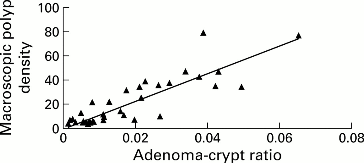
Adenoma:crypt ratio versus macroscopic polyp density for all patients. Macroscopic density is the total macroscopic polyp number divided by colon length.
Despite this strong association, a small number of patients showed discordance between macroscopic and microscopic counts. For example, a boy and girl, siblings aged 17 and 21 years (index Nos 18 and 19, respectively), had an almost twofold difference in macroscopic polyp count. Their adenoma: crypt ratio was the same, suggesting pathologist counting error rather than true discordance. Similarly, a 14 year old boy and 14 year old girl siblings (index Nos 25 and 26) had a threefold difference in macroscopic count but virtually identical adenoma:crypt ratios. On the other hand, two brothers aged 13 and 17 (index Nos 8 and 9, respectively) and siblings aged 18 and 17 (case Nos 22 and 23) had similar macroscopic polyp counts but more than twofold differences in their adenoma:crypt ratios. The data for mutation type, colonic polyposis severity, and adenoma:crypt ratio are summarised in table 1.
Summary data for colonic polyposis severity. Data for the 44 individuals on whom adenoma:crypt ratios were calculated. Individuals are numbered for identification (family No–pedigree No). All patients had prophylactic colectomy and ileorectal anastomosis. Macroscopic count is corrected for length of colectomy specimen
Further regional analysis of the slides was performed to see whether adenoma:crypt ratio varied according to location in the colon. Two factor analysis of variation was performed on 13 slide sets which contained sections from all regions of the colon (caecum, ascending, transverse, descending, and sigmoid). The results are summarised in table 2. Variation observed was attributable solely to between patient factors and not between region factors. The distribution of adenomas at the microscopic level throughout the large bowel did not deviate significantly from random.
Regional analysis of adenoma distribution in familial adenomatous polyposis colectomy specimens. Two factor analysis of variation is summarised
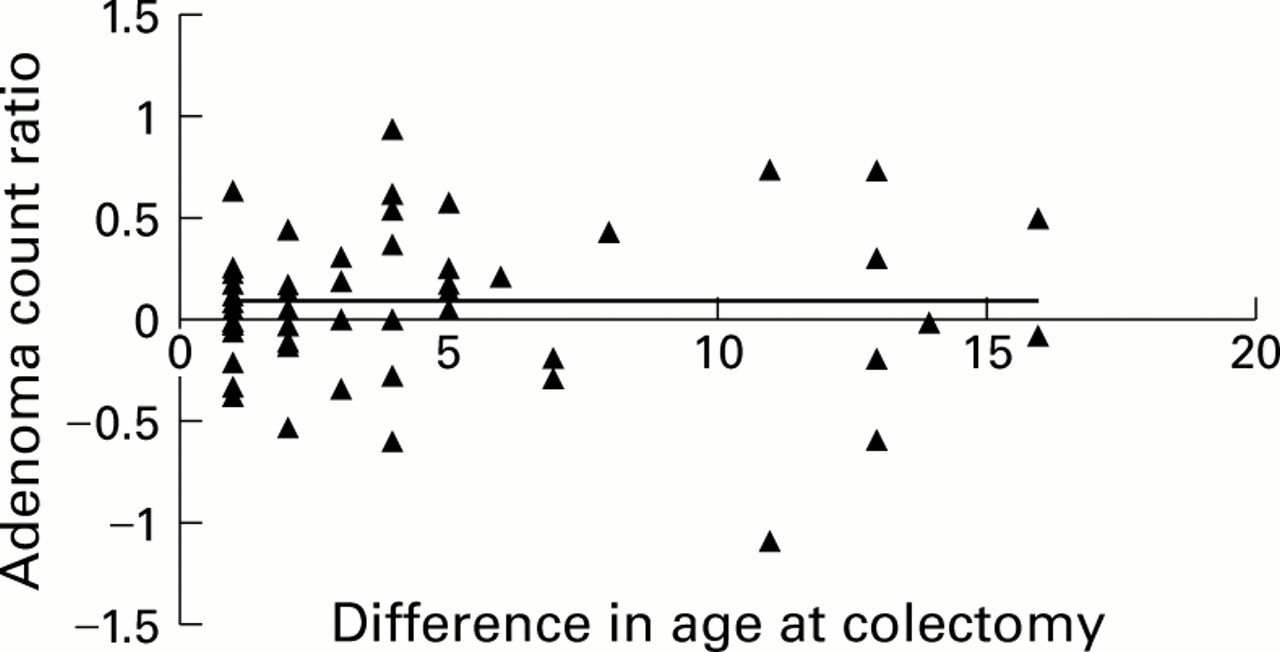
To determine the dependence of polyp counts on age (around the time of colectomy) the relationship between the difference in colonic polyposis severity and difference in age at colectomy was investigated. Figure 2displays the log of the ratio of macroscopic adenoma count between siblings plotted against the difference in age at colectomy. The Pearson product moment coefficient was 0.002 and the index of association 5.28×10⁻⁶. This was not significant. The residual variation was strongly significant (p<0.001).

Difference in age at colectomy correlated with difference in macroscopic polyp count. Difference in age at colectomy is (age at colectomy of older sibling−age at colectomy of younger sibling). The y axis shows the log (older sibling macroscopic count/younger sibling macroscopic count). This method allows comparison within families with the same mutation and between families with different APC mutations; it shows that there was no significant trend for increasing numbers of polyps in the sibling who was older at colectomy.
Discussion
The data presented showed a significant linear correlation between macroscopic and microscopic adenoma counts in the FAP colon. The bulk of the variation in macroscopic count was accounted for by variation in microscopic count (index of association 0.67)—that is, the linear relationship between the two parameters was close and there was no evidence for additional factors acting on the microadenomas to determine macroscopic polyp count. This strong association was present despite potentially confounding effects of non-random retrospective polyp counts performed, for example, in regions of denser macroscopic polyposis. These data are consistent with the hypothesis that progression from microadenomas to macroscopic size is essentially random. Variation in the severity of colonic FAP therefore results from differences in the numbers of microadenomas rather than in progression from microadenoma to microscopic lesion. Thus if different APC mutations provide different selective advantages, and if modifier genes act on colonic FAP, their main effects appear to be on tumour initiation rather than progression.
Our failure to find an association between age and severity of colonic polyposis around the time of colectomy is in accordance with previous models6 which have proposed that at least from the time of clinical presentation in the early teens, the number of polyps increases only slowly. These data are also consistent with results in the mouse in which susceptibility to chemical carcinogens was greatest in a “window” between 5 and 14 days post partum.8
There are potentially some confounding factors which may have affected the observed relationship between age and macroscopic adenoma count. Firstly, the decision on when to perform colectomy is not a random one. The usual practice at St Mark's is for colectomy to be performed in late adolescence, with the exact date tailored to an individual's needs, for example school terms and examination timetables. This is reflected in the young age at colectomy in our sample. Secondly, as the age difference of colectomy increases, it becomes increasingly likely that the older sibling had a delayed diagnosis or a less severe type of disease. Thirdly, for some siblings sociological factors may delay both age at diagnosis and operation age. The likely net effect of these factors would be to lower the observed difference in adenoma ratio with age.
Data from the regional analysis of adenoma: crypt ratio suggest that for a population of colons of a given germlineAPC mutation, the distribution of adenomas probably conforms to a Poisson distribution (data not shown). We found no statistically significant change in adenoma:crypt ratio as the colon was traversed although there was a weak trend to more severe disease in the distal large bowel. This finding is consistent with the hypothesis that initiation of both sporadic and FAP adenomas are spontaneous events, although weak environmental effects cannot be excluded.
Our data relating microscopic and macroscopic FAP will have practical value for researchers into FAP and colorectal cancer for a number of reasons. Firstly, macroscopic data may now be compared between centres allowing data validation using microscopic counts. This is not currently possible, as most whole colon specimens are usually destroyed once pathological examination is complete. Secondly, from the error range of the data presented, minimum difference criteria can now be estimated for measuring the severity of the colonic polyposis phenotype for each genotype. This will be important for quantitation of the disease and the search for modifier genes.9 Thirdly, it will be possible to estimate the severity of colonic polyposis on colectomy specimens where macroscopic polyp counts have not been performed. Fourthly, we have shown that age is not a major confounding factor in terms of assessment of colonic FAP polyposis severity. This is very important, as it is not infrequent for sibling pairs with the same mutation to undergo colectomy at quite widely differing ages. Finally, microscopic adenoma counts may be useful indicators of the validity of polyp regression, as is sometimes seen after colectomy, sulindac, and the COX-2 inhibitor celecoxib, indicating whether the phenomenon is one of real polyp loss or simply an alteration in polyp size so that the clinical observer no longer notices some of the polyps. The risk of cancer may be reduced if only a reduction in polyp size was achieved, but an even greater preventative effect might be achieved if actual polyp loss occurred.
Abbreviations used in this paper
- FAP
- familial adenomatous polyposis