Article Text

Abstract
Background: Coeliac disease (CD) is an enteropathy mediated by gluten specific T cells which secrete interferon γ (IFN-γ) when stimulated by gluten peptides presented by HLA-DQ2 or DQ8 molecules. Residues 62–75 of α2 gliadin have been proposed as the immunodominant epitope in the majority of CD patients. Deamidation by tissue transglutaminase (tTG) of the glutamine (Q) at position 65 to glutamic acid (E) is essential for T cell stimulation.
Aims: To investigate the antigenicity of this peptide and to establish whether its T cell activating properties can be downregulated by the formation of altered peptide ligands.
Patients: Individuals with known CD.
Methods: Peptide G4 corresponding to α2 gliadin residues 62–75, Q-E65 and analogues, substituting each amino acid, except E65, in turn for alanine residues, were synthesised. Small intestinal biopsies were obtained from patients. Biopsies were cultured overnight with a peptic/tryptic digest of gliadin (PTG). Lymphocytes were cultured and restimulated with tTG treated PTG. A T cell line was cloned and clones tested for stimulation and IFN-γ production in response to G4 and its analogues.
Results: Some high activity clones were isolated with, for example, a stimulation index (SI) of 15 to G4 and secreting 327 pg/ml of IFN-γ. Substitution of amino acids at several positions abolished or downregulated stimulation and IFN-γ production.
Conclusions: Peptide G4 is highly immunogenic. Certain amino acid substitutions in peptide G4 abolish T cell reactivity while others are partial agonists which may have potential in immunomodulation in this condition.
- APC, antigen presenting cells
- IL, interleukin
- OVA, ovalbumin
- CD, coeliac disease
- PBMC, peripheral blood mononuclear cells
- CPM, counts per minute
- PTG, peptic/tryptic gliadin
- FFIII, Frazer’s fraction III
- HLA, human leucocyte antigen
- SI, stimulation index
- IFN-γ, interferon γ
- TCR, T cell receptor
- tTG, tissue transglutaminase
- TNF-α, tumour necrosis factor α
Statistics from Altmetric.com
- APC, antigen presenting cells
- IL, interleukin
- OVA, ovalbumin
- CD, coeliac disease
- PBMC, peripheral blood mononuclear cells
- CPM, counts per minute
- PTG, peptic/tryptic gliadin
- FFIII, Frazer’s fraction III
- HLA, human leucocyte antigen
- SI, stimulation index
- IFN-γ, interferon γ
- TCR, T cell receptor
- tTG, tissue transglutaminase
- TNF-α, tumour necrosis factor α
The gluten dependent enteropathy characteristic of coeliac disease (CD) is mainly induced by production of interferon γ (IFN-γ) by gluten specific T cells.1,2 Approximately 95% of CD patients in the UK express the human leucocyte antigen (HLA) class II molecule DQ2. The majority of the remainder of patients express the rarer antigen DQ8.3 All small intestinal gluten sensitive T cell clones identified to date are DQ restricted, reflecting the critical role of these molecules in gliadin antigen presentation.4 It has been suggested that the T cell response may be dominated by two overlapping immunodominant epitopes.5,6 However, a significant body of literature suggests other gluten peptides may also be involved in the disease process.7–16
Peptides corresponding to positions 57–68 and 62–75 of wheat α gliadins stimulated all small intestinal gluten sensitive T cell clones from four adult Norwegian coeliac patients who carried HLA class II DQ2.6 Deamidation of glutamine residues at position 65 to glutamic acid in both molecules was essential for optimal HLA class II binding and subsequent T cell activation. This may be mediated by the enzyme tissue transglutaminase (tTG), either in vivo17 or in vitro.18 A single gluten sensitive clone from a DQ8 positive Norwegian patient did not respond to either of these peptides nor did clones from the majority of Dutch children with CD.6 However, studies of peripheral blood lymphocytes from HLA DQ2 adult coeliacs suggested immunodominance of a similar peptide.5
We wished to (i) establish the antigenicity of these peptides in activating small intestinal T cells from British adult coeliac patients and (ii) to investigate the possibility of immunomodulation by single point amino acid substitutions of these putative immunodominant peptides.
METHODS
Antigens
Peptic/tryptic gliadin (PTG) and Frazer’s fraction III (FFIII), a peptic/tryptic digest of whole gluten, were prepared.19
Gliadin peptides, including alanine point substitutions, were synthesised by Fmoc chemistry and a solid phase peptide synthesiser (Applied Biosystems 431 A, Wieterstadt, Germany).
The crude peptides were roughly purified by reversed phase liquid chromatography using C18 silica gel Lichroprep (Merck No 109303, Poole, Dorset, UK). The second purification step included reversed phase high pressure liquid chromatography. The identity of peptides was checked by electrospray mass spectrometry (LCQ, Finnigan mat). All purified peptides were chromatographically pure (>99 %) and had masses corresponding to the theoretical values.
The sequences of the peptides are shown in table 1.
Amino acid sequences of synthetic peptides and substituted peptides used in this study (single letter code)
tTG deamidation
Deamidation mixes were set up as follows: 100 μg/ml guinea pig liver tTG (Sigma, Poole Dorset, UK), 2 mM CaCl2, 400 μg/ml PTG, FFIII, or ovalbumin (OVA). Incubation was for four hours at 37°C.
Patients
Patient A was a 39 year old female with CD who had been on a gluten free diet for six months.
Patient B was a 49 year old male who was antiendomysial antibody positive. Biopsy revealed partial villous atrophy which responded to a gluten free diet, confirming a diagnosis of CD.
Patient C was a 62 year old man with CD who had been on a gluten free diet for eight months.
Patient D was a 29 year old woman who was on a gluten containing diet with a finding of positive antiendomysial antibodies. Biopsy revealed partial villous atrophy which responded to a gluten free diet, confirming the diagnosis of CD.
All patients gave written informed consent to the study which had been passed by the St Thomas’ Hospital ethics committee.
DR/DQ typing
Where possible, low resolution DQ typing of subjects was performed using polymerase chain reaction-single strand conformational polymorphism, as described previously.20
Establishment of small intestinal T cell lines
Methods were based on those of Molberg and colleagues.21 In later experiments, Plasmocin (2.5 μg/ml; Invivogen Ltd, Cayla, Toulouse-Cedex, France) was used at all stages to prevent infection with mycoplasma. Small intestinal biopsies obtained from coeliac patients were incubated overnight in the presence of 5 mg/ml FFIII or PTG. Biopsies were then disrupted to release lymphocytes and passed through a 70 μm cell filter (Falcon; Becton Dickson Ltd, Cowley, Oxford, UK). The collected cells were cultured in RPMI medium with 10% heat inactivated autologous plasma plus 1×10⁶/ml irradiated autologous peripheral blood mononuclear cells (PBMC) and 10 U/ml human recombinant interleukin 2 (IL-2) (Amersham, High Wycombe, Bucks, UK). Cells were restimulated every seven days with FFIII or PTG, both pretreated with tTG, at a final concentration of 200 μg/ml. Autologous irradiated (22 Grey) PBMC acted as antigen presenting cells (APC).
T cell transformation assays
T cell transformation assays were performed a minimum of seven days after antigenic stimulation. The antigens tested were tTG deamidated PTG or FFIII (200 μg /ml) and peptides G9 (10 μg/ml: 3.7 μmol/ml), G8 (10 μg/ml: 3.7 μmol/ml), G4 (3.7 μmol/ml), or G5 (3.7 μmol/ml). tTG treated OVA (200 μg /ml) was the negative control.
Complex antigens were incubated overnight with APC (5×10⁴) prior to addition of T cells (5×10⁴); peptide antigens were incubated for four hours only. Following incubation for 18–48 hours, tritiated thymidine was added for 18 hours prior to harvesting and counting of thymidine incorporation. The stimulation index (SI) was calculated by dividing the mean counts per minute (cpm) for T cells plus APC plus antigen by the mean cpm for T cells plus APC. An SI of 2 or more was considered positive.
In some cases, culture supernatants from the above assays were collected and the production of IFN-γ and interleukin 4 (IL-4) were measured using a kit, according to the manufacturer’s instructions (R&D systems, Minneapolis, USA). In later experiments, high sensitivity IL-4 and IL-10 detection kits, supplied by Amersham (High Wycombe, Bucks, UK) were used.
Cloning
Gluten antigen specific T cell lines were cloned at 1 cell per well in Terasaki plates in the presence of 1 μg/ml phytohaemagglutinin (Abbott Laboratories, Queenborough, Kent, UK), 1×10⁶/ml irradiated allogenic PBMC, and IL-2 10 U/ml. After 7–10 days the plates were screened. Clones were expanded using the same stimulation mix and then tested for reactivity with tTG treated PTG. PTG specific clones were tested with peptides and analogues.
RESULTS
T cell lines
A line stimulated by gliadins only
Results for patient B (DQ8) are shown in table 2. The biopsies were cultured overnight in the presence of PTG—that is, gliadins only. T cells were grown up and the line tested after seven days. The line displayed strong reactivity to both PTG and FFIII, with stimulation indices of 9.5 and 8.8, respectively.
Reactivity of a T cell line developed by incubating biopsies overnight with PTG—that is, gliadins only
Lines stimulated by gliadins and glutenins
Table 3 shows the responses of a T cell line derived from biopsies (patient A) cultured overnight with FFIII—that is, gliadins and glutenins. After one restimulation with phytohaemagglutinin, stimulation indices of 30 to FFIII and 6 to PTG were obtained.
Reactivity of a T cell line stimulated with a mix of gliadins and glutenins
The line from patient C (table 4), biopsies again having been cultured overnight with FFIII, was tested after four subsequent restimulations with FFIII. The line had an SI of 80 for PTG, 94 for FFIII, and 39 for peptide G9.
Reactivity of a T cell line with FFIII as the stimulating antigen
Attempts to clone these three lines failed; this was thought to be due to mycoplasma infection. For patient D, Plasmocin was added to eradicate this problem.
Changing reactivity of a line during culture
Biopsies from patient D were stimulated overnight with PTG and restimulated once with PTG. One week later, the T cell line had an SI of 3 for PTG and 4 for FFIII and peptide G9 (table 5). After a further two restimulations, the response to FFIII had doubled to 8 whereas the response to peptide G9 had increased eightfold to 37 (table 6).
Reactivity of a T cell line from patient D stimulated with PTG after two weeks in culture
Reactivity and cytokine secretion of a T cell line from patient D stimulated with PTG after four weeks in culture
T cell clones
The above line was cloned and 30 clones resulted. Eleven clones with strong growth were tested initially against tTG treated PTG.
Cytokine profile
The three clones with the highest stimulation to PTG were screened for their reaction to peptides G9, G4, and G5. Results are shown in table 7. In each case there was a high level of reactivity to G9 and G4, with somewhat lower levels to peptide G5. IFN-γ activity to some extent mirrored these results. Whereas clone 6 secreted only IFN-γ, a Th1 profile, clones 9 and 8 secreted IFN-γ and IL-4 in a ratio 4:1—that is, a Th0 profile (see tables 8 and 9).
Reactivity and interferon γ (IFN-γ) secretion of clones 6, 8, and 9
Comparison of reactivity of clone 9 with deamidated and non-deamidated peptide (peptide G9 v G8)
Reactivity of clones 6 and 8 with alanine substituted analogues of peptide G4
Effect of deamidation at Q65 on T cell stimulation
To confirm the importance of deamidation of the glutamine residue at position 65, we tested the response of clone 5 with peptides G8 and G9—that is, the combined G4 and G5 peptides with (G9) and without (G8) deamidation at position 65. SI to G9 was 56 while SI to G8 was 61. The difference between the means of triplicate results was not significantly different using the Student’s t test (p=0.45) (data not shown). We tested clone 9 with various concentrations of peptides G9 and G8. The results are shown in table 8. At various concentrations of peptide, stimulation of T cells with peptide G8 was consistently 50% less than that with G9. Interestingly, at 10 μg/ml of peptide, IFN-γ secretion was 50% lower in response to G8, mirroring T cell stimulation whereas IL-4 secretion was abolished. In a later experiment we showed that clone 9 could be stimulated (SI 2.5) by peptide G9 at 0.1 μg/ml. With peptide G8 at 0.1 μg/ml, the SI of 1.97 fell below the cutoff for positivity but the mean of triplicate counts was significantly different to that without antigen (p=0.012) (data not shown).
HLA class II restriction
Stimulation of clones 5 and 6 by peptide G9 could be blocked by 58% and 59%, respectively, by preincubation of antigen pulsed autologous PBMC with the anti-DQ2 antibody SPV-L3 (data not shown). Similarly, stimulation of T cell clone 5 could be achieved using human bare lymphocytes transfected with human DQ2,22 albeit with a relatively low SI of 3.2 (data not shown). Patient D was formally typed as DR3, DQ2.
Effect of amino acid substituted peptides on T cell stimulation and cytokine secretion
The SI of T cell clones with peptide G4 was compared with that of amino acid substituted peptides. Initially, we used T cell clone 6. In the case of testing peptides G4-13A and G4-14A, there were insufficient cells from clone 6 available and therefore clone 8 was used. Table 9 demonstrates that while substitutions G4-11A through G4-14A had no effect on T cell stimulation or IFN-γ production, other substitutions had a profound influence. Substitutions at positions G4-3A through G4-10A abolished T cell stimulation and IFN-γ production, with the exception of position 9, where there was residual IFN-γ production, equivalent to that without antigen. Substitutions at positions 1 and 2 led to partial downregulation of T cell stimulation and IFN-γ secretion. Clone 6 did not secrete IL-4. The results were confirmed in clones 4 and 9 from the same individual (see fig 1A, B). Again, G4-1A and G4-9A appeared to afford slight T cell stimulation and/or IFN-γ secretion but G4-2A was inactive with both of these clones. With clone 9, IL-4 secretion (60 pg/ml) fell below the detection limit of the assay (30 pg/ml) when any of the substituted peptides was used and therefore we could not calculate the ratio of the pro:anti-inflammatory cytokines. In the case of clone 4, we measured IL-10 secretion and showed that substitutions G4-1A and G4-9A allowed partial secretion of IL-10 compared with that with the native peptide G4 (fig 1C).

Effect of alanine point substitutions of peptide G4 on stimulation of clone 4 (A) and clone 9 (B). Bars represent the mean (SEM) of triplicate counts per minute (CPM) obtained for each test. Curves shows interferon γ (IFN-γ) secretion (detection limit of the assay was 15 pg/ml in (A) and 30 pg/ml in (B)). (C) Effect of alanine point substitutions of peptide G4 on interleukin (IL)-10 and IL-4 secretion of clone 4. The lower detection limit of the IL-10 assay was 1 pg/ml and for IL-4, 1.25 pg/ml. APC, antigen presenting cells.
DISCUSSION
Gliadins and glutenins as stimulating antigens
Early experiments show that the antigen used for prestimulation of biopsies and subsequent stimulation and expansion of a line may affect its specificities. This suggests that there may be T cell antigenic determinants present in FFIII (a whole gluten fraction) which are not present in PTG (gliadins only). Van de Wal and colleagues15, 16 used peptic tryptic gluten to stimulate the growth of intestinal T cells and isolated two clones which recognised glutenin peptides. However, Arentz-Hansen and colleagues6 used PTG to stimulate T cells overnight in biopsy explants and produced a set of clones which responded to a particular gliadin epitope. Our data support the notion that the glutenins, comprising a heterogeneous group of proteins, high and low molecular weight subunits of glutenin, and glutenin bound gliadin, may contain T cell antigens although these differ between protein types. The range of T cell antigenic determinants may also vary between individuals.
Immunogenicity of the peptides
Two groups have published evidence of immunodominant epitopes within the α gliadins.5,6 In the two patients that were tested by us—that is, patients C and D—the corresponding peptides appeared to be highly immunogenic. In patient D we examined the response after two and four weeks in culture. Although there was some response to peptide G9 in the early culture, the very high response was not seen until later. This patient was on a gluten containing diet when biopsied—that is, a mixture of gliadins and glutenins. When the T cell line was repeatedly stimulated in vitro with gliadins only, the response became directed towards peptides G9 and G4. It is possible that the very high level of response to this peptide may not be fully representative of the situation in vivo but to some extent an artefact of persistent culture with gliadins only, leading to epitope focusing in vitro. This result, obtained in one patient only, needs confirmation before such a conclusion could be drawn.
T cell clones
For patient D, the first three clones, selected only on the basis of good growth and high reactivity with PTG, were highly reactive with peptides G9, G4, and G5, the putative major and minor immunodominant epitopes.6 Additionally, there was high production of IFN-γ by these clones. The data again indicate that these peptides are highly immunogenic and are cross reactive at the T cell clone level with the larger combined peptide G9. Our unpublished data demonstrate that peptide G8—that is, native undeamidated G9—causes the histological lesion characteristic of CD.23
Effect of deamidation at position Q65
A peptide without deamidation at position Q65 (G8) was of similar activity to the deamidated peptide G9 when used at high concentration with clone 5. We repeated the experiment with clone 9 and found that with decreasing concentration of antigen the response to non-deamidated peptide was consistently approximately 50% of that with deamidated peptide. Our data, while slightly different, do not contradict those of Molberg and colleagues18 who reported the necessity for deamidation in T cell recognition of gliadin. In the latter case a minority of highly reactive clones gave good stimulation even without tTG treatment although the SIs were always less than with tTG deamidation. Our results with clone 9 suggest that although the DQ2 binding cleft has a preference for an amino acid with a negatively charged side chain24—that is, glutamic acid rather than glutamine at position 65—this is not essential for binding, and given suitably reactive clones a high level of stimulation may be achieved even in the absence of optimal DQ2 binding. The level of T cell stimulation was mirrored by IFN-γ production although IL-4 production was abolished, suggesting a subtle change in T cell function, secondary to a change in DQ2 binding characteristics caused by lack of a negative side chain at position 65. We suggest that clone 5 may have a T cell receptor (TCR) which is highly promiscuous with respect to the tightness/confirmation of binding of peptide by DQ2.
Effect of amino acid substituted peptides
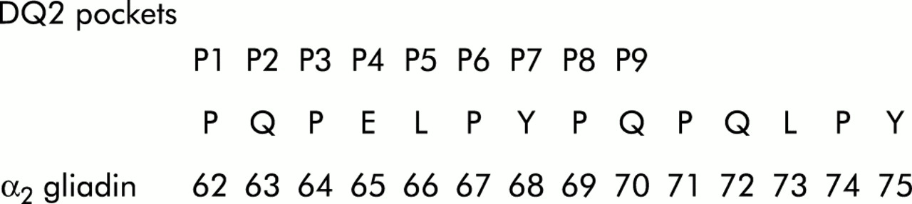
We used peptides with sequential amino acid substitutions to examine the specificity of the clones. Alanine was used as this is a small neutral amino acid. Substitutions G4-3A through G4-10A abolished T cell stimulation and IFN-γ production by clones 4, 6, and 9, with the exception of peptide G4-9A which generated a low stimulation and/or cytokine production from all clones. Substitution at position 1 caused a partial downregulation of T cell stimulation, IFN-γ and, in one case, IL-10 secretion. Our data support those of others6 who, using lysine residues to substitute three positions in α2-62-75 (G4), surmised that amino acids 62–70 lay within the binding cleft of DQ2 (see fig 2). We found that substituting any of these residues with neutral alanine profoundly affected T cell activation, suggesting that either DQ2 binding or TCR contact was affected. Whereas Arentz-Hansen et al noted that substitution of the neutral amino acid leucine with a positively charged one (lysine) at position 66 did not affect DQ2 binding or T cell activation, we found that T cell activation was obliterated by the substitution of a neutral alanine residue, peptide G4-5A. This is interesting because this residue is thought to point towards the TCR when a peptide is positioned within the DQ2 binding cleft.24 As the substitution in peptide G4-5A involves changing only one neutral amino acid (leucine) for another smaller one (alanine), interaction of this part of the peptide with the TCR may be exquisitely sensitive to change. According to the model of Vartdal and colleagues24 and Arentz-Hansen and colleagues,6 the key amino acid residues for DQ2 binding lie at positions 1, 7, and 9, with preferential residues at positions 4 and 6 (fig 2). We found that substitutions at positions 2, 3, 5, and 8 also profoundly affected T cell stimulation indicating that these residues may all interact with the TCR. Similarly, the residue at position 10 lies outside the DQ2 binding cleft but appears to be essential for T cell stimulation.

Proposed binding motif for peptide G4 in HLA-DQ2.6
Amino acid substituted peptides as partial agonists
Our finding that substitutions at positions 1 and 9 and possibly 2 lead to a partial downshift in T cell stimulation and cytokine secretion implies that altered binding within the DQ2 binding cleft could lead to reduced affinity and/or conformational changes in the gliadin/DQ2 complex, affecting TCR contact. Peptides G4-1A and G4-9A appear to be partial agonists which might act as altered peptide ligands in immunotherapy.
Altered peptides ligands in immunotherapy
Mouse experimental allergic encephalomyelitis has been studied and is a Th1 disease, dominated by the production of the proinflammatory cytokine tumour necrosis factor α (TNF-α). Alteration of a single amino acid within the immunodominant epitope within myelin basic protein led to a large reduction in the production of the proinflammatory cytokine TNF-α and a smaller decrease in the production of the anti-inflammatory cytokine IL-4 by antigen specific T cell clones. This constituted an overall decrease in the ratio of pro:anti-inflammatory cytokine.25 Additionally, this altered peptide ligand caused a switch in cytokine production by T cells directed against minor epitopes, a phenomenon known as bystander suppression. In order to generate this effect it appears essential to have a partial agonist. Any altered peptide which causes total abolition of T cell stimulation and cytokine production will not act as an immunomodulatory peptide.26–28
The parallels with CD are obvious. The T cell response in CD is of the Th1/Th0 type, with IFN-γ as the main cytokine and co-secretion of small quantities of IL-4 and IL-10 by some gluten sensitive clones.1 There appears to be an immunodominant epitope, and we have shown a similar pattern of secretion by our clones in response to this. We have shown that our potential altered peptide ligands cause a partial downregulation of IFN-γ, and in one case IL-10 production. It is not possible to comment at this stage on the pro/anti-inflammatory cytokine ratio; further experiments with other clones will be required.
The experiments described here confirm the observations of Anderson and colleagues5 and Arentz-Hansen and colleagues6 that highly antigenic peptides reside in the N terminal regions of α gliadins, although our experiment did not address the subject of immunodominance. Preliminary results suggest that modification of putative immunodominant epitopes within wheat gliadin might form the basis for immunotherapy in CD.
Acknowledgments
HJE, ELP, WE, HW, and PJC would like to thank the German Federal Ministry of Education and Research (project No 0312246D) for financial support. The authors also wish to thank Coeliac UK and Action Research for financial support. Additionally, we thank Professor Adrian Hayday for helpful discussions.