Article Text

Abstract
The occurrence of strictures as a complication of Crohn's disease is a significant clinical problem. No specific antifibrotic therapies are available. This systematic review comprehensively addresses the pathogenesis, epidemiology, prediction, diagnosis and therapy of this disease complication. We also provide specific recommendations for clinical practice and summarise areas that require future investigation.
- Crohn's colitis
- Fibrogenesis
- Fibrosis
- IBD Clinical
Statistics from Altmetric.com
Search strategy and study selection
A comprehensive literature search was conducted to identify all relevant citations. The electronic exploration involved keyword searches in Embase, Medline (service of the US National Library of Medicine and the National Institutes of Health) and the Cochrane library, supplemented by manually reviewing the reference list of included studies as well as relevant review articles. The search included studies from 1960 to October 2012 and the following search criteria were used (all fields): (‘Crohn's disease (CD)’ OR ‘Crohn's’ OR ‘Ulcerative colitis’ OR ‘Inflammatory Bowel Disease (IBD)’) AND (‘stricture’ OR ‘fibrosis’ OR ‘stenosis’ OR ‘complication’ OR ‘surgery’). References from those articles were examined for additional studies meeting these criteria. FR, EMZ, FHR and WJS assessed the articles and their relevance to the above topic. The date from these articles as well as our own experiences form the basis of this review article.
Pathogenesis: molecular and gross histopathology
The modern understanding of the pathogenesis of Crohn's disease (CD) has emerged from research on the lymphocyte interactions and regulation of cytokine expression, genetics, gut barrier function, and the relationships between the mucosal immune system and the microbiota. Less well understood are mechanisms driving the development of complications of the disease including the formation of fibrotic strictures.
In the presence of an intestinal wound, mesenchymal cells (fibroblasts, myofibroblasts and smooth muscle cells—in this review termed ‘mesenchymal cells’) accumulate in the area of the defect and secrete extracellular matrix (ECM) components, such as collagens and fibronectins, to close the defect1–3 (figure 1). This explains the remarkable ability of the gastrointestinal tract for self-regeneration following short-lived and mild insults, as in peptic ulcer disease, infectious enteritis or mild diverticulitis. However, if inflammation becomes chronic and severe, as in CD, inflammatory mechanisms drive the excessive production of ECM components. This attempt to repair tissue damage can result in a reduction in the diameter of the lumen, intestinal stenosis and ultimately obstruction.4 In this process inflammation and fibrosis are intimately intertwined mechanisms and coexist in intestinal stenoses to varying degrees. Intestinal mesenchymal cells were commonly believed to be a passive bystander of immune cell activation that reacts to its inflamed local environment by proliferation and ECM secretion. This view has proved to be overly simplistic. Fibrosis is a consequence of the pleiotropic actions of inflammatory mediators activated in the process of chronic inflammation (table 1). Growth factors such as transforming growth factor β1,5 ,6 insulin-like growth factor,7 ,8 platelet-derived growth factor9 ,10 basic fibroblast growth factor9 or cytokines, such as interleukin (IL)-13 or IL-1711 ,12 are all known to drive the changes in tissue architecture and function that can ultimately impact the structure and function of the small intestine and colon and result in clinical symptoms.3 ,13 An imbalance of tissue degradation through matrix metalloproteinases or cathepsins and their tissue inhibitors of metalloproteinases is likely to be involved.4 ,14–16 Mesenchymal cells themselves are highly motile and versatile cell types. Once the body senses an intestinal wound it recruits mesenchymal cells from the intestine through migration from adjacent tissue areas,17 proliferation of existing local mesenchymal cells,9 and differentiation from intestinal epithelial or endothelial cells through a process called epithelial or endothelial to mesenchymal transition.18 ,19 Circulating mesenchymal cell precursors and bone marrow stem cell-derived mesenchymal cells can be attracted to expand the pool of cells available for local repair of the intestinal mucosa.20 ,21 To enhance the complexity even further, the intestinal microbiota can act in a profibrogenic fashion.22 ,23 The factors that activate mesenchymal cells are the same ones that recruit them and therefore activation and expansion of mesenchymal cell numbers is likely to happen simultaneously. A detailed discussion of all mechanisms is beyond the scope of this review and can be found elsewhere.1–3
Profibrotic mediators in intestinal fibrosis

Interplay between tissue damage of the intestinal bowel wall, caused by recruited and activated leukocytes and tissue repair, exerted by intestinal mesenchymal cells. Chronic, severe inflammation and tissue damage leads to excessive repair and untimately intestinal fibrosis. ECM, extracellular matrix; MMP, matrix metalloproteinase; ROS, reactive oxygen species.
In chronic intestinal inflammation in humans as well as animal models a change from a predominantly inflammatory T helper 1 marker profile to a predominantly T helper 2 milieu, with an increase in profibrotic cytokines, such as IL-4, IL-13 and transforming growth factor β1, over time can be noted,24 ,25 further fuelling the excessive matrix deposition. It has also become apparent that fibrosis can progress independently of inflammation. Once matrix accumulates in the bowel wall it enhances tissue stiffness, which in itself acts as a mesenchymal cell activator via integrin-mediated mechanisms.26
It is widely accepted that stricture formation, given the transmural nature of CD, affects all layers of the bowel wall with histomorphological thickening, caused by ECM accumulation and mesenchymal cell expansion. However, to date no validated or even commonly accepted histopathological scoring system is available to grade the severity of fibrosis. Most studies propose their own semiquantitative or gross histopathological parameters.27–30 These include factors such as the severity of fibrosis and the thickness of the muscle layers. In some studies a ‘predominant stricture phenotype’ has been proposed. Due to the lack of controlled data the authors of this review cannot make any recommendation for a specific histopathological scoring system. Clinical scoring systems, such as the CD activity index, correlate to some degree with tissue inflammation, but not fibrosis. In fact, the occurrence of strictures can confound the CD activity index. This lack of any standardised scoring system for histological or clinical fibrosis makes comparisons between studies impossible.
Epidemiology, natural history and risk factors
At diagnosis, most CD patients present with predominantly inflammatory pathology. Then, over time, a majority of patients experience disease progression to complications such as strictures and fistulae. Using the Vienna classification 77% of patients had pure inflammatory disease at diagnosis, whereas strictures had already occurred in 11% and fistulae in 16%.31 These percentages shifted over time from inflammation to complications. In population-based cohorts 19–36% of patients newly diagnosed with CD present with complications.32–35 The cumulative rates of complication in patients with CD have been reported to range from 48% to 52% at 5 years and 69–70% at 10 years after diagnosis, with approximately half of the patients developing a stricture.31 ,36 Disease complications of stricture, fistula and abscess are the main indications for surgery in CD, and population-based cohort studies describe a cumulative risk of surgery between 40% and 71% within 10 years after diagnosis.34 ,37–42 Concepts describing the natural history of CD have evolved. The above described concept of different disease phenotype categories, that is, inflammatory, stricturing or fistulising is considered too rigid. The subsequent epidemiological natural history studies emphasised the progressive nature of CD leading to the modern concept of chronically accumulating bowel damage in CD variably manifesting as the disease complications of stricture, fistula and abscess.
In years past, patients who had strictures were believed to have a more indolent pre- and post-operative disease course compared to patients who had penetrating complications of fistula and abscess,43 ,44 even though only limited evidence for this concept exists. This is also reflected in the Vienna as well as the Montreal CD classification systems, as patients with fistulae are scored as having the highest level of disease complication, irrespective of the presence of strictures. Patients are only classified as having a stricture if this is the only complication that is present.45 Therefore, the incidence of strictures in studies using the Vienna or Montreal classifications is very likely to be underestimated. Pathogenetically, fistulae and abscess are thought to develop in regions of full thickness bowel wall inflammation in a high-pressure region upstream from a stricture.46 ,47 In one study the positive predictive value for fistulae predicting strictures was 86.2%.48 It is also widely believed that strictures, once present, are gradually progressive over time, but longitudinal data to confirm this belief do not exist.
The most common location of de novo strictures is the ileum and the ileocolonic region, presumably due to the smaller diameter of the ileum relative to the colon.49 ,50 However, strictures can appear at any site affected by CD, including the upper gastrointestinal tract, the colon and rectum. The frequency and location of de novo strictures probably resembles the distribution of inflammation—40–55% terminal ileum and colon, 15–25% colon alone, 25–40% exclusively ileum and up to 10% in the upper gastrointestinal tract, but data supporting this hypothesis are lacking.51 ,52 The National Cooperative Crohn's Disease Study reported at least one small bowel stricture in 25% and at least one colonic stricture in 10% of patients.53 After intestinal resection for the complication of a stricture, postoperative recurrence of CD at the anastomosis occurs commonly, particularly in patients with an ileocolonic anastomosis.41 ,54
Biomarkers for strictures could allow for an increased understanding of fibrogenesis and patient stratification. This knowledge could help to determine follow-up schedules and guide earlier intervention with highly effective therapy regimens in selected patients. The most commonly studied risk factors to date are clinical, environmental or endoscopic parameters55 (table 2). It has to be noted, however, that most of the following discussed biomarkers have not been shown to be specific for fibrostenosis per se, but rather represent ‘complicated’ or ‘debilitating’ CD courses, including stricture formation. Therefore, the presently available markers are at best only partly related to intestinal fibrosis.
Predictors of fibrostenosing Crohn's disease
The most commonly used clinical parameters for predicting a more serious course of CD are age of disease onset less than 40 years of age, perianal disease or the need for steroids during the first flare.56 ,57 If a patient carries two out of the three parameters the positive predictive value for disabling CD in the future is approximately 90%. A history of smoking is another risk factor for complicated CD58 ,59 and a faster rate of progression from diagnosis to first stricture.60 Patients with active colonic or ileocolonic CD with deep and extensive mucosal ulcerations have a higher risk of subsequent surgical intervention.61 Location of inflammation to the small bowel, rather than the colon, has also been identified as predictive of a patient's progression to stricturing disease and a higher rate of surgery.60 The commonly used classification systems, in particular the Montreal classification,45 only identify a stricture after it has become clinically apparent, and therefore using this classification to perform risk factor studies has substantial limitations.
Genetic markers have been proposed to predict stricture formation in CD. NOD2/CARD15, the first described gene to be linked to CD, confers a mild risk increase for stricturing CD. Carrying at least one out of the three common variants enhanced the relative risk by 33%.62 The more variants a patient carries, the higher the risk. Surgery for stricturing disease was significantly more frequent and earlier in CD patients with NOD2/CARD15 variants. In addition, NOD2/CARD15 variants conferred a higher risk of earlier surgical recurrence.63 However, as NOD2/CARD15 is also a risk factor for ileal disease location, it is difficult to separate the association of ileal disease location and stricture formation from the association of NOD2/CARD15 and stricture formation. Several single genes and their association with either stricture formation or time to stricture have been found. The 5T5T genotype in the MMP3 gene as well as a gene encoding a hypothetical protein near the IL-12B locus (rs1363670 GG homozygosity) are independently linked to fibrostenosis.64 ,65 The prevalence of the above genetic variants in the CD population is low and so is their penetrance, prohibiting their routine use in clinical practice.
A dysregulated immune response towards the luminal microbiota is a pathogenic hallmark of CD and gives rise to antimicrobial antibodies that can be found in the circulation of patients with inflammatory bowel diseases, in particular CD.66 These include the first discovered and best-described antibody anti-Saccaromyces cervisiae antibody, but recently several others, such as the antiglycan antibodies, CBir1, anti-I2 or OmpC, have been identified.66 The presence and concentration of these serological markers are quantitatively and qualitatively linked to more complicated CD, including strictures.66–68 However again, this predictive value is not specific for strictures, but rather a more complicated disease course. In addition, these markers are not specific for CD and can be found in other diseases,66 and their presence and concentrations are not stable over time.69
Taken together, clinical parameters are currently the most accurate predictor of fibrostenosing CD, but they are not specific for this phenotype and rather reflect a tendency towards developing complications of CD in general. The field is rapidly evolving and genetic or serological predictors might become relevant in the near future.
Diagnosis
In the later stages of intestinal fibrosis, patients can develop clinical symptoms of intestinal obstruction, which should trigger a diagnostic work-up. Several imaging modalities are available for assessing the intestinal tract. Barium small bowel follow-through can determine the extent and severity of narrowing. Endoscopy techniques, such as colonoscopy, enteroscopy or double-balloon endoscopy, are very sensitive to investigate changes in the superficial layers of the mucosa and can detect severe luminal narrowing by visualisation or inability to pass the scope, but cannot assess transmural disease and extraintestinal complications. In recent years, most data are available on cross-sectional imaging techniques, such as CT or MRI. Even in these powerful techniques, defining strictures is not straightforward. Bowel narrowing can be confused for underdistension or motility. In modern protocols, the use of glucagon and neutral oral contrast has minimised these effects. Furthermore, the definition of a ‘stricture’ is not uniform across studies, including luminal narrowing and bowel wall thickening with and without prestenotic dilatation,70 to small bowel narrowing with prestenotic dilatation,71 to lesion-causing narrowing below 1 cm.72 The presence of upstream dilatation, used in the most rigorous radiographic definition of a ‘stricture’, is not uniformly applied and may be too limiting to be a clinically useful definition.
CT and MRI
CT is gaining increasing acceptance in the evaluation of patients with small bowel disorders and has almost completely replaced traditional barium small bowel follow-through studies. Computed tomography enterography (CTE) is the most commonly used technique to assess CD, at least in the USA.73 CTE has the advantage of high spatial resolution, wide availability and rapid diagnosis. New dose-reduction techniques and algorithms for enhancing image quality have resulted in decreased radiation exposure,74 at least partly addressing the potential harmful long-term effects of ionising radiation.75
CTE is superior to conventional CT for the detection of small bowel pathology including strictures.76 Studies evaluating accuracy for the detection of small bowel stenosis differ widely in study design, inclusion criteria and gold standard and are hard to compare. CTE has been shown to have sensitivity for small bowel stenosis of 85–93% and specificity of 100%.71 ,72 ,77 ,78 While the presence of a single stricture was correctly determined in 100% of patients, the accuracy for the number of strictures was only 83%. CTE overestimated or underestimated the extent of complicated disease in 31% of CD patients.79 CTE altered clinical management in nearly half of patients with CD.80
MRI eliminates the ionising radiation exposure, but carries higher costs, takes more time and is not as readily available. Progress in this field was initially hampered by low spatial resolution and motion artefacts, but a newer generation of scanners and programmes with higher resolution and faster imaging sequences have increased its use in CD. MRI has a high diagnostic accuracy for diagnosis of CD (sensitivity 78% and specificity 85%). The sensitivity for MRI in the detection of stenoses ranges from 75% to 100% with a specificity of 91–100%.71 ,81–87 A comparison of magnetic resonance enterography (MRE) and enteroclysis revealed superior bowel distension with enteroclysis, but a comparable sensitivity and specificity for the detection of stenosis.87 A direct comparison of CT and MRI for diagnosis of stenosis showed generally comparable sensitivity (85% vs 92%) and specificity (100% vs 90%).71
Using surgical pathology/histology as a reference,27 standard CTE findings of inflammation including abnormal mucosal enhancement, mesenteric hypervascularity, mesenteric inflammatory stranding and bowel wall thickening correlated well with the histological grade of inflammation in CD strictures. However, the absence of CTE signs of inflammation in an involved segment of bowel did not predict fibrosis.27 Equivalent to CTE, mucosal enhancement and hypervascularity on MRE correlated with histological inflammation.88 ,89 On the contrary, MRI findings of bowel wall thickening have a negative association with response to medical therapy and a positive association with small bowel fibrosis.90 In a different study, CTE was considered accurate in differentiating between inflammatory and fibrostenotic lesions (77% and 79% accuracy, respectively).28 Inter-observer and inter-modality agreement between CTE and MRE was found to be high.91 Once therapy has been implemented CTE or MRE could be used to monitor response,92 ,93 but further studies need to address if in fact intestinal healing occurs in relation to the findings on cross-sectional imaging.
Currently, no cross-ectional imaging modality specifically detects fibrosis. Promise exists in a MRI sequence that is sensitive to stiff tissue, such as collagen, and relatively insensitive to tissue oedema and inflammation. This technique, termed magnetisation transfer MRI, is able to measure fibrosis semiquantitatively in a rat model of CD and in early studies when applied to patients with CD.94
In most investigations of the sensitivity and specificity of cross-sectional imaging techniques for identifying diseased bowel regions, surgery has been used as the gold standard. However, significant selection bias exists, because the prevalence of stricturing disease in this population is higher than the general disease population. Furthermore, as CD is a chronic progressive and destructive disease the currently available techniques to measure bowel damage are insufficient as they only depict one specific point in time. A longitudinal tool, the Lehman score has been proposed, which is currently being developed and validated prospectively.95 This score measures the progressive nature or cumulative structural bowel damage, independent of the current and fluctuating disease activity. This score could serve as a future methodology to assess fibrostenosing CD as it combines different diagnostic modalities (CTE, MRE, endoscopy) depending on the location of disease. Finally, existing cross-sectional imaging techniques do not specifically detect fibrosis. Fibrosis is often assumed when a thickened bowel segment lacks CT or magnetic resonance evidence of mucosal enhancement or hypervascularity. However, studies have shown that tissue inflammation and fibrosis are closely linked, such that a clear distinction based on standard CT or magnetic resonance criteria is not possible.27 ,30 In the future, unique magnetic resonance sequences,94 combined techniques96 or the application of techniques that exploit fibrotic tissue characteristics, such as ultrasound elastrography,97 will help us guide our therapeutic approaches.
Therapy
Up to 80% of CD patients require at least one surgical resection within 10 years of diagnosis.40 A large portion of these interventions are due to stricturing disease. To avoid surgery, medical as well as endoscopic treatment options have been described. In addition, stricturoplasties are employed to preserve bowel length.
Medical therapy
Traditionally, intestinal strictures in CD have been treated by oral and intravenous corticosteroids, bowel rest, and in the case of intractable symptoms, bowel resection. With the emergence of immunosuppressants, such as azathioprine/6-mercaptopurine and biological agents (anti-tumour necrosis factor α (TNF)) additional medical therapy options are available.98 In spite of this evolution, to date no specific intestinal antifibrotic therapy exists. Strikingly, the incidence of stricture formation and rate of surgery in CD did not decrease in past decades,99 despite the use of azathioprine.99 Data are emerging that earlier and prolonged use of immunosuppressive medication, including anti-TNF agents, can reduce the need for surgical interventions and hospitalisations.100 ,101 These results are encouraging, but have either been retrospective or in the case of prospective studies follow-up times were short. Nevertheless, these investigations suggest that our current ability to control inflammation is not sufficient consistently to prevent stricture formation. Whether earlier intervention with combination therapy with azathioprine and an anti-TNF agent would be more effective in the prevention of stricture formation is unknown.102 ,103
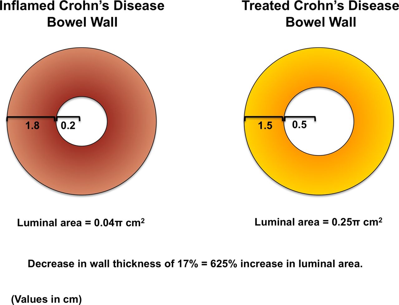
At the current time a variety of modalities are used to try and discriminate medically responsive strictures from those that will require surgery, such as imaging (CTE, MRE), laboratory biomarkers (C-reactive protein, erythrocyte sedimentation rate) and stool biomarkers (fecal calprotectin). Yaffe and colleagues104 reported their experience with non-operative management of acute small bowel obstruction in 26 CD patients. In all but one patient the obstruction was relieved within 72 h using a regimen that included clear liquid diet, small bowel tube, total parenteral nutrition, prednisone, intravenous fluids and intravenous crystalline adrenocorticotrophic hormone. Seventy-five per cent of patients experienced at least a second episode during a mean follow-up of 52 months, all of which again responded to medical management. Forty-six per cent of patients eventually underwent elective surgery. If the patients remained free of obstruction after the initial episode for at least 8 months the risk of surgery thereafter was only 17%, indicating that medical therapy can ultimately prevent surgery in a clinically meaningful proportion of patients.104 A theoretical example of an oedematous bowel before and after medical therapy is illustrated in figure 2.

Reduction of transmural oedema significantly affects the luminal cross-sectional area (adapted from Yaffe et al).104
There was initially some concern regarding the use of infliximab in patients with established strictures, based on two retrospective reports.105 ,106 Subsequently, this idea was challenged by a study of 20 CD patients, 15 of whom had obstructive symptoms, treated with infliximab. Small intestinal contrast ultrasound was performed. In no case was progression of strictures or the occurrence of new strictures seen. In 80% of the patients responding to infliximab the stenosis completely regressed.107 Most importantly, data in large numbers of patients from the TREAT registry and the ACCENT I infliximab maintenance trial did not show an increased risk of the clinical occurrence of strictures.108 A recent review on this topic came to the same conclusion.109
Currently, no specific medical therapy exists to treat fibrotic intestinal strictures. The dogma is that once fibrosis exists, it cannot be reversed. Data from hepatic cirrhosis and pulmonary fibrosis challenge this notion. Finding a specific antifibrotic therapy is an intense area of investigation in pharmaceutical companies and academic centres. A wide variety of mechanisms and platforms are being explored. Our hope is that in the next iteration of this or similar reviews, promising medical therapies for the treatment of fibrotic intestinal strictures will have emerged.
Endoscopic dilation
If medical therapy fails to improve obstructive symptoms, endoscopic dilation (ED) has become an accepted modality for the treatment of selected CD strictures. Main applications are short and isolated strictures within reach of a standard colonoscope, with many amenable strictures localised at the site of the ileocecal anastomosis after ileocecal resection.110 Most commonly through-the-scope balloons (TTS) are used to reach and pneumatically dilate strictures (figure 3). In general, the available reports are highly heterogeneous in respect of techniques used, follow-up times and endpoints applied.
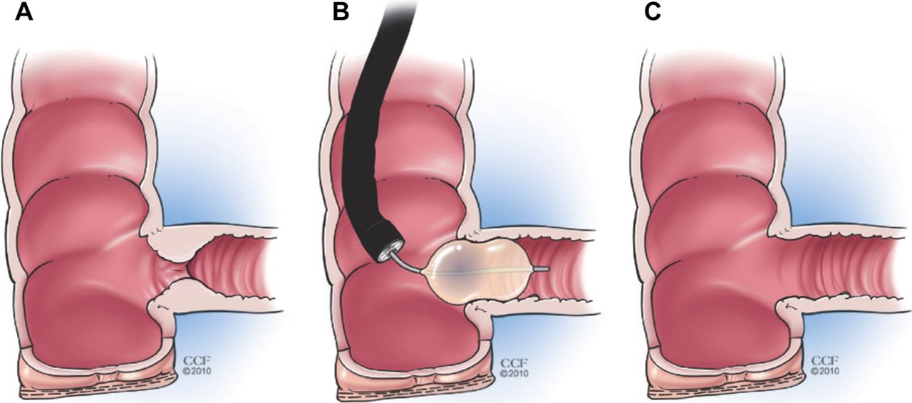
Through-the-scope endoscopic pneumatic balloon dilation of an anastomotic ileocecal stricture.
In a systematic review and descriptive pooled data analysis of 13 retrospective studies from 1990 to 2007, including 347 CD patients, the mean time from diagnosis of CD to occurrence of stricture was 13 years and the mean stricture length 2.7 cm (0.5–20 cm). This analysis mainly included postsurgical strictures and all were dilated with TTS. ED was technically successful in 86% of cases; 89% of the unsuccessful attempts had to undergo surgery. Long-term clinical efficacy (mean follow-up was 33 months), defined as being free of surgery was achieved in 58% of the patients. The mean interval between ED and surgery was 15 months in the remaining 42% of CD patients. A stricture length of 4 cm or less was associated with a surgery-free outcome in a multivariate analysis (OR 4, 95% CI 1.16 to 13.8). In subsequent dilations the estimation of clinical efficacy remained unchanged.111 In a single-centre study assessing 776 dilations in 178 CD patients (80% anastomotic) at 5-year follow-up 52% of patients had no further intervention or one dilation only and 36% of patients had surgery.112 Thienpont and colleagues113 reported the need for repeated dilation in 46% and surgery in 24% of CD patients during a mean follow-up of 5.8 years.
Factors influencing outcome after endoscopic balloon dilation in fibrostenotic CD are largely unknown. Technically successful dilation,114 ,115 stricture length of 4 cm or less111 and the absence of ulcer in the stricture116 are positively associated with successful dilatation. The data on smoking are inconsistent.116 ,117 In contrast, neither C-reactive protein concentrations, endoscopic disease activity nor medical treatment after dilation influenced the subsequent disease course in a different study.113 The majority of the observations were made with anastomotic strictures. No difference was noted when comparing the dilation efficacy or the probability of surgery-free survival of native versus postsurgical strictures.111 ,112 Small bowel adenocarcinoma is rare, but if overlooked can be fatal.118 In the CD-affected colon malignancy is more frequent and the incidence is comparable to ulcerative colitis.119 ,120 The endoscopist should therefore have a low threshold for taking a biopsy before ED. There is no convincing evidence that such mucosal biopsies increase the risk of perforation with subsequent balloon dilatation.121 The availability of deep enteroscopy, including double-balloon endoscopy makes dilatations in the more proximal upper small bowel feasible.50 ,122 ,123
When mechanically dilating the intestine, perforation is a valid concern. Only few studies with low patient numbers addressed the safety of this procedure. In a randomised controlled trial with 29 paediatric patients with ED and intralesional steroid injection no complications were reported.124 A systematic review including 13 studies reported a major complication rate (defined as bleeding, perforation, infection or other event leading to hospitalisation) of 2%, with this value being up to 11% and 18% in two series.115 ,125 In 776 dilations in 178 patients a complication rate of 5.3% has been reported, which included bowel perforation, major bleeding, minor bleeding and abdominal pain or fever.112 To our knowledge no death related to the procedure has ever been reported.
Adjuvant techniques to endoscopic dilation
Intralesional injection of steroids has been successfully used in other stricturing gastrointestinal conditions, such as peptic, corrosive or anastomotic strictures or fibrosis post-radiotherapy.126–129 Triamcinolone is considered an appropriate agent given its prolonged local effect, believed to last for 3–4 weeks.130
In CD strictures most available evidence is retrospective and uncontrolled. Intralesional steroid injection in 13 CD patients led to a 100% immediate success rate. However, no follow-up data were provided.131 In a small retrospective series assessing anastomotic strictures, steroid injection delayed re-stricturing and reduced re-dilation rate.132 Singh et al125 found a lower stricture recurrence rate in the steroid group compared to placebo. In a systematic review the use of steroid injection appeared to be related to dilatation efficacy.111 In a single-centre prospective randomised controlled trial with 29 paediatric CD subjects intralesional triamcinolone injections after ED, led to a longer time to re-dilation and to surgery in the steroid group compared to placebo. However, sample size was small and follow-up time short.124 It should be noted that a prospective study in 13 adult CD patients was terminated early after reporting that triamcinolone injection led to an earlier need for re-dilation compared to placebo.133 In this series, however, only anastomotic strictures were examined, the strictures were possibly long-standing (8–30 years after surgery) and the multicentre design could have influenced different endoscopic procedures among different centres. Small, non-controlled, case reports and series assessed the use of intralesional TNF inhibitor therapy with encouraging results.134 ,135
Endoscopic metallic stent insertion has been tried in a few patients. The initial success rate was reported to be 100%, but major complications, such as migration, perforation or fistulisation were frequent (67% of patients).136 In a prospective cohort study with 11 patients the authors concluded that the complication rate is too high to make this a routine treatment option, even when extractable stents are used.137 ,138 Biodegradable stents might be an emerging alternative.139 ,140 Finally, carving the stricture with a sphincterotome supplementing ED has been reported in one study with no increase in complications,141 and this technique has been successfully combined with steroid injections.142 A preliminary report indicates that patients receiving budesonide after dilation as opposed to dilation alone have a better outcome.143
Surgery
Resection
After failure of medical therapy and ED or inability to perform ED, surgical resection of the affected segment is currently the most commonly used treatment strategy. The problem with this approach arises when performing multiple bowel resections over time and the subsequent development of loss of gastrointestinal function and ultimately short bowel syndrome. Therefore, resection should be as limited as possible. Supporting this notion neither the microscopic presence of inflammation at the resection margin nor the type of anastomosis (end-to-end vs side-to-side) influences postoperative recurrence.144–146 In our clinical practice a gross examination of normal-looking bowel, complemented by a manual examination to evaluate for tissue thickening, in which the index finger opposes the thumb at the mesenteric–intestinal border, is sufficient to determine the resection margin in the operating room (figure 4).

Manual examination for evaluation of tissue thickening and definition of the resection margin: the index finger opposes the thumb at the mesenteric–intestinal border.
Stricturoplasty
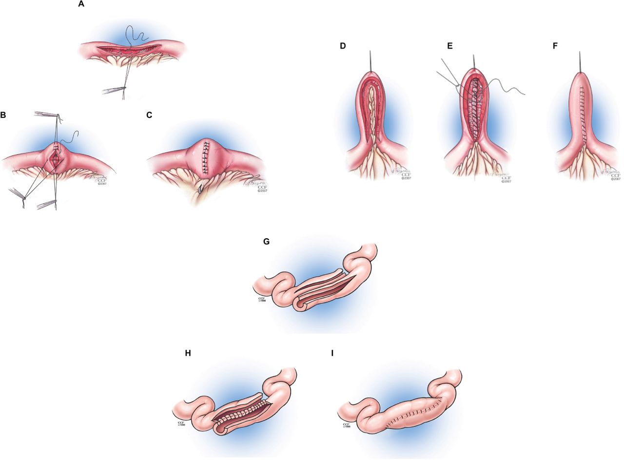
To preserve the bowel length and to lower the risk of anastomotic leakage stricturoplasty emerged as an option to widen the strictured area without shortening the bowel. Commonly performed techniques can be grouped into three categories: the Heineke–Mikulicz (HM)-like technique (commonly used for strictures <10 cm)147 (figure 5A–C); the intermediate procedures (eg, Finney procedure, for strictures >10 and <25 cm)148 (figure 5D–F); or the enteroenterostomies for strictures greater than 25 cm in length (eg, isoperistaltic side-to-side stricturoplasty by Michelassi)149 (figure 5G–I).

Surgical techniques for stricturoplasty. (A–C) Heineke–Mikulicz-like procedure; (D–F) intermediate procedure (eg, Finney procedure); (G–I) enteroenterostomies (eg, isoperistaltic side-to-side strictureplasty by Michelassi).
Indications for stricturoplasty include the presence of multiple strictures over an extensive length of bowel, previous significant small bowel resection (>100 cm), short bowel syndrome, strictures without phlegmon or septic fistula, duodenal strictures and anastomotic strictures.150 ,151 Contraindications have been reported, such as associated abscess or phlegmon, perforation with diffuse peritonitis, suspicion for carcinoma in the stricture or poor nutritional status. Strictureplasty can be performed safely in active disease.150 The overall complication rate of stricturoplasties is reported to range between 0% and 57%, with an average of 13%,152 with the incidence of major complications (eg, anastomotic leakage, abscess, fistula, sepsis) being approximately 6%.153 The median incidence of perioperative complications has been reported as high as 11%.153 ,154
Procedure-related recurrence rates have been published for the most commonly used techniques. The recurrence rates for Finney and HM stricturoplasties was between 28% and 41%.152 Michelassi side-to-side isoperistaltic stricturoplasty was shown to have a 5-year recurrence rate of 23%.155 In a meta-analysis, the HM technique had a lower morbidity, but a higher recurrence rate and re-operative rate compared to Finney.156 Disease recurrence at the site of stricturoplasty was reported to be lower157 or unchanged155 ,158 ,159 compared to intestinal resection sites. Suspicion of small bowel cancer should be high and a low threshold should exist for obtaining tissue biopsies for frozen sections.160 ,161 No direct comparison of ED and stricturoplasty exists.
Practical approach to stricture therapy
How can we use this information in clinical practice? Surgery and ED should not be viewed as opposed options in the therapy of strictures. Technically successful ED has the potential to postpone or avoid surgery in naive and previously operated patients. Possibly repeated recurrence of patients symptoms in between dilations should be taken into consideration when making the decision for this approach. Within the group of technically non-successful dilations a large portion of patients had endoscopically non-reachable strictures or angulated strictures with inability to pass a balloon.111 Available studies do not provide control groups, making direct comparison between different ED techniques and outcomes impossible, but most centres have positive experiences with TTS ED.
There seems to be agreement in the field that patients who are not acutely ill and who have a symptomatic, short (≤4 cm), straight, endoscopically accessible, benign and fibrotic (rather than inflammatory) stricture will benefit most from ED. These features are most commonly found in postoperative strictures at the anastomotic site. Careful patient selection is paramount. As most ileal or anastomotic strictures cannot be traversed before dilatation, cross-sectional imaging before ED is particularly useful to determine the length of the stricture and/or angulation proximal to the stricture. It is common for native strictures to be longer than expected and for anastomotic strictures to be shorter than anticipated, based on endoscopic evaluation alone. Fluoroscopy is not generally needed for ED, except when angulation or other factors preclude adequate assessment of the stricture characteristics. Full bowel preparation is recommended before all ED even when the stricture is in the rectosigmoid region in case perforation should occur. The immediate success of dilation can be measured endoscopically by the ability to pass the endoscope and clinically by the alleviation of symptoms of obstruction. Repeated ED is an option, with the limits being the procedure and sedation-related risk, duration of the symptom-free interval and the prospect of possibly reduced quality of life due to symptom recurrences.
If ED is not feasible and the patient needs to undergo surgery, stricturoplasty can be performed whenever indicated and technically possible, because it is effective and the complication rate is acceptably low. This is especially true in cases of multiple previous resections and concern for impending short bowel syndrome. Disease recurrence at the surgery site is similar or reduced compared to intestinal resection. The European Crohn's and Colitis Organization (ECCO) guidelines specify stricturoplasty as a safe alternative to resection in jejuno-ileal CD, including ileocolonic recurrence, for strictures less than 10 cm in length. In longer strictures non-conventional stricturoplasties can be considered. Stricturoplasty in the colon can be technically feasible, but is not routinely recommended, because of the concern of an increased chance of cancer.162 ,163
Given the lack of strong scientific evidence in general and prospective data in particular, the development of a management plan for patients with stricturing complications of CD remains a challenge and at this time relies heavily on expert opinion. The European Panel on the Appropriateness of CD Therapy (EPACT II) developed appropriateness criteria for the management of CD strictures.164 We have modified and extended these suggestions (figure 6). The major differences between the recommendations include: (1) medical treatment in case of signs of active CD, as this has been shown to prevent surgery in a clinically meaningful proportion of patients with intestinal obstruction;104 (2) influence of stricture length on therapeutic decision (≤4 cm instead of ≤7 cm) based on Hassan et al;111 (iii) non-hierarchical approach to resection and stricturoplasty in the small bowel, dependent on indications and contraindications for both;150 ,151 (iv) negative recommendation for stricturoplasty in the colon, because of concern over an increased risk for cancer.162 ,163

Suggested approach to stricture therapy. *If endoscopically reachable, technically feasible and indicated. #Indications for stricturoplasty include: presence of multiple strictures over extensive length of bowel, previous significant small bowel resection (>100 cm), short bowel syndrome, strictures without phlegmon or septic fistula, duodenal strictures and anastomotic strictures.150 ,151 Contraindications include: associated abscess or phlegmon, perforation with diffuse peritonitis, suspicion of carcinoma in the stricture or poor nutritional status. Stricturoplasty can be performed safely in active disease.150 §Need to rule out malignancy. ED, endoscopic dilation.
Unanswered questions
Important new insights into the pathogenesis, epidemiology and diagnosis of intestinal stricture formation are beginning to emerge.
Our inability to determine which patients develop critical strictures and which will have rapid disease progression remains a significant knowledge gap. Furthermore, detailed information on the ability of modern biological therapies to alter the natural history of CD remains a fundamental question in our field. The pathogenesis of intestinal fibrosis is more complicated than previously thought and the answer to preventing fibrogenesis is more complex than simply the early treatment of inflammation.
In the area of therapeutic intervention controlled trials are needed to help guide the optimal treatment approach. The lack of unified definitions for intestinal strictures and thus the lack of available trial endpoints for the prevention of stricture formation poses a problem. Pathologists, endoscopists and radiologists all use their own group or centre-specific scoring systems, making comparisons of different studies difficult to impossible, and none of these scores has been validated.
Significant promise exists in new or refined imaging modalities and in the extension of our current disease activity indices to include information about strictures and fibrosis. Longitudinal studies are necessary in patients with already present strictures. It remains unclear if existing strictures have the ability to regress (either spontaneously or with medical therapy) or if fibrosis is a one-way process. A validated biomarker to monitor disease progression would allow study of medications to treat fibrosis specifically and would enable controlled trials to address many unanswered questions such as the optimal management of asymptomatic strictures. A biomarker would help us better determine the role and timing of endoscopic and surgical therapies as well as the impact of novel strategies such as manipulation of stress pathways and the microbiome.
The future of CD care is in the understanding and anticipation of disease progression and designing strategies to impact the natural history of the disease for better long-term patient outcomes.
Conclusion
Significant progress has been made in our understanding of fibrosis and our ability to detect strictures in CD. We are only scratching the surface of the therapeutic options for preventing and treating strictures. Progress will be facilitated by adopting uniform definitions of strictures and consistency in adopting clinical and pathological scoring systems that incorporate elements of fibrosis. The future of CD care is in harnessing new medical therapies and novel biomarkers to predict clinical outcomes and utilise medical and surgical therapies more effectively.
References
Footnotes
-
Contributors All authors were involved in literature search, structuring the article, writing the text and editing the manuscript. All authors approved the final version of the manuscript.
-
Funding EMZ (R01 DK073992) and FR (1T32DK083251–01A1) are funded by the National Institutes of Health.
-
Competing interests FR is receiving consulting fees from GA Generic Assays GmbH, Germany. EMZ is receiving research grants from UCB Pharmaceuticals and Abbott Laboratories. FHR has no disclosures to report. WJS has received consulting fees from Abbott, ActoGeniX NV, AGI Therapeutics Inc, Alba Therapeutics Corp, Albireo, Alfa Wasserman, Amgen, AM-Pharma BV, Anaphore, Astellas, Athersys Inc, Atlantic Healthcare Ltd, Aptalis, BioBalance Corp, Boehringer-Ingelheim, Bristol-Myers Squibb, Celgene, Celek Pharmaceuticals, Cellerix SL, Cerimon Pharmaceuticals, ChemoCentryx, CoMentis, Cosmo Technologies, Coronado Biosciences, Cytokine Pharmasciences, Eagle Pharmaceuticals, EnGene Inc, Eli Lilly, Enteromedics, Exagen Diagnostics Inc, Ferring Pharmaceuticals, Flexio Therapeutics Inc, Funxional Therapeutics Ltd, Genzyme Corp, Gilead Sciences, Given Imaging, GlaxoSmithKline, Human Genome Sciences, Ironwood Pharmaceuticals, KaloBios Pharmaceuticals, Lexicon Pharmaceuticals, Lycera Corp, Meda Pharmaceuticals, Merck Research Laboratories, Merck Serono, Millennium Pharmaceuticals, Nisshin Kyorin Pharmaceuticals, Novo Nordisk, NPS Pharmaceuticals, Optimer Pharmaceuticals, Orexigen Therapeutics Inc, PDL Biopharma, Pfizer, Procter and Gamble, Prometheus Laboratories, ProtAb Ltd, Purgenesis Technologies Inc, Relypsa Inc, Roche, Salient Pharmaceuticals, Salix Pharmaceuticals, Santarus, Schering Plough, Shire Pharmaceuticals, Sigmoid Pharma Ltd, Sirtris Pharmaceuticals, SLA Pharma UK Ltd, Targacept, Teva Pharmaceuticals, Therakos, Tilliotts Pharma AG, TxCell SA, UCB Pharma, Viamet Pharmaceuticals, Vascular Biogenics Ltd, Warner Chilcott UK Ltd and Wyeth; research grants from Abbott, Bristol-Myers Squibb, Genentech, GlaxoSmithKline, Janssen, Millennium Pharmaceuticals, Novartis, Pfizer, Procter and Gamble, Shire Pharmaceuticals and UCB Pharma; payments for lectures/speakers bureaux from Abbott, Bristol-Myers Squibb and Janssen; and holds stock/stock options in Enteromedics.
-
Provenance and peer review Commissioned; externally peer reviewed.