Article Text

Abstract
Introduction a1AT, synthesised predominantly in the liver, is the archetypal inhibitor of the serpin protein family. a1AT deficiency is a common inherited disorder; Glu342Lys substitution causes abnormal folding of mutant protein, which may polymerise and aggregate in the endoplasmic reticulum. a1AT aggregates are the histological hallmark of a1AT-related liver disease but it is unclear how aggregates induce liver injury.
Aim To determine whether hepatocytes containing polymerised-a1AT (pa1AT) had accelerated ageing manifest as shortened telomeres.
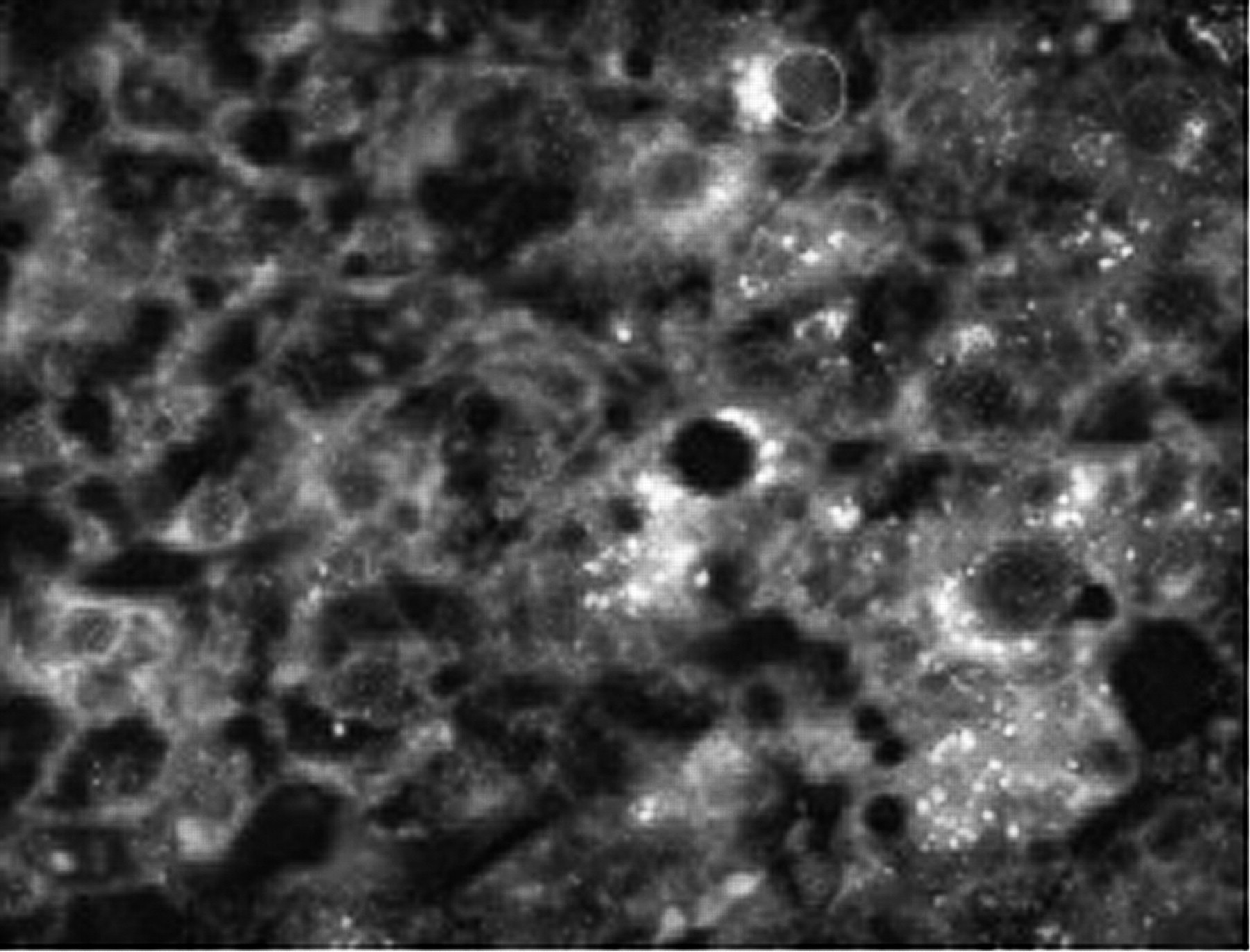
Method Liver biopsy sections were studied from 60 patients with a1AT- related liver disease with a broad spectrum of fibrosis, recruited from the Cambridge metabolic liver clinic (30 were homozygous and 30 heterozygous). Comparison was made with sections from 20 age and sex matched time zero biopsies obtained at liver transplant. Mean hepatocyte telomere length, a reflection of age, was measured by quantitative fluorescent in situ hybridisation (QFISH) with a PNA Cy5 probe. Nuclei were identified with DAPI, hepatocytes with antibody against hepar-1 and pa1AT with specific mouse monoclonal antibody (2C1). Images were obtained and analysed using the Olympus ScanR software system (Abstract P90 figure 1). Statistical analysis used Graph Pad Prism.

QFISH image which highlights the a1AT polymers (shown as bright white speckles), surrounding some of the affected hepatocytes.
Results Hepatocyte nuclei were larger in patients with both homozygous and heterozygous a1AT deficiency (p=0.002) and had shorter telomeres (p<0.0001) than age and sex matched controls. Homozygous patients had shorter hepatocyte telomeres than heterozygous patients (p=0.003). Hepatocyte nuclei in both homozygous and heterozygous a1AT deficiency were larger in cells with pa1AT compared to neighbouring cells without pa1AT (p=0.002). Hepatocyte telomeres were far shorter in cells that contained pa1AT than neighbouring hepatocytes without pa1AT (p<0.0001, Abstract P90 figure 2). Hepatocytes with pa1AT showed additional telomere shortening with increased age (p=0.0002). Fibrosis stage was related to telomere shortening- telomeres shortened as the stage of fibrosis increased.

Hepatocyte telomere length in patients with a1AT deficiency with or without polymerised a1AT.
Conclusion Senescence, characterised by increased nuclear size and shortened telomeres, was present in hepatocytes from patients with both homozygous and heterozygous a1AT related liver disease. The novel 2C1 antibody showed that these markers of senescence were even more marked in cells expressing pa1AT. The steps between pa1AT expression and accelerated senescence are a clear target for therapeutic intervention.