Article Text

Statistics from Altmetric.com
Luminal GI
Intra-tumoural stroma predicts survival in gastric cancer
Gastric adenocarcinoma (gastric cancer, GC) is a major global killer. Identifying molecular programmes contributing to GC patient survival may improve our understanding of GC pathogenesis, highlight new prognostic factors and reveal novel therapeutic targets. In this landmark paper from Singapore, Wu et al produced a comprehensive inventory of gene expression programmes expressed in primary GCs, and identified those expression programmes significantly associated with patient survival. They found that stromal gene expression predicted GC patient survival in multiple independent cohorts, and might be closely related to the intra-tumoural stroma proportion, a specific morphological GC phenotype (see figure 1). These findings suggest that therapeutic approaches targeting the GC stroma, such as TGF-b signalling, may merit evaluation in GC (see page 1100).⇓

Kaplan-Meier analysis demonstrates that patients with GCs exhibiting a high ITS proportion (blue line) have poorer cancer-specific survival compared to patients with low ITS proportion GCs (green line, p=0.006, log-rank test).
The future of IBD surveillance colonoscopies
Research in endoscopic imaging is currently being developed into two directions: delineation of very fine details up to the cellular level (called endoscopic histology) and visualisation of lesions otherwise undetected (called red flag techniques). The latter is dealt with in this Japanese animal study by Mitsunaga et al which uses a topically applied fluorescent probe (gGlu-HMRG) highlighting the cancer-associated enzyme γ-glutamyltranspeptidase. In an azoxymethane/dextran sodium sulfate–induced inflammatory colorectal cancer mouse model, lesions were generated and endoscoped 30 min after topical application of the probe. Cancers ≥1 mm were reliably and repeatedly detected; the fluorescence intensity decreased in flat and adenomatous lesions. In tissue microarrays from human colon tumour lesions, the strongest positivity was achieved from cancer, with some positivity detected in adenomatous lesions. Endoscopic techniques including staining, contrast imaging (such as narrow band and others) and endomicroscopy have had variable success in human IBD studies. New agents (eg, gGlu-HMRG) could be administered topically before colonoscopy by means of enemas or as part of bowel preparation. Whether they are safe and effective for human IBD surveillance remains to be seen (figure 2) (see page 1180).⇓

Fluorescence endoscopic detection of colitis associated cancer. (A) White light image of colon tumours. (B) gGlu-HMRG detection by fluorescence endoscopy.
What controls colon cancer metastases?
Adhesion molecules play an important role in tumour metastasis. The liver is one of the most common organs to which cancers metastasise. However, the reason for the predilection of metastases to the liver is not clear but of obvious clinical importance. This led Zuo and colleagues to determine the role of liver sinusoidal endothelial cell lectin (LSECtin), a protein involved in cell adhesion in colon carcinoma metastasis to the liver. Using mouse models and colon cancer cell lines, they found that LSECtin adhered to colon cancer cells and that blocking LSECtin significantly decreased hepatic metastases of these cells. They also found that primary colon cancer cells from patients also exhibited remarkably low rates of hepatic metastasis in LSECtin knockout mice. They also found that serum sLSECtin was detected at significantly higher levels in colon cancer patients with or without hepatic metastases compared to healthy controls and was also increased in colon cancer patients with metastases compared to those without metastases. Their results indicate that LSECtin plays an important role in colorectal carcinoma liver metastasis and may be a promising new target for intervention in metastasis formation (figure 3) (see page 1170).
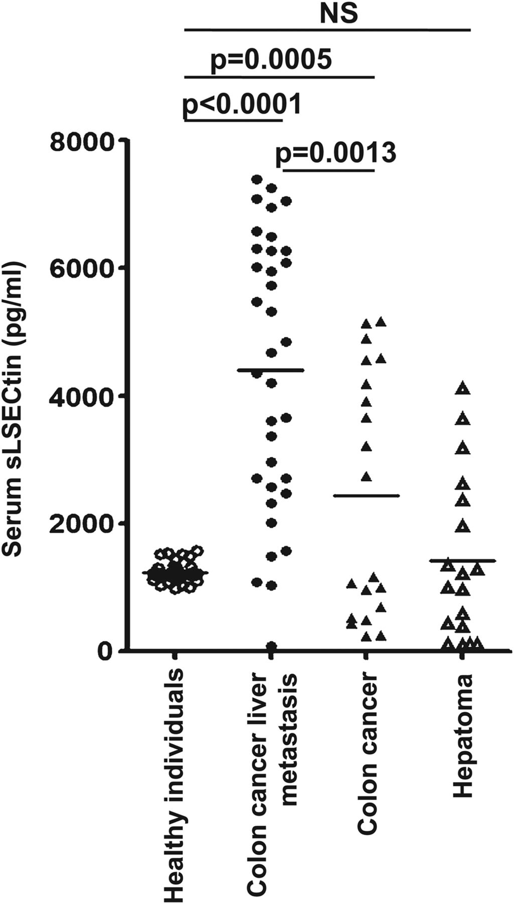
Serum levels of sLSECtin are significantly elevated in colon cancer patients with hepatic metastases. The graph displays a scatter plot of sLSECtin levels in serum samples from 73 patients and 34 healthy blood donors, as determined by ELISA. The horizontal lines indicate the mean levels.
Hepatology
A novel approach to treat fulminant viral hepatitis
MHV-3, a murine coronavirus, is an established animal model for fulminant viral hepatitis. It involves a major role for macrophages and thus is different from viruses replicating predominantly in hepatocytes such as HBV and HCV. This interesting study from China investigated the role of BTLA (B and T attenuator protein) containing an immunoglobulin domain. In mice infected with MHV-3 BTLA enhanced fulminant hepatitis and increased mortality (figure 4). Therefore targeting BTLA may be a novel strategy for the treatment of fulminant viral hepatitis (see page 1205).⇓

Blocking B and T lymphocyte attenuator (BTLA) signalling prolonged the survival of wild-type (WT) mice after murine hepatitis virus strain-3 (MHV-3) infection.
Elucidating the role of HCV lipo-viro particles
Previous studies suggest that lipoprotein lipase (LPL) can suppress HCV. This paper from Taiwan investigates the role and regulation of LPL in HCV. In vitro LPL favoured HCV infection in an apoC III dependent manner. The clinical relevance of their findings is supported by correlations of human plasma viral load with apoC III concentration and an inverse correlation with LPL activity. Thus, LPL is an anti-HCV factor while apoC III reduces LPL activity (see figure 5). These findings could be the basis for novel anti-HCV strategies (see page 1194).⇓

A model of LPL-mediated inhibition of HCV infection that was modulated by VLDL and HCV LVPs.
Footnotes
-
Competing interests None.
-
Provenance and peer review Not commissioned; internally peer reviewed.
Linked Articles
- Hepatology
- Hepatology
- Colon
- Colon
- Stomach