Article Text

Abstract
Objective Extracellular ATP mediates mast cell-dependent intestinal inflammation via P2X7 purinoceptors. We have previously shown that CD300f (also called the leucocyte mono-immunoglobulin-like receptor 3 (LMIR3)) suppresses immunoglobulin E-dependent and mast cell-dependent allergic responses by binding to ceramide. The aim of the present study was to clarify the role of ceramide–LMIR3 interaction in the development of IBD.
Design The dextran sodium sulfate (DSS)-induced colitis model was used in wild-type (WT), LMIR3−/−, mast cell-deficient KitW-sh/W-sh, KitW-sh/W-shLMIR3−/− or KitW-sh/W-sh mice engrafted with WT or LMIR3−/− bone marrow-derived mast cells (BMMCs). The severity of colitis was determined by clinical and histological criteria. Lamina propria cell populations were assessed by flow cytometry. Production of chemical mediators from lamina propria cells was measured by real-time reverse transcription PCR. Production of chemical mediators from ATP-stimulated BMMCs in the presence or absence of ceramide was measured by ELISA. The severity of DSS-induced colitis was assessed in mice given either an Fc fusion protein containing an extracellular domain of LMIR3, and anticeramide antibody, or ceramide liposomes.
Results LMIR3 deficiency exacerbated DSS-induced colitis in mice. KitW-sh/W-sh mice harbouring LMIR3−/− mast cells exhibited more severe colitis than those harbouring WT mast cells. Ceramide–LMIR3 interaction inhibited ATP-stimulated activation of BMMCs. DSS-induced colitis was aggravated by disrupting the ceramide–LMIR3 interaction, whereas it was suppressed by treating with ceramide liposomes.
Conclusions LMIR3-deficient colonic mast cells were pivotal in the exacerbation of DSS-induced colitis in LMIR3−/− mice. Ceramide liposomes attenuated DSS-induced colitis by inhibiting ATP-mediated activation of colonic mast cells through ceraimide–LMIR3 binding.
- IBD MODELS
- INFLAMMATORY BOWEL DISEASE
- EXPERIMENTAL COLITIS
- GASTROINTESTINAL IMMUNE RESPONSE
This is an Open Access article distributed in accordance with the Creative Commons Attribution Non Commercial (CC BY-NC 4.0) license, which permits others to distribute, remix, adapt, build upon this work non-commercially, and license their derivative works on different terms, provided the original work is properly cited and the use is non-commercial. See: http://creativecommons.org/licenses/by-nc/4.0/
Statistics from Altmetric.com
Significance of this study
What is already known on this subject?
Mast cells are involved in the pathogenesis of IBD.
Extracellular ATP mediates mast cell-dependent intestinal inflammation via P2X7 purinoceptors.
An inhibitory receptor LMIR3/CD300f suppresses immunoglobulin E-dependent and mast cell-dependent allergic responses in mice.
What are the new findings?
LMIR3 deficiency exacerbated dextran sodium sulfate (DSS)-induced colitis in mice.
Mast cell-deficient KitW-sh/W-sh mice transplanted with LMIR3−/− mast cells exhibited more severe colitis than those with wild-type mast cells.
Ceramide–LMIR3 interaction inhibited ATP-stimulated activation of bone marrow-derived mast cells.
DSS-induced colitis was aggravated by disrupting the ceramide–LMIR3 interaction, whereas it was suppressed by treating with ceramide liposomes.
How might it impact on clinical practice in the foreseeable future?
The present study provided evidence that the ceramide–LMIR3 interaction inhibits ATP-mediated activation of colonic mast cells, thereby suppressing the development of experimental colitis. LMIR3-targeted ceramide liposomes would provide novel therapeutic strategies for IBD.
Introduction
IBD is characterised by dysregulated intestinal inflammation. The incidence and prevalence of IBD, including UC and Crohn's disease, have increased worldwide. IBD is a complex multifactorial disease regulated by the interplay between immunity, environmental factors and genetic susceptibility.1–4 To define the underlying mechanisms, a number of chemically induced mouse models of IBD have been developed. Among them, the dextran sodium sulfate (DSS) or 2,4,6-trinitrobenzene sulfonic acid (TNBS)-induced colitis models have similarities to human UC or Crohn's disease, respectively.5–7 Extensive research has revealed that together with intestinal epithelial cells, a variety of colonic innate immune cells, including mast cells, neutrophils, eosinophils and CD11b+CX3CR1int mononuclear cells, release an array of chemical mediators (eg, cytokines, chemokines, proteases and lipid mediators) in inflammatory cites of the colon.2–4 ,8–12 Dysregulated inflammatory mediators exacerbate acute colitis, although several cytokines promote tissue repair to maintain intestinal homeostasis.2–4 Studies with mast cell-deficient mice and with P2X7-deficient mast cells have recently demonstrated that ATP-mediated mast cell activation plays a critical role in the initiation and development of experimental colitis induced by DSS and by TNBS; extracellular ATP produced in injured colons activates colonic mast cells via the P2X7 purinoceptor, which release chemical mediators, including neutrophil chemoattractants.8 Accordingly, we aimed to identify a negative regulator of ATP-stimulated mast cell activation that would lead to a new therapeutic target for IBD. One of the possible candidates is an inhibitory receptor expressed in mast cells,13–16 which suppresses mast cell activation through binding to its specific ligand.
A variety of paired activating and inhibitory receptor families regulate the immune system.14–18 The inhibitory receptor CD300f (also called leucocyte mono-immunoglobulin-like receptor 3 (LMIR3), CMRF35-like molecule-1 or myeloid-associated immunoglobulin-like receptor-V) harbours two immunoreceptor tyrosine-based inhibitory motifs (ITIMs) and a single immunoreceptor tyrosine-based switch motif (ITSM).14 ,17 ,18 LMIR3 is expressed in myeloid cells, including mast cells. We have recently identified ceramide as a ligand for LMIR3.15 Ceramide–LMIR3 interaction inhibits immunoglobulin E (IgE)-dependent and mast cell-dependent allergic responses via the two ITIMs and single ITSM;14 however, its role in other settings of inflammation, such as colitis, remains elusive.
In this study, we demonstrate that LMIR3−/− mice are highly susceptible to experimental colitis. Analyses of mast cell-deficient mice adoptively transferred with wild-type (WT) or LMIR3−/− bone marrow-derived mast cells (BMMCs) underscore the importance of mast cells in the exacerbation of DSS-induced colitis in LMIR3−/− mice. In addition, our results provide evidence that the ceramide–LMIR3 interaction inhibits ATP-mediated mast cell activation, thereby suppressing the development of experimental colitis.
Materials and methods
Mice
Female, LMIR3−/− mice aged 10 to 12 weeks on the C57BL/6 background (12 generations backcrossed) or on the BALB/c background (eight generations backcrossed), C57BL/6 or BALB/c mice (Charles River Laboratories Japan), KitW-sh/W-sh mice and KitW-sh/W-shLMIR3−/− mice were used as described.14 ,19 All mice were housed under specific pathogen-free conditions. All the procedures were approved by an institutional review committee of the University of Tokyo (approval no 20-8).
Cells
BMMCs and BMMC transfectants were generated as described.14 ,20 Lamina propria mononuclear cells (LPMNCs) were isolated from the colon as described.8 ,21 Briefly, the colon was digested with 1 mg/mL collagenase type IV (Sigma-Aldrich, St Louis, Missouri, USA) in RPMI-1640 medium. After filtering, the cells were washed, resuspended in the 40% Percoll (GE Healthcare, Munich, Germany) and overlaid on top of the 75% Percoll. After centrifugation, the cells at the interface were collected as LPMNCs.
Induction of experimental colitis
Sibling or non-sibling mice were cohoused for 1 month before induction of experimental colitis. For DSS-induced colitis, mice were given DSS (molecular weight 36–50 kDa; MP Biologicals, Solon, Ohio, USA) supplemented in sterile, distilled water for 7 days followed by normal drinking water until the end of the experiment, as previously described.5 ,6 ,8 For mice on the C57BL/6 background, 1.5% or 2.5% (wt/vol) DSS was used to induce colitis. In the case of mice on the BALB/c background, 3% (wt/vol) DSS was used to induce colitis. For treatment with antibody (Ab) or Fc fusion protein, mice on the C57BL/6 background were intraperitoneally injected with 5 μg of anticeramide Ab or control Ab on day 2 after 1.5% DSS treatment or with 300 μg of an extracellular domain of LMIR3 fused to an Fc domain of human IgG1 (LMIR3-Fc) or of control Fc on days 2 and 6 after 1.5% DSS treatment. For treatment with liposomes, mice on the C57BL/6 background were intravenously injected with 100 μg of ceramide or phosphatidylcholine (PC) liposomes on days 2 and 6 after 2.5% DSS treatment. For TNBS-induced colitis, anaesthetised mice on the C57BL/6 background were sensitised with 2.5% TNBS (Sigma-Aldrich) together with acetone and olive oil. One week after sensitisation, the mice were intrarectally administered with 100 μg of 1.5% TNBS in 50% ethanol, as described.5 ,7 ,8 A Disease Activity Index (DAI) score was determined by daily assessment of weight loss, stool consistency and bleeding.5–8 For histopathological analysis, the entire colon was fixed in 10% formalin after measuring the colon length on day 7 after DSS treatment. Paraffin-embedded sections were stained with H&E.
Statistical analyses
Results are expressed as mean±SEM. An unpaired Student's t test was used to compare the differences between groups. The Kaplan–Meier method and log-rank tests were used to analyse survival studies. *p<0.05, **p<0.01 or ***p<0.001 was taken as statistically significant.
Results
LMIR3−/− mice are highly susceptible to DSS-induced colitis
To explore the role of the inhibitory receptor LMIR3 in the development of colitis, we used the DSS-induced colitis model in WT and LMIR3−/− mice on the C57BL/6 background. The mice were given 1.5% DSS in the drinking water for 7 days. The clinical signs of colitis, including body weight loss, diarrhoea and rectal bleeding, were monitored daily. On day 7 after DSS treatment, colon length was measured before histological examination. WT mice developed only mild colitis in this model (figure 1A–D). Conversely, LMIR3−/− mice exhibited greater weight loss and colon shortening as well as higher DAI scores as compared with WT mice (figure 1A–C). No histological difference was observed in the colonic sections between WT and LMIR3−/− mice prior to DSS treatment, whereas we found a massive destruction of colonic epithelial structures with robust inflammatory cell infiltrates in the mucosa in DSS-treated LMIR3−/− mice compared with WT mice (figure 1D). Notably, 2.5% DSS treatment induced more severe colitis in WT mice than 1.5% DSS treatment did (figure 1E; see online supplementary figure S1). WT mice were still protected against DSS-induced lethal colitis during the observation period (figure 1F). In contrast, >70% of LMIR3−/− mice died of aggressive colitis with prominent histological alterations and clinical features, including rapid weight loss, within 11 days after 2.5% DSS treatment (figure 1E, F; see online supplementary figure S1). Similarly, LMIR3−/− mice on the BALB/c background were more susceptible to DSS-induced colitis than their WT counterparts (see online supplementary figure S2). Similar to DSS treatment, TNBS treatment induced more severe colitis with greater weight loss and colon shortening in LMIR3−/− mice than in WT mice (see online supplementary figure S3). Thus, LMIR3 deficiency exacerbated acute experimental colitis in mice.

Leucocyte mono-immunoglobulin-like receptor 3 (LMIR3)−/− mice are highly susceptible to dextran sodium sulfate (DSS)-induced colitis. (A)–(F) Wild-type (WT) or LMIR3−/− mice on the C57BL/6 background were subjected to (A–D) the 1.5% DSS-induced colitis model (each, n=15) or to (E and F) the 2.5% DSS-induced colitis model (each, n=10). (A and E) Body weight and (B) Disease Activity Index (DAI) score. (C) Colon length on day 7. (D) Representative H&E-stained colonic sections are shown. Scale bars represent 100 μm. (F) Mice were monitored for survival. (A–C and E) Data are expressed as the mean±SEM. **p<0.01, ***p<0.001 (Student's t test). (A–F) The data are from one experiment which is representative of the other one experiment performed (C) or of the other two experiments performed (A, B, D–F).
DSS-treated LMIR3−/− mice exhibit a robust increase in degranulated mast cells in the colon
For further analysis, we induced colitis using 1.5% DSS to prevent DSS-induced death in LMIR3−/− mice. We then examined LMIR3 expression in myeloid cells within the colonic lamina propria of mice before and after DSS exposure. Surface expression of LMIR3 was detected in the high-affinity IgE receptor (FcεRI)+c-kit+ mast cells, CD11b+Gr-1high neutrophils, CD11b+Siglec-F+ eosinophils and CD11b+F4/80+ cells, but not in CD11b+CD11c+ cells, within the colonic lamina propria of WT mice (figure 2A, B). DSS treatment upregulated LMIR3 in colonic lamina propria mast cells, while the same treatment did not influence LMIR3 expression in other myeloid cells tested (figure 2A, B). In light of the fact that the ATP-P2X7 signalling in mast cells contributes to colonic inflammation,8 ,22 we investigated P2X7 expression in colonic lamina propria mast cells of WT or LMIR3−/− mice. Neither LMIR3 deficiency nor DSS treatment altered surface expression levels of P2X7 in colonic lamina propria mast cells (figure 2A). We then determined the percentage of FcεRI+c-kit+ mast cells in CD45+ LPMNCs of WT or LMIR3−/− mice before and after DSS exposure. The percentages of colonic lamina propria mast cells were higher in DSS-treated LMIR3−/− mice than in WT counterparts, although those were increased in both types of mice after DSS exposure (figure 2C). In addition, DSS-treated LMIR3−/− mice, but not WT mice, exhibited elevated percentages of CD63+ degranulated mast cells in the colonic lamina propria (figure 2D).8 ,23 Thus, LMIR3 deficiency caused the increase of activated mast cells in the colonic lamina propria of 1.5% DSS-treated mice, which was correlated with the severity of DSS-induced colitis.

Dextran sodium sulfate (DSS) treatment induces a robust increase in degranulated mast cells in the colon of leucocyte mono-immunoglobulin-like receptor 3 (LMIR3)−/− mice. (A–D) Wild-type (WT) or LMIR3−/− mice on the C57BL/6 background (each, n=4) were subjected to the 1.5% DSS-induced colitis model. Flow cytometric analysis was performed on days 0 and 7 after DSS treatment. (A) Surface expression of LMIR3 or P2X7 in colonic lamina propria mast cells. (B) Surface expression of LMIR3 in CD11b+Gr-1high neutrophils, CD11b+Siglec-F+ eosinophils, CD11b+CD11c+ cells or CD11b+F4/80+ cells within the colonic lamina propria. (C) Percentages of mast cells in CD45+ lamina propria mononuclear cells. (D) Percentages of CD63+ colonic lamina propria mast cells. (C and D) Data are expressed as the mean±SEM. **p<0.01 (Student's t test). (A–D) The data are from one experiment which is representative of the other two experiments performed.
DSS-treated LMIR3−/− mice exhibit enhanced colonic inflammation with a massive infiltration of inflammatory cells into the colon
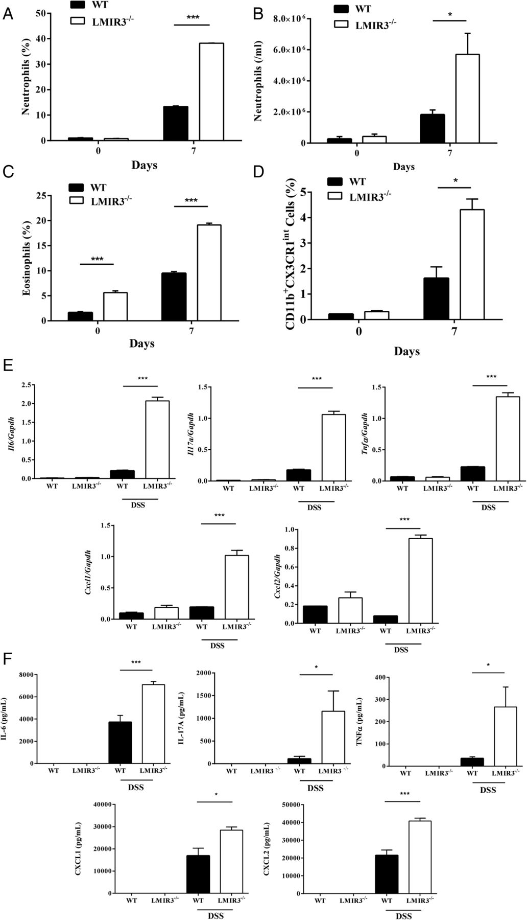
We then examined the infiltration of inflammatory cells into the colon of DSS-treated mice. Consistent with histological observations, 1.5% DSS treatment increased the percentage of neutrophils in CD45+ LPMNCs, which were markedly higher in LMIR3−/− mice than in WT mice (figure 3A). Consistent with these results, we found a marked increase in myeloperoxidase activity related to neutrophil number in the colon of DSS-treated LMIR3−/− mice (figure 3B). As reported,24 the percentages of eosinophils in CD45+ LPMNCs were higher in LMIR3−/− mice on the C57BL/6 background than in WT mice before DSS exposure (figure 3C). Although 1.5% DSS treatment increased the percentage of colonic lamina propria eosinophils in both types of mice, DSS-treated LMIR3−/− mice exhibited a remarkable increase in eosinophil populations (figure 3C). Notably, colonic lamina propria eosinophil numbers were not different between WT and LMIR3−/− mice on the BALB/c background under steady-state conditions, whereas DSS treatment strongly increased these eosinophil populations in LMIR3−/− mice as compared with WT mice (see online supplementary figure S4). These results implied that the observed inflammatory differences between WT and LMIR3−/− mice were independent of the difference in eosinophils. Analysis of CD11b+CX3CR1int cells in CD45+ LPMNCs showed that the percentages of these cell populations were also increased by DSS treatment in both types of mice, while those were higher in LMIR3−/− mice than in WT mice (figure 3D).11 Consistent with a robust infiltration of inflammatory cells in the colon of DSS-treated LMIR3−/− mice, real-time reverse transcription PCR analysis of mRNA levels displayed higher amounts of inflammatory cytokines (interleukin-6 (IL-6), IL-17A or tumour necrosis factor α) and chemokines (CXCL1 or CXCL2) in colonic LPMNCs of DSS-treated LMIR3−/− mice compared with WT mice (figure 3E). Similarly, ex vivo colon punch biopsy cultures of DSS-treated LMIR3−/− mice exhibited remarkably increased levels of inflammatory cytokines and chemokines at the protein levels as compared with those of DSS-treated WT mice (figure 3F). Thus, 1.5% DSS treatment caused remarkably more severe colonic inflammation with an enhanced infiltration of inflammatory cells in LMIR3−/− mice.

Leucocyte mono-immunoglobulin-like receptor 3 (LMIR3) deficiency enhances colonic inflammation with a massive infiltration of inflammatory cells in dextran sodium sulfate (DSS)-treated mice. (A–F) Wild-type (WT) or LMIR3−/− mice on the C57BL/6 background (each, n=4) were subjected to the 1.5% DSS-induced colitis model. (A, C and D) Percentages of (A) CD11b+Gr-1high neutrophils, (C) CD11b+Siglec-F+ eosinophils or (D) CD11b+CX3CR1int cells in CD45+ lamina propria mononuclear cells (LPMNCs) or (B) colonic tissue neutrophils estimated by measuring myeloperoxidase activity from the mice on days 0 and 7. (E and F) Transcript levels of cytokines and chemokines quantified by real-time reverse transcription PCR analysis in colonic LPMNCs (E) or protein levels of cytokines and chemokines measured by ELISA in colon explant culture supernatants (F) from the mice on days 0 and 7. (A–F) Data are expressed as mean±SEM. *p<0.05, **p<0.01 and ***p<0.001 (Student's t test). The data are from one experiment which is representative of the other three experiments performed. IL, interleukin; TNF, tumour necrosis factor.
LMIR3−/− mast cells contribute to an exacerbation of DSS-induced colitis
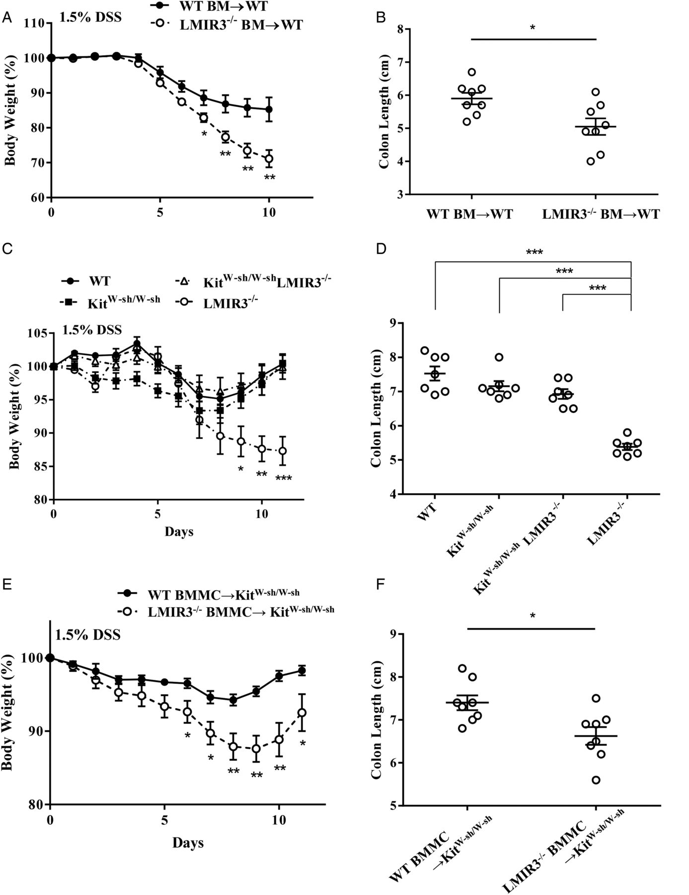
We then asked which cell populations were responsible for the high susceptibility of LMIR3−/− mice to DSS-induced colitis. First, we used 1.5% DSS to induce colitis in lethally irradiated WT mice adoptively transferred with WT or LMIR3−/− bone marrow (BM). The results show that LMIR3−/− BM-transplanted mice suffered from more severe colitis with greater weight loss and colon shortening compared with WT BM-transplanted mice (figure 4A, B), implicating LMIR3−/− BM-derived cells in the exacerbation of colitis of LMIR3−/− mice. We next used the same model in mast cell-deficient KitW-sh/W-sh and KitW-sh/W-shLMIR3−/− mice. KitW-sh/W-sh mice exhibited mild body weight loss and colon shortening at levels comparable with those seen in WT mice (figure 4C, D). LMIR3 deficiency did not influence the severity of DSS-induced colitis in KitW-sh/W-sh mice (figure 4C, D). We further compared the severity of 1.5% DSS-induced colitis between KitW-sh/W-sh mice adoptively transferred with WT or LMIR3−/− BMMCs. Colonic lamina propria mast cells were detected at comparable levels in these two types of BMMC-transplanted mice (see online supplementary figure S5). Transplantation with LMIR3-deficient mast cells, but not with WT mast cells, remarkably increased the severity of colitis (causing body weight loss and colon shortening) in KitW-sh/W-sh mice (figure 4E, F). Consistently, KitW-sh/W-sh with LMIR3-deficient mast cells, but not with WT mast cells, exhibited a marked increase in neutrophils and eosinophils in CD45+ LPMNCs (see online supplementary figure S6). In contrast, we found that WT mice were more susceptible to 2.5% DSS-induced colitis than KitW-sh/W-sh mice (data not shown), which is in agreement with a previous report.8 In this model, KitW-sh/W-shLMIR3−/− mice developed only mild colitis with phenotypes similar to KitW-sh/W-sh mice (see online supplementary figure S7). Transplantation of BMMCs, irrespective of LMIR3 expression, into KitW-sh/W-sh mice increased the severity of 2.5% DSS-induced colitis; however, KitW-sh/W-sh mice with LMIR3-deficient mast cells exhibited more severe colitis with a higher lethality than those with WT mast cells did (see online supplementary figure S8). Thus, LMIR3-deficient mast cells seemed to play an important role in the exacerbation of DSS-induced colitis in LMIR3−/− mice.

Leucocyte mono-immunoglobulin-like receptor 3 (LMIR3)−/− mast cells contribute to an exacerbation of dextran sodium sulfate (DSS)-induced colitis. (A–F) Mice on the C57BL/6 background were subjected to the 1.5% DSS-induced colitis model. (A and B) Wild-type (WT) mice transplanted with WT or LMIR3−/− bone marrow (BM; each, n=8), (C and D) WT, KitW-sh/W-sh, KitW-sh/W-shLMIR3−/− or LMIR3−/ − mice (each, n=7) or (E and F) KitW-sh/W-sh transplanted with WT or LMIR3-deficient bone marrow-derived mast cells (BMMCs; each, n=8) were used. (A, C and F) Body weight. (B, D and F) Colon length on day 7 after DSS treatment. Data are expressed as the mean±SEM. *p<0.05, **p<0.01 and ***p<0.001 (Student's t test). The data are from one experiment which is representative of the other one experiment performed.
Ceramide–LMIR3 interaction inhibits ATP-stimulated mast cell activation
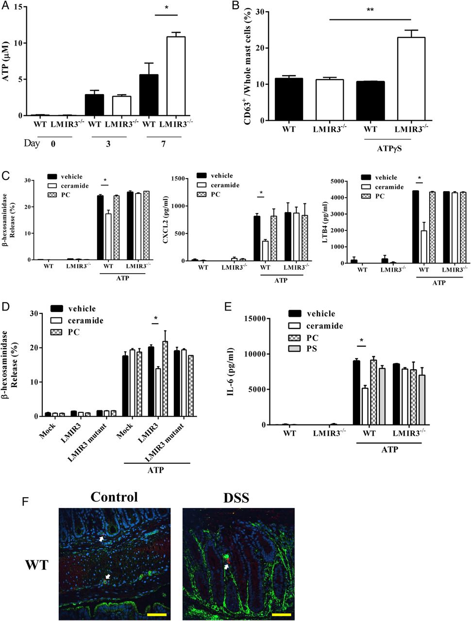
To clarify the role of the ATP-P2X7 signalling axis in the exacerbation of colitis in LMIR3−/− mice, we measured ATP levels in the colon of WT or LMIR3−/− mice after 1.5% DSS exposure. DSS treatment gradually increased the ATP content of the colon of mice on days 3–7; ATP levels were comparable between both groups of mice on day 3, but were higher in LMIR3−/− mice than in WT mice on day 7 (figure 5A). When mice were intrarectally injected with non-hydrolyzable adenosine 5′-O-(3-thio) triphosphate (ATPγS), this treatment increased the percentage of CD63+ colonic lamina propria mast cells of LMIR3−/− mice, but not of WT mice, under our experimental conditions (figure 5B). We then asked whether the ceramide–LMIR3 interaction suppresses ATP-stimulated mast cell activation. WT or LMIR3−/− BMMCs were stimulated with ATP on plates coated with ceramide, PC or vehicle. Surface expression levels of P2X7 as well as FcεRI and c-kit were comparable between WT and LMIR3−/− BMMCs (see online supplementary figure S9). ATP stimulation induced the release of β-hexosaminidase, a marker of degranulation, and the production of neutrophil chemoattractants, CXCL2 and leucotriene B4 (LTB4), at equivalent levels in WT and LMIR3−/− BMMCs in the absence of ceramide (figure 5C). In contrast, ceramide–LMIR3 binding specifically inhibited β-hexosaminidase release as well as CXCL2 and LTB4 production of WT BMMCs, but not of LMIR3−/− BMMCs, in response to ATP (figure 5C). To test whether LMIR3 inhibitory motifs are involved in LMIR3-mediated inhibition of ATP-stimulated mast cell activation, LMIR3−/− BMMCs were transduced with LMIR3 WT, LMIR3 (Y241F-Y289F-Y325F) mutant or empty vector. In this mutant, three tyrosine residues in the two ITIMs and a single ITSM of LMIR3 were replaced with phenylalanine residues. These BMMC transfectants exhibited equivalent levels of FcεRI, c-kit and P2X7 (data not shown). Surface expression levels of the transduced LMIR3 were comparable between BMMCs transduced with LMIR3 WT or its mutant (data not shown). Measurement of β-hexosaminidase release showed that ATP-stimulated mast cell degranulation was inhibited only in LMIR3 WT-transduced BMMCs, but not in LMIR3 (Y241F-Y289F-Y325F) mutant-transduced BMMC, on ceramide-coated plates (figure 5D). Similarly, the production of proinflammatory cytokine IL-6 in WT BMMCs was suppressed only on plates coated with ceramide, but not with phosphatidylserine (PS), PC or vehicle (figure 5E). However, such inhibition in the presence of ceramide was not observed in LMIR3−/− BMMCs stimulated by ATP (figure 5E). Confocal microscopic analysis showed that extracellular ceramide was distributed in the surroundings of mast cells in colonic tissues and tended to be increased by DSS treatment (figure 5F). Thus, DSS treatment strongly enhanced ATP-mediated activation of colonic mast cells in LMIR3−/− mice presumably due to the lack of inhibitory signals via the ceramide–LMIR3 interaction.

Ceramide–leucocyte mono-immunoglobulin-like receptor 3 (LMIR3) interaction inhibits ATP-stimulated mast cell activation. (A) Wild-type (WT) or LMIR3−/− mice on the C57BL/6 background (each, n=3) were subjected to 1.5% dextran sodium sulfate (DSS)-induced colitis model. The concentrations of ATP released by colonic tissues from the mice on days 0, 3 or 7. (B) WT or LMIR3−/− mice on the C57BL/6 background (each, n=3) were intrarectally administered with ATPγS. Percentages of CD63+ colonic lamina propria from the mice on days 0 and 3 after administration. (C) WT or LMIR3−/− bone marrow-derived mast cells (BMMCs) were stimulated with 3 mM ATP on plates coated with ceramide, phosphatidylcholine (PC) or vehicle. Release of β-hexosaminidase (left) or production of CXCL2 (middle) or LTB4 (right). (D) LMIR3−/− BMMCs transduced with LMIR3 WT, LMIR3(Y241F-Y289F-Y325F) mutant, or empty vector (mock) were stimulated with 3 mM ATP on plates coated with ceramide, PC or vehicle. Release of β-hexosaminidase. (E) WT or LMIR3−/− BMMCs were stimulated with 3 mM ATP on plates coated with ceramide, PC, phosphatidylserine (PS) or vehicle. Production of interleukin (IL)-6. (A–E) Data are expressed as mean±SEM. *p<0.05, **p<0.01 and ***p<0.001 (Student's t test). The data are from one experiment which is representative of the other two experiments performed. (F) Frozen colon sections of WT mice on days 0 and 7 after 1.5% DSS treatment were stained with an anticeramide Ab (ceramides; green) and an anti-mast cell tryptase Ab (mast cell; red). The nuclei were counterstained with 4′,6-diamidino-2-phenylindole (blue). White arrow indicates mast cell. The data are from one experiment which is representative of the other four experiments performed. Scale bars represent 100 μm.
DSS-induced colitis is aggravated by disrupting ceramide–LMIR3 binding, whereas it is suppressed by administration of ceramide liposomes
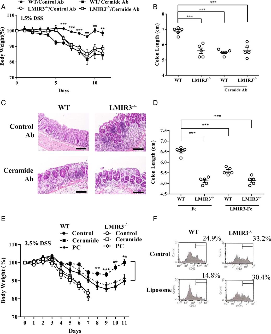
To validate the protective role of the ceramide–LMIR3 interaction in DSS-induced colitis, WT or LMIR3−/− mice were treated with an anticeramide Ab or with a control Ab during 1.5% DSS exposure. Notably, treatment with anticeramide Ab, but not with a control Ab, increased the severity of colitis in WT mice at levels comparable with that found in LMIR3−/− mice (figure 6A–C).Conversely, treatment with the anticeramide Ab did not influence DSS-induced colitis in LMIR3−/− mice (figure 6A–C). Similarly, WT or LMIR3−/− mice were administered with LMIR3-Fc or with a control Fc during 1.5% DSS exposure. LMIR3-Fc-treated WT mice developed more severe colitis with greater weight loss than control Fc-treated WT mice (figure 6D). We confirmed that LMIR3-Fc treatment failed to exacerbate DSS-induced colitis in LMIR3−/− mice (figure 6D). Thus, disrupting the ceramide–LMIR3 interaction aggravated DSS-induced colitis in WT mice. We finally tested whether treatment with ceramide liposomes improves DSS-induced colitis. To this end, we used 2.5% DSS to induce severe colitis in mice. WT mice treated with ceramide liposomes compared with control liposome exhibited an attenuated DSS-induced colitis with less weight loss (figure 6E). Neither treatment reversed the lethality of DSS-induced colitis in LMIR3−/− mice (figure 6E). Consistent with these results, treatment with ceramide liposomes substantially inhibited colonic mast cell degranulation in WT mice, but not in LMIR3−/− mice, after DSS exposure (figure 6F). These results indicate that the ceramide liposome treatment inhibited ATP-mediated mast cell activation, thereby improving DSS-induced colitis in WT mice.

Dextran sodium sulfate (DSS)-induced colitis is aggravated by disrupting ceramide–leucocyte mono-immunoglobulin-like receptor 3 (LMIR3) binding, while that is suppressed by administration of ceramide liposomes. (A–C) Anticeramide Ab or control Ab-treated wild-type (WT) or LMIR3−/− mice on the C57BL/6 background (each, n=5) were subjected to the 1.5% DSS-induced colitis model. (A) Body weight. (B) Colon length on day 7. (C) Representative H&E-stained colonic sections were shown. Scale bars represent 100 μm. (D) LMIR3-Fc-treated or control Fc-treated WT or LMIR3−/− mice on the C57BL/6 background (n=5 per genotype) were subjected to the 1.5% DSS-induced colitis. Colon length on day 7. (E and F) Ceramide or phosphatidylcholine (PC) liposomes or phosphate buffer saline-treated WT or LMIR3−/− mice on a C57BL/6 background (n=10 per genotype) were subjected to the 2.5% DSS-induced colitis. (E) Body weight. (F) Percentages of CD63+ colonic lamina propria mast cells on day 7. (A, B, D and E) Data are expressed as the mean±SEM. **p<0.01, ***p<0.001 (Student's t test). (A–F) The data are from one experiment which is representative of the other one experiment performed.
Discussion
The development of colitis is temporally and spatially regulated by a variety of immune cells in concert with colonic epithelial cells.2–4 ,8–12 However, the key cellular signals that trigger exacerbation of colitis remained unclear until the recent finding that the ATP-P2X7 signalling axis in mast cells is pivotal to intestinal inflammation in experimental colitis.8 Based on this knowledge, we demonstrate in the present study that the ceramide–LMIR3 interaction suppresses experimental colitis by inhibiting ATP-mediated mast cell activation.
Comparative analysis of DSS-induced colitis between WT and LMIR3−/− mice demonstrated that LMIR3−/− mice were more prone to colonic inflammation than WT mice under the same experimental conditions including genetic background and the dose of DSS administration: low-dose (1.5%) DSS treatment caused only mild colitis in WT C57BL/6 mice, but induced severe colitis in LMIR3−/− mice; high-dose (2.5%) DSS treatment induced lethal colitis only in most LMIR3−/− C57BL/6 mice. Moreover, the high susceptibility of LMIR3−/− mice to TNBS-induced colitis underscored the importance of LMIR3 as a negative regulator of experimental colitis. Interestingly, treatment with low-dose DSS mildly increased neutrophils, eosinophils and CD11b+CX3CR1int cells, which were involved in the development of colitis,8–11 in the colonic lamina propria of WT mice; however, the same treatment failed to significantly induce activation of colonic lamina propria mast cells expressing LMIR3 as well as P2X7. Consistent with these results, the severity of low-dose DSS-induced colitis was influenced neither by mast cell deficiency in mice nor by reconstitution with WT mast cells in mast cell-deficient KitW-sh/W-sh mice. Thus, low-dose DSS treatment induced a mild infiltration of inflammatory cells into the colonic lamina propria, independently of mast cell activation. However, we found a prominent increase in activated mast cells in the colonic lamina propria of LMIR3−/− mice after low-dose DSS treatment. Importantly, similarly induced colitis in KitW-sh/W-sh mice was profoundly enhanced by reconstitution with LMIR3−/− BMMCs, but not by crossing with LMIR3−/− mice. Since LMIR3 deficiency did not affect P2X7 expression both in colonic lamina propria mast cells and in BMMCs, it is therefore plausible that LMIR3 deficiency in mast cells exacerbates colitis induced by low-dose DSS. In view of previous results,8 it is also possible to speculate that LMIR3 dampens colonic mast cell activation in response to relatively low levels of ATP produced in the colon injured by low-dose DSS. These assumptions may be supported by two of our observations: low-dose DSS treatment gradually elevated colonic ATP levels in proportion to the severity of colonic inflammation in mice; an intrarectal administration of ATPγS activated colonic lamina propria mast cells only in LMIR3−/− mice. Meanwhile, in agreement with a previous report,8 we confirmed that high-dose DSS-treated KitW-sh/W-sh mice compared with WT mice developed remarkably more mild colitis, which was aggravated by reconstitution with WT mast cells. At the same time, we found that high-dose DSS-induced colitis in KitW-sh/W-sh mice was more profoundly aggravated by reconstitution with LMIR3−/− mast cells compared with WT mast cells, while it was not significantly influenced by LMIR3 deficiency. Collectively, it is possible to conclude that colonic mast cells contribute to the development of high-dose DSS-induced colitis in WT mice; severe injuries to the colon was followed by the release of high levels of ATP in the colon where ATP-mediated mast cell activation cannot be completely blocked by LMIR3. However, LMIR3-deficient colonic mast cells are pivotal in the exacerbation of colitis induced by DSS, irrespective of its dosage, presumably because those cells are fully activated due to the lack of LMIR3-mediated inhibitory signals. Consequently, LMIR3-deficient colonic mast cells released profoundly high levels of soluble factors that recruit inflammatory cells as well as of inflammatory mediators that directly promote tissue damage, all of which amplify intestinal inflammation. To clarify the possible role of LMIR3 deficiency in myeloid cells except mast cells in experimental colitis, further investigation will be required; a recent study reported that eosinophil-mediated signalling attenuates experimental colitis.25
Analysis of WT or LMIR3−/− BMMCs demonstrated that, like high-affinity IgE receptor signalling, P2X7 signalling via exogenous ATP was inhibited by the binding of ceramide to LMIR3 in BMMCs.15 Note that other lipids tested, including PS which was reported to bind to CD300f/LMIR3, did not act as a functional ligand for CD300f/LMIR3 in this setting.26 In addition, the LMIR3(Y241F-Y289F-Y325F) mutant failed to inhibit ATP-stimulated activation of the transduced BMMCs in the presence of ceramide. These results therefore suggest that ceramide–LMIR3 binding inhibited ATP-mediated mast cell activation through tyrosine-phosphorylation of the two ITIMs and a single ITSM, which was able to induce recruitment of tyrosine phosphatases.14 As revealed by histochemical examination, extracellular ceramide was distributed in the surroundings of colonic mast cells. Moreover, administration of an anticeramide Ab or LMIR3-Fc, both of which disrupt ceramide–LMIR3 binding in colonic mast cells, aggravated DSS-induced colitis in WT mice, but not in LMIR3−/− mice. These in vitro and in vivo data highlight the importance of ceramide–LMIR3 binding in colonic mast cells in suppressing experimental colitis. Intriguingly, DSS treatment upregulated LMIR3 in colonic mast cells, while the same treatment tended to increase extracellular ceramide in colonic tissues possibly in association with tissue damage. These two events may co-operate to prevent excessive activation of colonic mast cells in this model. Most importantly, intravenous administration of ceramide liposomes, which was thought to possibly increase extracellular ceramide in colonic tissues, attenuated DSS-induced colitis, although more effective and safe treatment against colitis with ceramide liposomes, including the dose, route and frequency, remains to be determined. Given that intestinal mast cell activation via extracellular ATP is implicated in the pathogenesis of certain types of human IBD,8 LMIR3-targeted ceramide liposomes would provide novel therapeutic strategies for IBD.
In conclusion, LMIR3 inhibits ATP-mediated colonic mast cell activation through its interaction with ceramide, thereby attenuating experimental colitis. Accordingly, treatment with ceramide liposomes ameliorates colonic inflammation in mouse models of IBD.
Acknowledgments
We thank Yosuke Kurashima (The University of Tokyo) for helpful discussions.
References
Supplementary materials
Supplementary Data
This web only file has been produced by the BMJ Publishing Group from an electronic file supplied by the author(s) and has not been edited for content.
Files in this Data Supplement:
- Data supplement 1 - Online supplement
Footnotes
Contributors TM performed all the experiments and participated in writing the manuscript. KI, MI, MT, AM, YY and AK assisted with the experiments. KO and TT analysed the data. TK and JK conceived the project, analysed the data and actively participated in manuscript writing.
Funding This study was supported by JSPS KAKENHI Grant Number 23390257 and 26293231 and in part by Grant-in-Aid for JSPS Fellows (26000014).
Competing interests None.
Provenance and peer review Not commissioned; externally peer reviewed.