Article Text

Abstract
Clinical presentation A middle-aged man was admitted for episodes of fresh per-rectal bleeding, which were not associated with defecation. He was recently investigated for macrocytic anaemia in the outpatient haematology clinic. Examination of the perineum revealed grade 1 internal haemorrhoids with no signs of bleeding.
Initial laboratory tests revealed macrocytic anaemia (haemoglobin 10.5 g/dL, normal 12.9–17.0 g/dL; mean corpuscular haemoglobin 95.3 fL, normal 80.0–95.0 fL). Peripheral blood film showing blasts, dysplastic neutrophils, nucleated red blood cells and hypogranular platelets.
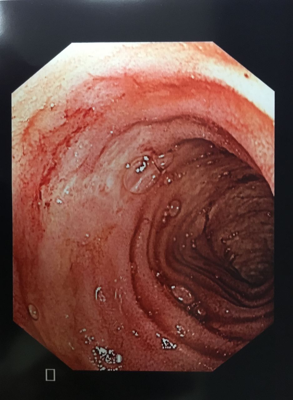
The patient underwent a sigmoidoscopy and rubber band ligation of the internal haemorrhoids after persistent fresh per-rectal bleeding. The bleeding persisted with the development of hypotension and a significant drop of haemoglobin to 4.8 g/dL requiring blood transfusions and intensive care monitoring. Repeated endoscopy, including intubation of the terminal ileum, revealed uncomplicated right-sided diverticulosis. CT mesenteric angiography performed during an episode of significant bleeding revealed extravasation of contrast in the ileum, but mesenteric angiography was unsuccessful, possibly due to a temporary cessation of bleeding. Bleeding subsequently recurred and in light of the persistent bleeding with no clear source and with a total of 12 units of packed cell transfused, exploratory laparotomy, on-table enteroscopy (figure 1) with small bowel resection was performed. Histopathological examination of the specimen was performed (figures 2⇓–4).

Multiple ileal lesions with stigmata of recent bleed.

Area of ulceration associated with atypical mononuclear infiltrate.

Atypical mononuclear infiltrate composed of cells with enlarged, irregular nuclei containing variably prominent nucleoli.

Atypical cells displayed cytoplasmic expression of myeloperoxidase.
Question What is the diagnosis?
- GASTROINTESTINAL BLEEDING
Statistics from Altmetric.com
Footnotes
Contributors FHK: study concept and design, acquisition of data, drafting manuscript and critical revision. FP: acquisition of data and critical revision. CSC: study concept and design and critical revision.
Competing interests None.
Ethics approval Approved by the National Healthcare Group Institutional Review Board.
Provenance and peer review Not commissioned; externally peer reviewed.