Article Text

Abstract
Common bile duct stones (CBDS) are estimated to be present in 10–20% of individuals with symptomatic gallstones. They can result in a number of health problems, including pain, jaundice, infection and acute pancreatitis. A variety of imaging modalities can be employed to identify the condition, while management of confirmed cases of CBDS may involve endoscopic retrograde cholangiopancreatography, surgery and radiological methods of stone extraction. Clinicians are therefore confronted with a number of potentially valid options to diagnose and treat individuals with suspected CBDS. The British Society of Gastroenterology first published a guideline on the management of CBDS in 2008. Since then a number of developments in management have occurred along with further systematic reviews of the available evidence. The following recommendations reflect these changes and provide updated guidance to healthcare professionals who are involved in the care of adult patients with suspected or proven CBDS. It is not a protocol and the recommendations contained within should not replace individual clinical judgement.
- HEPATOBILIARY DISEASE
- BILIARY OBSTRUCTION
- ACUTE PANCREATITIS
- ENDOSCOPIC RETROGRADE PANCREATOGRAPHY
- GALLSTONE DISEASE
Statistics from Altmetric.com
- HEPATOBILIARY DISEASE
- BILIARY OBSTRUCTION
- ACUTE PANCREATITIS
- ENDOSCOPIC RETROGRADE PANCREATOGRAPHY
- GALLSTONE DISEASE
Summary of recommendations
Where recommendations from the 2008 guidelines1 are obsolete, they are omitted. Where recommendations are prefaced by ‘2008’ there has been no new evidence found since the last guideline and no change in the recommendation; ‘2008, amended 2016’ indicates that while no new evidence has been found since the last guideline there has been a change in wording that effects the meaning of the recommendation; ‘2016’ indicates that new evidence has been found and no change in the recommendation is necessary; ‘New 2016’ indicates that new evidence has resulted in a new or amended recommendation.
General principles in management of common bile duct stones
New 2016
It is recommended that patients diagnosed with common bile duct stones (CBDS) are offered stone extraction if possible. Evidence of benefit is greatest for symptomatic patients. (Low-quality evidence; strong recommendation)
Identifying individuals with CBDS
New 2016
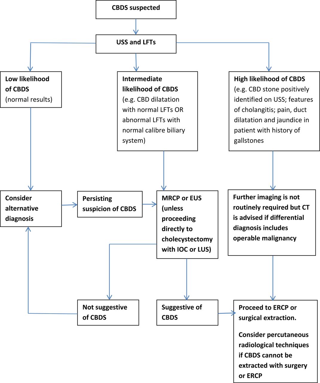
Trans-abdominal ultrasound scanning (USS) and liver function tests (LFTs) are recommended for patients with suspected CBDS. Normal results do not preclude further investigation if clinical suspicion remains high. (Low-quality evidence; strong recommendation)
New 2016
Magnetic resonance cholangiopancreatography (MRCP) and endoscopic ultrasound (EUS) are both recommended as highly accurate tests for identifying CBDS among patients with an intermediate probability of disease. MRCP predominates in this role, with choice between the two modalities determined by individual suitability, availability of the relevant test, local expertise and patient acceptability. (Moderate quality evidence; strong recommendation)
New 2016
It is suggested that patients with suspected CBDS who have not been previously investigated should undergo USS and LFTs. For patients with an intermediate probability of stones, MRCP or EUS is recommended as a next step unless the patient is proceeding directly to cholecystectomy supplemented by intraoperative cholangiography (IOC) or laparoscopic ultrasound (LUS). Endoscopic retrograde cholangiopancreatography (ERCP) should be reserved for patients in whom preceding assessment indicates a need for endoscopic therapy. (Low-quality evidence; weak recommendation)
Endoscopic management of CBDS
New 2016
It is suggested that the British Society of Gastroenterology (BSG) national standards framework for ERCP is implemented by service providers. (Very low-quality evidence; weak recommendation)
New 2016
For selected patients, tolerability and likelihood of therapeutic success is higher if ERCP is performed with propofol sedation or general anaesthesia. It is recommended that hospitals looking after patients with CBDS should have ready and prompt access to anaesthesia supported ERCP. This can be an on-site service or provided by another ERCP unit as part of a clinical network. (Low-quality evidence; strong recommendation)
2008
It is suggested that patients should be managed in accordance with the BSG guidelines on antibiotic prophylaxis during endoscopy. (Very low-quality evidence; weak recommendation)
New 2016
To reduce the risk of post-ERCP pancreatitis (PEP) it is recommended that diclofenac or indomethacin (at a dose of 100 mg) should be administered rectally at the time of ERCP to all patients who do not have a contraindication to non-steroidal anti-inflammatory drugs (NSAIDs). (Moderate-quality evidence; strong recommendation)
New 2016
In patients with a high risk of PEP arising from repeated pancreatic duct cannulation, insertion of a pancreatic stent is suggested in addition to administration of rectal NSAID. (Moderate-quality evidence; weak recommendation)
2008, amended 2016
It is recommended that patients undergoing biliary sphincterotomy for ductal stones have a full blood count (FBC) and international normalised ratio or prothrombin time (INR/PT) performed prior to their ERCP. If deranged clotting or thrombocytopenia is identified, subsequent management should conform to locally agreed guidelines. (Low-quality evidence; strong recommendation)
New 2016
It is recommended that ERCP patients taking warfarin, antiplatelet treatment or a direct oral anticoagulant (DOAC) should be managed in accordance with the combined BSG and European Society of Gastrointestinal Endoscopy (ESGE) guidelines for patients undergoing endoscopy. (Low-quality evidence; strong recommendation)
2008, amended 2016
Competency in access papillotomy is suggested for all endoscopists who perform ERCP. Training and subsequent mentorship should facilitate this. (Very low-quality evidence; weak recommendation)
New 2016
As an adjunct to biliary sphincterotomy, endoscopic papillary balloon dilation (EPBD) is recommended as a technique to facilitate removal of large CBDS. (High-quality evidence; strong recommendation)
New 2016
EPBD without prior biliary sphincterotomy is associated with an increased risk of PEP but may be considered as an alternative to biliary sphincterotomy in selected patients, such as those with an uncorrected coagulopathy or difficult biliary access due to altered anatomy. If EPBD is performed without prior biliary sphincterotomy, use of an 8 mm diameter balloon is recommended. (Moderate-quality evidence; strong recommendation)
New 2016
It is recommended that cholangioscopy-guided electrohydraulic lithotripsy (EHL) or laser lithotripsy (LL) be considered when other endoscopic treatment options fail to achieve duct clearance. (Low-quality evidence; strong recommendation)
Surgical management of CBDS
New 2016
IOC or LUS can be used to detect CBDS in patients who are suitable for surgical exploration or postoperative ERCP. Although not considered mandatory for all patients undergoing cholecystectomy, IOC or LUS is suggested for those patients who have an intermediate to high pre-test probability of CBDS and who have not had the diagnosis confirmed preoperatively by USS, MRCP or EUS. (Low-quality evidence; weak recommendation)
2016
It is recommended that, in patients undergoing laparoscopic cholecystectomy, transcystic or transductal laparoscopic bile duct exploration (LBDE) is an appropriate technique for CBDS removal. There is no evidence of a difference in efficacy, mortality or morbidity when LBDE is compared with perioperative ERCP, although LBDE is associated with a shorter hospital stay. It is recommended that the two approaches are considered equally valid treatment options. (High-quality evidence; strong recommendation)
New 2016
It is suggested that training of surgeons in LBDE is to be encouraged in order to decrease the number of interventions required to manage CBDS. (Low-quality evidence; weak recommendation)
Management of ‘difficult’ ductal stones
New 2016
Laparoscopic duct exploration and ERCP (supplemented by EPBD with prior sphincterotomy, mechanical lithotripsy or cholangioscopy where necessary) are highly successful in removing CBDS. It is recommended that percutaneous radiological stone extraction and open duct exploration should be reserved for the small number of patients in whom these techniques fail or are not possible. (Low-quality evidence; strong recommendation)
New 2016
When endoscopic cannulation of the bile duct is not possible with standard techniques including access papillotomy, it is recommended that percutaneous or EUS-guided procedures can be considered as a means of facilitating subsequent ERCP. (Low-quality evidence; strong recommendation)
2016
It is important that endoscopists ensure adequate biliary drainage is achieved in patients with CBDS that have not been extracted. The short-term use of a biliary stent followed by further endoscopy or surgery is recommended. (Moderate-quality evidence; strong recommendation)
2016
The use of a biliary stent as sole treatment for CBDS should be restricted to a selected group of patients with limited life expectancy and/or prohibitive surgical risk. (Moderate-quality evidence; strong recommendation)
Management of CBDS in specific clinical setting
New 2016
Cholecystectomy is recommended for all patients with CBDS and gall bladder stones unless there are specific reasons for considering surgery inappropriate. (High-quality evidence; strong recommendation)
Where operative risk is deemed prohibitive, biliary sphincterotomy and endoscopic duct clearance alone is suggested as an acceptable alternative. (Low-quality evidence; weak recommendation)
2008
Biliary sphincterotomy and endoscopic stone extraction is recommended as the primary form of treatment for patients with CBDS post cholecystectomy. (Low-quality evidence; strong recommendation)
New 2016
Patients with acute cholangitis who fail to respond to antibiotic therapy or who have signs of septic shock require urgent biliary decompression. Endoscopic CBDS extraction and/or biliary stenting are recommended in this setting. If ERCP is not possible, percutaneous radiological drainage can be considered as an alternative. (Moderate-quality evidence; strong recommendation)
New 2016
Patients with pancreatitis of suspected or proven biliary origin who have associated cholangitis or persistent biliary obstruction are recommended to undergo biliary sphincterotomy and endoscopic stone extraction within 72 hours of presentation. (High-quality evidence; strong recommendation)
New 2016
It is recommended that following gallstone pancreatitis early laparoscopic cholecystectomy should be offered to all patients on whom it is safe to operate as the most effective means to prevent recurrent episodes. (Moderate-quality evidence, strong recommendation)
New 2016
In cases of mild acute gallstone pancreatitis, it is advised that cholecystectomy should be performed within 2 weeks of presentation and preferably during the same admission. (Moderate-quality evidence; weak recommendation)
New 2016
It is recommended that patients with gallstone pancreatitis who do not require ERCP within 72 hours of presentation should be considered for elective ERCP and endoscopic sphincterotomy if there is evidence of retained CBDS on imaging or the patient is unsuitable for definitive treatment in the form of cholecystectomy. (Moderate-quality evidence; strong recommendation)
New 2016
ERCP for CBDS extraction can be successfully performed in patients with Billroth II anatomy. Where ERCP with a duodenoscope is difficult, use of a forward viewing endoscope is recommended. (Moderate-quality evidence; weak recommendation)
In cases where biliary sphincterotomy cannot be safely completed, a limited sphincterotomy supplemented by EPBD is suggested as an alternative. (Low-quality evidence; weak recommendation)
New 2016
Patients with Roux-en-Y gastric bypass (RYGB) and CBDS should be referred to centres that are able to offer the advanced endoscopic and surgical treatment options that are necessary for stone extraction. (Low-quality evidence; weak recommendation)
Members of guideline development group and acknowledgements
The guideline development group (GDG) comprised of the following members:
Earl Williams. Consultant hepatologist, Royal Bournemouth Hospital, representing BSG. Chair of GDG, Editor and lead for introductory and concluding sections; section on general principles in the management of CBDS and section on identification of individuals with CBDS.
Peggy and Hannah Anderson. Patient representatives, approached via British Liver Trust.
Ian Beckingham, Consultant HPB surgeon, Nottingham University Hospitals, representing Association of Upper Gastrointestinal Surgeons of Great Britain and Ireland (AUGIS) and Royal College of Surgeons. Lead for section on surgical management of CBDS.
Ghassan El Sayed. ERCP fellow, Royal Bournemouth Hospital. Representing GI trainees. Responsible for literature search.
Kurinchi Gurusamy, Reader in Surgery, University College London and member of European Association for the Study of the Liver guidelines panel for management of gallstones. Co-author of sections on development process for guideline; identifying individuals with CBDS and surgical management of CBDS.
Richard Sturgess, Consultant hepatologist, Aintree Hospital Liverpool, representing BSG. Lead for sections on management of “difficult” ductal stones and management of CBDS in specific clinical settings.
George Webster. Consultant gastroenterologist, University College Hospital, representing BSG. Lead for section on endoscopic management of CBDS.
Tudor Young, Consultant GI Radiologist, The Princess of Wales Hospital, Bridgend. Representing Royal College of Radiologists and British Society of Gastrointestinal and Abdominal Radiology. Co-author of section on identifying individuals with CBDS.
The GDG would like to acknowledge the following individuals and organisations:
Jonathon Green, Rowan Parks, Derrick Martin and Martin Lombard; co-authors of the 2008 BSG guidelines on management of CBDS.
Andrew Langford, Chief Executive, British Liver Trust.
Ashley Guthrie, President of the British Society of Gastrointestinal and Abdominal Radiology.
Development process for current guideline
The updated guideline was commissioned by the BSG in 2014. The purpose of the updated guideline was to provide guidance to healthcare professionals who are involved in the care of adult patients with suspected or proven CBDS. The chair convened a GDG, consisting of clinicians and patients with experience in this area. Members of the GDG were selected to ensure relevant professional bodies and specialities were represented. Authors were required to declare any interests. The AGREE II instrument2 was used as a framework to assist in guideline development. Key questions were derived from the content of the previous guideline and can be summarised as
When should investigation and treatment for CBDS be considered? (General principles in the management of CBDS)
What is the best way of identifying patients with CBDS? (Identifying individuals with CBDS)
When undertaking ERCP for CBDS, what can be done to improve success rates and minimise risk? (Endoscopic management of CBDS)
What is the role of surgery in managing CBDS? (Surgical management of CBDS)
In patients with CBDS that are difficult to treat, what are the management options? (Management of “difficult” ductal stones)
How should CBDS be managed in the most commonly encountered clinical settings? (Management of CBDS in specific clinical settings)
A literature search was performed using PubMed and Medline. The search terms employed were common bile duct stones, gallstones, choledocholithiasis, laparoscopic cholecystectomy, ERCP, sphincteroplasty and cholangioscopy. The search was restricted to English-language articles published 6 months before the last BSG guideline or later (ie, June 2007 onwards).
Articles were selected by title and their relevance confirmed by review of the corresponding abstract. Systematic reviews and full-length reports of prospective design were sought. Retrospective analyses and case reports were also retrieved if the topic had not been addressed by prospective study. Guidelines published by national and international bodies were automatically included for review. Data published in abstract form only were considered if full-length papers addressing the same issue were lacking.
The GDG corresponded with one another to identify the principal clinical developments since publication of the 2008 guideline. The topics that would need to be addressed in order to answer the key questions were agreed at this point and each section of the guideline was assigned a lead author. Upon completion of the literature search, section leads drafted preliminary recommendations linked to a referenced narrative. As part of this, they were asked to search the reference lists of retrieved papers for missing articles and were also free to suggest additional references for consideration. The GDG met at University College Hospital London on 13 December 2014. The output from each section lead was reviewed and each recommendation contained within the 2008 guidelines was considered and judged as being still valid, in need of revision, obsolete or no longer valid. A new set of recommendations were generated at this meeting. Evidence was graded for each recommendation by discussion and consensus among the GDG members, based on the group's confidence in the effect of an intervention and whether further research was likely to alter confidence in the estimate (table 1). The GDG took account of the principles of the GRADE working group3 and considered risk of bias in the included studies, inconsistency, indirectness, imprecision and publication bias. However, given the large number of interventions examined the group did not attempt to produce outcome tables with pooled estimates of effect. Recommendations were graded as either strong or weak (table 2).
Grading of evidence4
Grading of recommendations5
The revised output from the group was reviewed by the BSG Endoscopy Committee on 13 May 2015. A draft document and was then forwarded to the Royal College of Surgeons, Royal College of Radiologists, AUGIS and the British Liver Trust. Comments from the professional and patient groups were received and considered by the GDG at a meeting held on the 27 September 2015. In a number of areas, it was recognised that while evidence was weak there was clear consensus among members of the GDG regarding the optimal clinical approach, and in this situation it was agreed by the contributors to make a strong recommendation. In keeping with BSG policy, the guideline was then reviewed by the Society's clinical services and standards committee, prior to submission for publication.
Additional references were incorporated into the guideline following anonymised international peer review and the finalised recommendations were ratified by the GDG.
General principles in the management of CBDS
New 2016
It is recommended that patients diagnosed with CBDS are offered stone extraction if possible. Evidence of benefit is greatest for symptomatic patients. (Low-quality evidence; strong recommendation)
Primary ductal stones form de novo within the intrahepatic and extrahepatic ducts. They are most prevalent in Asian populations and give rise to the distinct clinical entity of recurrent pyogenic cholangitis.1 ,6 ,7 Secondary CBDS originate in the gall bladder and migrate into the bile duct via the cystic duct. They account for the majority of CBDS that occur in European patients. The following guideline focuses on the diagnosis and management of secondary CBDS.
Data suggest the prevalence of CBDS in patients with symptomatic gallstones lies between 10% and 20%,8–13 although it should be noted that among patients where there is no clinical suspicion of ductal stones prior to surgery the incidence is significantly lower and is typically reported to be <5%.14–20
Two to four per cent of individuals with stones within the gall bladder will develop symptoms over the course of a year.21 ,22 In comparison to gall bladder stones, the natural history of CBDS is less well understood. Complications of CBDS are potentially life threatening and include pain, partial or complete biliary obstruction leading to obstructive jaundice, cholangitis, hepatic abscesses, pancreatitis and secondary biliary cirrhosis. Such problems can occur without warning,23 but not all patients will experience difficulties secondary to CBDS. Studies confirm that a number of patients will spontaneously pass ductal stones into their duodenum before or after laparoscopic cholecystectomy.14 ,24 ,25 That small unsuspected stones can have a benign natural history is also supported by trials of selective IOC, where the incidence of CBDS-related complications in patients who do not undergo cholangiography is reported to be low.17–20 ,26 This contrasts with a recent national cohort study that examined the outcomes of patients with proven CBDS at the time of cholecystectomy. In the GallRiks study,27 34 200 patients underwent an IOC and 3969 (11.6%) were found to have one or more CBDS. Of the 3828 patients for whom there were adequate follow-up data, 594 (15.5%) received conservative treatment of their CBDS, while those remaining were recommended a treatment strategy that involved CBDS removal. Over a follow-up period that varied from 0 to 4 years 25.3% of patients in whom CBDS were left in situ experienced an unfavourable outcome (which was defined as pancreatitis, cholangitis, obstruction of the bile duct within 30 days of surgery or subsequent symptoms in association with proven CBDS on investigation with ERCP). Only 12.7% of patients for whom some form of stone extraction was scheduled experienced an unfavourable outcome (OR 0.44, 95% CI 0.35 to 0.55). The benefits of active treatment persisted for patients with CBDS <4 mm in diameter, where risk of unfavourable outcome with planned stone extraction was 8.9% versus 15.9% for patients treated conservatively (OR 0.52, 95% CI 0.34 to 0.79).
Therefore, in keeping with recent National Institute for Health and Care Excellence (NICE) guidelines,28 patients with CBDS should be offered stone extraction, assuming that they are fit enough to undergo treatment. It should be noted that there are no controlled studies examining the natural history of CBDS that are found incidentally in asymptomatic patients being investigated for other medical problems. Patients should be made aware that advice to undergo stone extraction in this setting is based on evidence from symptomatic patients and expert opinion.
Identifying individuals with CBDS
Introduction
Clinical presentations that warrant investigation for CBDS include epigastric or right upper quadrant pain,29 especially if associated with jaundice30 and/or fever.31 CBDS should also be considered in patient with acute pancreatitis, where gallstones migrating to the CBD are estimated to be a causal factor in up to 50% of cases.32 ,33 A minority of patients do not present with classical symptoms. As a consequence, further tests are sometimes needed in patients with atypical abdominal symptoms that persist despite alternative forms of management.28
The following section examines the performance of the various tests available to the clinician and suggests an algorithm for investigation of patients with suspected CBDS.
Role of trans-abdominal ultrasound and liver function tests
New 2016
Trans-abdominal USS and LFTs are recommended for patients with suspected CBDS. Normal results do not preclude further investigation if clinical suspicion remains high. (Low-quality evidence; strong recommendation)
USS and LFTs are cheap, widely available and safe. They are therefore potentially useful tests for patients who have not undergone previous assessment for possible CBDS. In recent years, a number of studies have examined the performance of one or other investigation. Measuring diagnostic accuracy is difficult as many such studies are subject to bias.34 In addition, the reference standards for patients identified as being at high risk of having ductal stones (ie, endoscopic or surgical exploration) are rarely employed in patients thought to be at low risk of the condition. This makes it difficult to accurately establish the incidence of false negative results. This is important if a normal test means the diagnosis of CBDS is discounted. However, a recent Cochrane analysis34 has been performed based on studies that incorporated at least six months of clinical follow-up for patients who did not undergo endoscopic or surgical exploration.35–39 Assuming a pre-test probability of 0.095 (9.5%), this analysis reported that 45 out of 100 patients with a positive USS, variously defined in studies as the presence of echogenic material in the CBD or CBD dilatation, will have CBDS, rising to 85 out of 100 if pre-test probability is 0.408 (40.8%). Conversely in patients with a negative USS, 3 out of 100 patients with a pre-test probability of 0.095 (9.5%) will have CBDS versus 17 out of 100 patients with a pre-test probability of 0.408 (40.8%). Analogous results for LFTs were dependent on the parameter and cut-off points used, but, if pre-test probability was 0.095 (9.5%), 32 out of 100 patients with an alkaline phosphatase of >125 IU/L would have CBDS versus 2 out of 100 patients with an alkaline phosphatase that was <125 IU/L (noting the average alkaline phosphatase level in an adult population is between 50 and 170 IU/L). The performance of both USS and LFTs according to the pre-test probability of CBDS is summarised in table 3.
Performance of ultrasound scanning and liver function tests according to pre-test probability34
These results are helpful in formulating guidance, although it is important to note that clinicians routinely use both LFTs and USS together having first taken into account the pre-test probability of stones, based on clinical history. This strategy is likely to be more effective than the isolated use of any one parameter.40–44
When there is a persistent suspicion of CBDS and results of LFTs and USS are non-diagnostic, further investigation may be necessary as both USS and LFTs can be normal in people with CBDS.
Magnetic resonance cholangiopancreatography and endoscopic ultrasound
New 2016
MRCP and EUS are both recommended as highly accurate tests for identifying CBDS among patients with an intermediate probability of disease. MRCP predominates in this role, with choice between the two modalities determined by individual suitability, availability of the relevant test, local expertise and patient acceptability. (Moderate-quality evidence; strong recommendation)
MRCP is produced by a heavily T2-weighted scan sequence that displays fluid, such as bile, as a high-intensity bright signal on the resulting images. Solid material such as CBDS will appear as well-defined, dark-filling defects within the CBD. An echo-endoscope when positioned in the duodenal bulb uses high-frequency sound waves to image the bile duct. When using EUS, CBDS appear as hyperechoic foci, with characteristic acoustic shadowing.
Studies that examine the performance of MRCP and/or EUS are heterogeneous with regard to patient selection and reference standards used. The potential for bias is also a concern (when, for example, researchers are aware of index test results when interpreting the reference standard). Nonetheless, when analysis is restricted to published data that incorporate at least six months clinical follow-up for patients who do not undergo duct exploration,45–62 it is possible to demonstrate that both MRCP and EUS perform well. Specifically at a median pre-test probability for CBDS of 0.41 (41%), Cochrane systematic review data63 indicate that the summary sensitivity of EUS is 0.95 compared with 0.93 for MRCP, while summary specificity is 0.97 for EUS compared with 0.96 for MRCP. These results are consistent with other published reviews.64 ,65 It is important to note that the performance quoted does not apply to patients at low pre-test probability of stones (where the incidence of false positives can be expected to be higher) or patients with high pre-test probability of stones (where the clinician needs to be mindful of false negative results).
In keeping with the above observations, studies that subject the same group of patients to both EUS and MRCP51 ,53 ,66 do not demonstrate clear superiority of one test over the other in relation to diagnosis of CBDS.
Factors that favour EUS over MRCP are that it can be performed in the presence of intracranial metallic clips, cardiac pace makers, mechanical heart valves, claustrophobia and morbid obesity. Factors that favour MRCP over EUS include its wide availability, minimally invasive nature, ability to image the intrahepatic ducts, cost effectiveness67 and suitability for patients with altered gastric or duodenal anatomy. In addition, all images can be captured allowing for review by other clinicians at a later date. For these reasons, current NICE guidelines28 suggest that in most cases MRCP represents the safest and most acceptable test for patients, while acknowledging that appropriately skilled clinicians may choose to use EUS instead and a minority of patients may need both investigations to ensure an accurate diagnosis.
CT
CT plays an important role in the identification and staging of malignant biliary obstruction but is not routinely used for the express purpose of detecting CBDS. Formal CT cholangiography, using excreted biliary contrast, is a useful and accurate diagnostic tool68–71 for ductal stones but the required contrast agent has not been available in the UK since 2009. Recent studies using data from modern multislice scanners suggest that standard contrasted CT scanning can also achieve reasonable sensitivity (69–87%) and specificity (68–96%) for detecting CBDS,72–75 although diagnostic accuracy decreases considerably when calculi are small or of similar density to bile. In addition, CT exposes patients to the potential harm of ionising radiation and contrast injection.
In current clinical practice, CT is widely used to investigate patients who present with pain or other abdominal symptoms and it is inevitable that a proportion of CBDS will be diagnosed this way. Sensitivity is best when radiologists look specifically for the presence of CBDS.72 The available evidence favours EUS or MRCP as the investigations of choice for CBDS, but CT is an important and appropriate diagnostic test for patients in whom features of CBDS and malignancy coexist.
Suggested algorithm for investigation of suspected CBDS
New 2016
It is suggested that patients with suspected CBDS who have not been previously investigated should undergo USS and LFTs. For patients with an intermediate probability of stones, MRCP or EUS is recommended as a next step unless the patient is proceeding directly to cholecystectomy supplemented by IOC or LUS. ERCP should be reserved for patients in whom preceding assessment indicates a need for endoscopic therapy. (Low-quality evidence; weak recommendation)
The probability of CBDS may be established on history, LFTs and USS. For example, the American Society of Gastrointestinal Endoscopy (ASGE) indicates that in patients with symptomatic gall bladder stones there is a high likelihood of CBDS if a calculus is visible in the CBD on USS, there are features of cholangitis or the patient presents with a combination of CBD dilatation on USS and jaundice.76 Further investigation prior to scheduling endoscopic or surgical duct clearance is not mandated in this setting, although the need for CT to exclude pancreatobiliary malignancy should always be considered according to the clinical scenario. For other patients, the likelihood will either be considered low (on the basis of normal LFTs and USS in the absence of a preceding clinical predictor such as cholangitis or gallstone pancreatitis) or intermediate. Among the latter group, a common scenario is pain with abnormal LFTs in the absence of duct dilatation on USS or vice versa. Further investigation of patients with a low or intermediate likelihood of CBDS is recommended prior to undertaking endoscopic or surgical bile duct clearance. A suggested pathway for investigation of suspected CBDS is described in figure 1.
Investigation of suspected common bile duct stone (CBDS). ERCP, endoscopic retrograde cholangiopancreatography; EUS, endoscopic ultrasound; IOC, intraoperative cholangiography; LFT, liver function test; LUS, laparoscopic ultrasound; MRCP, magnetic resonance cholangiopancreatography; USS, ultrasound scanning.
Endoscopic management of CBDS
Introduction
New 2016
It is suggested that the BSG national standards framework for ERCP is implemented by service providers. (Very low-quality evidence; weak recommendation)
ERCP is a minimally invasive technique that is an effective treatment for CBDS.77 High rates of duct clearance are possible, although the potential for serious adverse events is also recognised.77–79 In a large observational study conducted in England in 2004, >5% of patients undergoing ERCP experienced some form of complication, including acute pancreatitis, bleeding, perforation and biliary sepsis.80 As such, it is essential that the UK offers high-quality training and that clinicians are able to maintain their skills in appropriately resourced facilities. Previous BSG guidelines made a number of recommendations in relation to this. These have recently been updated in the form of a national standards framework for ERCP,81 published in 2014. This describes the minimum standards that service providers should adhere to and also recommends a set of achievable standards that service providers should work towards implementing.
In addition, several important developments in ERCP practice have occurred in the last 10 years, which have the potential to improve success rates and minimise risk. These are described below.
Anaesthesia-supported ERCP
New 2016
For selected patients, tolerability and likelihood of therapeutic success is higher if ERCP is performed with propofol sedation or general anaesthesia. It is recommended that hospitals looking after patients with CBDS should have ready and prompt access to anaesthesia supported ERCP. This can be an on-site service or provided by another ERCP unit as part of a clinical network. (Low-quality evidence; strong recommendation)
The great majority of ERCPs in the UK are performed under conscious sedation (ie, intravenous benzodiazepine and opiate) and are generally well tolerated. However 14% of ERCPs performed under conscious sedation are reported to be poorly tolerated,82 and this is an important cause of unsuccessful therapeutic ERCP.83 In the setting of CBDS, this outcome almost always necessitates further procedures and delays in achieving clinical resolution. Anecdotally it may be an important cause of distress for individuals undergoing the procedure as was highlighted by the GDG's patient representatives. Failure to complete the procedure may also present a clinical risk. The duration and complexity of ERCP often necessitates doses of benzodiazepine that are higher than routine diagnostic endoscopy. The national BSG audit of ERCP in 2004 showed that 33% of patients received >5.5 mg of midazolam and approximately 8% of patients required the administration of reversal agents (flumazenil or naloxone).80 Although high-quality evidence on the optimal form of sedation for ERCP is lacking,84 most ERCP services in Western Europe and North America now use enhanced sedation (eg, with propofol) or general anaesthesia as standard. In 2011, the BSG issued guidance in conjunction with the Royal College of Anaesthetists regarding the use of propofol sedation without the need for tracheal intubation in patients undergoing ERCP and other complex endoscopic procedures.85 These guidelines highlighted the minimum requirements for all endoscopic units wanting to deliver this service. In contrast to other healthcare systems, there is a lack of support in the UK for propofol-anaesthesia at endoscopy to be administered by non-anaesthetists. In patients with CBDS who require long and complex endoscopic procedures (eg, cholangioscopy-assisted EHL), a lack of enhanced sedation/general anaesthesia has been correlated with lack of therapeutic success.86 Propofol-assisted ERCP in UK practice has recently been shown to be safe and to be associated with high rates of ERCP success and patient satisfaction.87
In summary, clinician and patient opinion is in favour of wider availability of anaesthetist-assisted ERCP in the UK. The demand for propofol-assisted ERCP is likely to increase and should be specifically considered for complex cases of CBDS (eg, intrahepatic ductal stones and cholangioscopy-assisted lithotripsy). General anaesthesia with endotracheal intubation is an alternative but is generally reserved for patients with anaesthetic issues independent of those related to ERCP per se (eg, morbid obesity, airway/ventilation problems).
Antibiotic use during endoscopic stone extraction
2008
It is suggested that patients should be managed in accordance with the BSG guidelines on antibiotic prophylaxis during endoscopy. (Very low-quality evidence; weak recommendation)
No changes have been made to the recommendation on antibiotic use published as part of the 2008 guidelines on CBDS.1 In the absence of specific risk factors for sepsis such as sclerosing cholangitis, communicating pancreatic cysts, hilar strictures, liver transplantation, cholangioscopy or a failed attempt to drain an opacified bile duct, it is suggested that prophylactic antibiotics can be safely avoided.
Prophylaxis of PEP
New 2016
To reduce the risk of PEP, it is recommended that diclofenac or indomethacin (at a dose of 100 mg) should be administered rectally at the time of ERCP to all patients who do not have a contraindication to NSAIDs. (Moderate-quality evidence; strong recommendation)
New 2016
In patients with a high risk of PEP arising from repeated pancreatic duct cannulation, insertion of a pancreatic stent is suggested in addition to administration of rectal NSAID. (Moderate-quality evidence; weak recommendation)
Acute pancreatitis is a well-recognised complication of ERCP. The frequency of PEP varies considerably in the literature (from <1% to >20%), with 2–5% commonly reported. ERCP for bile duct stones does not confer an inherent increased risk of PEP above the baseline rate described for all forms of therapeutic ERCP. However, the only way of definitively avoiding risk of PEP is by avoiding ERCP. This fact emphasises the necessity of reserving ERCP as a therapeutic procedure for patients with proven bile duct stones, with the diagnosis made through modalities carrying little or no risk of PEP (eg, USS, EUS or MRCP as described above).
In people who require ERCP, a number of prophylactic approaches may reduce the risks of PEP. The most important recent advance is in the use of prophylactic NSAIDs. High-quality randomised control trials (RCTs) have unequivocally demonstrated the benefit of rectal NSAIDs (100 mg indomethacin or diclofenac),88 ,89 and a recent ESGE practice guideline has recommended this in all patients undergoing ERCP, unless there is a contraindication.90 Short-term pancreatic duct stenting at ERCP reduces the risk of PEP in patients at increased risk of this complication by virtue of patient-specific factors (young age, female sex, suspected Sphincter of Oddi dysfunction) or procedure-specific factors (repeated pancreatic duct cannulation),91 but also in mixed-risk populations that include those undergoing ERCP for CBDS.92 Pancreatic duct cannulation or contrast-filling should be avoided at ERCP for CBDS wherever possible. If pancreatic duct cannulation repeatedly occurs (eg, > 1 pancreatic wire passage) while attempting to gain biliary access, insertion of a 5F pancreatic stent can be considered.90 ,93 This may both facilitate biliary access and reduce the risk of PEP. Importantly, failed attempts at stent placement may dramatically increase the risk of PEP, and so endoscopists who perform ERCP require appropriate training in this technique. The optimum duration of placement is unknown but likely to be hours to days. As such, ERCP units should reassess patients after pancreatic stent insertion to confirm spontaneous migration. A plain abdominal X-ray is the simplest method for demonstrating this. Where spontaneous migration does not occur, endoscopic removal is recommended.90 With the universal use of rectal NSAIDs, the additive benefit of pancreatic stents in the prevention of PEP is uncertain.94
Coagulopathy prior to sphincterotomy
2008, amended 2016
It is recommended that patients undergoing biliary sphincterotomy for ductal stones have an FBC and INR/PT performed prior to their ERCP. If deranged clotting or thrombocytopenia is identified, subsequent management should conform to locally agreed guidelines. (Low-quality evidence; strong recommendation)
New 2016
It is recommended that ERCP patients taking warfarin, antiplatelet treatment or a DOAC should be managed in accordance with the combined BSG and ESGE guidelines for patients undergoing endoscopy. (Low-quality evidence; strong recommendation)
Abnormal clotting is a feature of biliary obstruction and parenchymal liver disease. Portal hypertension and severe sepsis can also result in thrombocytopenia. A recognised complication of biliary sphincterotomy is GI haemorrhage but the point at which clotting abnormalities become an absolute contraindication to sphincterotomy cannot be asserted from the available evidence. Nonetheless, attempts should be made to correct coagulopathy (including severe thrombocytopenia) before performing sphincterotomy, and if this is not possible initial therapy should involve a procedure with an inherently lower risk of bleeding such as endoscopic stenting. It is therefore recommended that patients undergoing biliary sphincterotomy for ductal stones should have an FBC and INR/PT performed prior to their ERCP. If deranged clotting is identified, subsequent management should conform to locally agreed guidelines.
For patients taking warfarin or antiplatelet treatment, the previous BSG guideline95 has been incorporated into a new BSG and ESGE guideline,96 which includes advice on patients prescribed DOACs. This class of drugs include factor 10a inhibitors (rivaroxaban, apixiban) and the thrombin inhibitor dabigatran. They benefit from fewer drug interactions than warfarin and have shorter half-lives. However, they cannot be readily reversed and INR cannot be used to assess bleeding risk.97–99 In the context of ERCP, management of antiplatelet and oral anticoagulant therapy will vary depending on the medication prescribed, the reason for its use and on whether a high-risk procedure (sphincterotomy) or low-risk procedure (stenting) is being considered. For patients taking warfarin, antiplatelet treatment or DOAC, it is recommended that clinicians follow the management algorithms presented in the combined BSG and ESGE guidelines.96 These guidelines advise that for endoscopic stenting alone warfarin is continued and DOACs omitted on morning of procedure. For elective sphincterotomy, the guidelines suggest discontinuation of oral anticoagulation 2–5 days before intervention (depending on the anticoagulant used and patients renal function), with bridging therapy reserved for patients who have a high-risk condition that is being treated with warfarin. In patients taking clopidogrel for a high-risk heart condition, liaison with a cardiologist is advised prior to discontinuation.
Role of access papillotomy
2008, amended 2016
Competency in access papillotomy is suggested for all endoscopists who perform ERCP. Training and subsequent mentorship should facilitate this. (Very low-quality evidence; weak recommendation)
Access papillotomy (previously described as precut or needle knife papillotomy) is a useful adjunct to endoscopic biliary cannulation in cases where access is difficult. Previous guidance has stressed the need for this technique to be restricted to those who are expert in its use in view of a higher incidence of complication.1 The current guideline recognises that most clinicians performing ERCP will wish to employ access papillotomy in selected cases. It is therefore suggested that endoscopists who perform ERCP acquire sufficient experience during their period of training and mentorship to be able to identify when access papillotomy is indicated and safely perform the procedure.
Endoscopic papillary balloon dilation
New 2016
As an adjunct to biliary sphincterotomy, EPBD is recommended as a technique to facilitate removal of large CBDS. (High-quality evidence; strong recommendation)
New 2016
EPBD without prior biliary sphincterotomy is associated with an increased risk of PEP but may be considered as an alternative to biliary sphincterotomy in selected patients, such as those with an uncorrected coagulopathy or difficult biliary access due to altered anatomy. If EPBD is performed without prior biliary sphincterotomy, use of an 8 mm diameter balloon is recommended. (Moderate-quality evidence; strong recommendation)
Studies over the last decade confirm EPBD for larger stones may be a safe and effective technique provided that dilation is performed following prior sphincterotomy.100 ,101 Systematic review of meta-analyses suggests that, in patients with large stones, EPBD with sphincterotomy can reduce the need for mechanical lithotripsy and may be associated with a lower rate of overall complications compared with sphincterotomy alone.102 Technical aspects of its use are important. Balloons >10 mm in diameter are usually used, though it is generally accepted that endoscopists should avoid dilating the sphincter beyond the diameter of the bile duct above. Most practitioners also advise caution in dilating to >18 mm. In conjunction with balloon stone extraction and mechanical lithotripsy, EPBD with prior sphincterotomy has an important role to play in the management of large CBDS.76
EPBD without prior sphincterotomy has also been described in the management of CBDS. It fell out of general favour in view of an increased risk of pancreatitis and poorer rates of stone clearance (with higher requirements for mechanical lithotripsy) compared with sphincterotomy.79 ,103 ,104 Recently, its role has been reconsidered, based on new meta-analyses,105–108 with evidence of similar rates of success and overall complication for the removal of small (<8 mm) bile duct stones. Meta-analysis has also suggested relative risks of cholecystitis and recurrent CBDS may be lower in patients undergoing EPBD as opposed to biliary sphincterotomy.108 Most studies analysed used an 8 mm diameter balloon regardless of CBD diameter, with longer duration balloon dilation (>1 min to 5 min) being reported as the safest technique.106 It is important to note that the success rates quoted for EPBD in recent meta-analyses included patients randomised to EPBD who subsequently underwent rescue sphincterotomy. In addition, there are a number of accepted contraindications to EPBD without prior sphincterotomy, including biliary strictures or malignancy, previous biliary surgery (other than cholecystectomy), cholangitis, pancreatitis, prior access papillotomy and large CBDS (usually defined as >12 mm).105 The GDG felt that the increased risk of PEP remained an important limitation to recommending EPBD without prior sphincterotomy, but that it did have a role in routine clinical practice, and in particular could be considered where the risk of biliary sphincterotomy was increased, either because of coagulopathy that could not be readily corrected or anatomical factors such as a papilla within a diverticulum.
Role of cholangioscopy
New 2016
It is recommended that cholangioscopy-guided EHL or LL be considered when other endoscopic treatment options fail to achieve duct clearance. (Low-quality evidence; strong recommendation)
Per oral cholangioscopy allows endoscopic visualisation within the biliary tree and offers the potential to perform lithotripsy under direct vision using electrohydraulic or laser energy. Early studies used a ‘mother and baby’ system, which required two operators, was technically challenging and the cholangioscope broke easily. While it was clear that stones could be treated effectively,109 the above limitations restricted its widespread use and interest in the technique was limited.
The introduction of new technologies has rekindled interest in cholangioscopy. The SpyGlass Legacy (Boston Scientific, Natick, Massachusetts, USA) cholangioscope was introduced in 2006 and allows a single-operator cholangioscopy (SOC) to be performed using a disposable cholangioscope, incorporating a fibre optic visualisation system, passed through the duodenoscope. Insertion of accessories through the scope may be a challenge, and the fibre optic visualisation has also been criticised. These concerns may be addressed by a new Spyglass DS digital platform introduced in 2015. In direct per oral cholangioscopy, an ultra-slim video upper GI endoscope is steered through a biliary sphincterotomy and into the bile duct. While image quality is excellent, the major difficulty with this technique is stability of the endoscope within the bile duct due to the duodenal loop. When using this method, the air or CO2 supply is switched off while cholangioscopy is being performed to reduce the risk of gas embolism.
The principle of EHL is the generation of a shock wave following the rapid thermal expansion of a fluid caused by a high-voltage spark. A subsequent hydraulic pressure wave causes stone fragmentation. In LL, pulsed laser energy is focused on the stone. The thermal effect that is absorbed by the water contained in stones causes expansion and a shock wave that causes fragmentation. The delivery of such energy needs to be conducted under direct vision to ensure safety and precise targeting during fragmentation.
In patients in whom clearance of CBDS has been unsuccessful (despite the use of techniques including mechanical lithotripsy and EPBD with prior sphincterotomy), SOC-guided intraductal lithotripsy using both EHL and LL results in very high stone clearance rates (73–97%).110–112 Similarly, high rates of stone clearance have been reported for direct cholangioscopy, albeit in smaller studies.113 Cholangioscopy is safe but cholangitis has been reported to occur in up to 9% of patients,112 necessitating the use of prophylactic antibiotics. Otherwise complications are comparable to conventional ERCP.114 Cholangioscopy-guided lithotripsy is an important advance in the management of CBDS and is a useful strategy for patients in whom standard techniques fail.
Surgical management of CBDS
Introduction
Surgical extraction of CBDS at the same time as (laparoscopic) cholecystectomy offers the opportunity to definitively treat gallstone-related disease in a single-stage procedure. Operator, patient and procedure related factors all influence outcome.
Required facilities and personnel
Although in a minority of patients there remains an important requirement for open surgical treatment, laparoscopic cholecystectomy has superseded open cholecystectomy as the operation of choice for symptomatic gallstones.
Over 95% of gall bladders are now removed laparoscopically,115 and more recently the technique of LBDE has become more widely available. LBDE requires (in most cases) a flexible choledochoscope together with light source and camera, and disposable instrumentation similar to that required for ERCP (eg, baskets, balloons, stents). Although open bile duct exploration can be carried out without a choledochoscope, because of the risks involved with blind instrumentation of the bile duct (ie, perforation and traumatisation with increased risk of later stricture development), bile duct exploration should always be undertaken with a choledochoscope unless no alternative is available.
There is a significant learning curve for laparoscopic bile duct surgery, both among surgeons and nursing staff.116 In the UK, centralisation of hepatopancreatobiliary resectional surgery into a defined number of units (currently 22) has allowed for the development of LBDE not only within those specialised units but also among benign upper GI surgeons in non-resection centres.
Investigation of the CBD prior to surgical exploration
New 2016
IOC or LUS can be used to detect CBDS in patients who are suitable for surgical exploration or postoperative ERCP. Although not considered mandatory for all patients undergoing cholecystectomy, IOC or LUS is suggested for those patients who have an intermediate to high pre-test probability of CBDS and who have not had the diagnosis confirmed preoperatively by USS, MRCP or EUS. (Low-quality evidence; weak recommendation)
The standard way of imaging the CBD intraoperatively is by IOC, which involves transcystic cannulation of the CBD with a fine catheter and direct injection of non-ionic contrast into the bile duct. LUS is an alternative modality but is not as widely available. Both tests show high sensitivity. The IOC rate in the UK varies widely between surgeons but overall is around 10%.115 The advantages of routine or selective IOC have been extensively debated in the literature, and the reader is directed to the 2008 guidance on management of CBDS1 for a full description of the role of IOC at the time of laparoscopic cholecystectomy. RCTs of IOC versus no IOC in patients judged to be at low risk of CBDS17–20 ,26 suggest the use of preoperative results to select patients for further imaging is an acceptable strategy, although it is recognised that some clinicians may opt to perform an IOC in all patients undergoing cholecystectomy.
Surgical bile duct exploration versus endoscopic duct clearance
2016
It is recommended that, in patients undergoing laparoscopic cholecystectomy, transcystic or transductal LBDE is an appropriate technique for CBDS removal. There is no evidence of a difference in efficacy, mortality or morbidity when LBDE is compared with perioperative ERCP, although LBDE is associated with a shorter hospital stay. It is recommended that the two approaches are considered equally valid treatment options. (High-quality evidence; strong recommendation)
New 2016
It is suggested that training of surgeons in LBDE is to be encouraged in order to decrease the number of interventions required to manage CBDS. (Low-quality evidence; weak recommendation)
In patients undergoing laparoscopic cholecystectomy, LBDE allows for single-stage treatment of CBDS with removal of the gall bladder as part of the same procedure. There are now a sufficient number of studies to determine that there is no significant difference in clinical outcomes77 ,117 ,118 between LBDE and laparoscopic cholecystectomy combined with preoperative or postoperative ERCP. Studies have shown that single-stage LBDE is associated with a reduction in overall hospital stay and cost compared with the two-stage approach of ERCP and laparoscopic cholecystectomy.119 ,120 It should be noted that there is some evidence to suggest that endoscopic sphincterotomy and stone clearance at the time of laparoscopic cholecystectomy is also cost saving and may be associated with a lower incidence of complication compared with preoperative ERCP.28 ,121 The GDG recognised intraoperative ERCP as a valid treatment option for CBDS but acknowledged the logistic challenges of providing this service on a routine basis. The complications of surgical duct exploration are predominantly related to choledochotomy (bile duct leakage) and T-tube use (bile leakage, tube displacement). Pancreatitis is rare unless there has been antegrade instrumentation of the papilla.122
T-tubes were traditionally inserted in open bile duct exploration because of the risk of bile leakage from the choledochotomy, which arose as a result of uncertainty regarding duct clearance (in the absence of choledochoscopy), or because of the presence of oedema and inflammation as a result of blind instrumentation of the duct. LBDE with optical magnification, direct visualisation and more delicate instrumentation allows reduced trauma to the bile duct and has resulted in an increasing tendency to close the duct primarily. This avoids the morbidity associated with T-tubes, which includes the discomfort of managing 10–14 days with a T-tube through the abdominal wall, the risk of inadvertent early T-tube removal resulting in bile leakage, peritonitis and reoperation, and the need for postoperative T-tube cholangiograms. In addition, a small number of bile ducts leaks occur following planned removal of the T-tube and this can necessitate repeat laparotomy. Several studies have shown that primary duct closure without T-tube insertion is superior to planned T-tube insertion with reductions in hospital stay and a similar number of bile leaks and recurrent stones.123 In addition, primary duct closure is associated with a shorter operative time and faster return to work of around 8 days.124
In terms of operative technique, LBDE can be performed under image intensifier control or with the use of an ultra-thin choledochoscope (3 mm). It may involve a transcystic or transductal approach. The transcystic approach is more limited allowing retrieval of only small stones and poor access to the common hepatic duct. Consequently, the majority of surgeons use the transductal approach directly through the CBD. Regardless of exact technique used, the high rates of duct clearance reported with LBDE119 ,120 ,125–129 can be increased to near 100% with the availability of intraductal piezoelectric or LL.130 Long-term results also appear favourable.131 ,132 In patients undergoing laparoscopic cholecystectomy, transcystic or transductal exploration of the CBD is therefore considered an appropriate technique for CBDS removal. It is estimated that only 20% of bile duct explorations are performed laparoscopically at the present time,115 with findings from a 2005 survey of English hospitals suggesting less than one in three units treat patients using this technique.133 Given that ERCP and laparoscopic cholecystectomy involves two procedures (unless the former can be performed intraoperatively), it is suggested that surgeons are trained in LBDE in order to decrease the number of interventions required to manage CBDS.
Management of ‘difficult’ ductal stones
Introduction
New 2016
Laparoscopic duct exploration and ERCP (supplemented by EPBD with prior sphincterotomy, mechanical lithotripsy or cholangioscopy where necessary) are highly successful in removing CBDS. It is recommended that percutaneous radiological stone extraction and open duct exploration should be reserved for the small number of patients in whom these techniques fail or are not possible. (Low-quality evidence; strong recommendation)
Extraction of ductal stones via an endoscopic biliary sphincterotomy or laparoscopic route may be difficult for a variety of reasons. In most situations, size, shape and number of stones are the key determinants of whether extraction will be easy or not. The likelihood of successful extraction can also be reduced in patients who have altered anatomy as result of previous surgery (see section on stone extraction in patients with altered anatomy). Where standard stone extraction techniques supplemented by mechanical lithotripsy, EPBD with prior sphincterotomy and cholangioscopy (or, where available, extracorporeal shock wave lithotripsy) fail to remove stones, the patient can be considered to have difficult stone disease. For the small number of individuals in whom problems persist despite deploying the above techniques, percutaneous stone extraction and open duct exploration are sometimes necessary and should be considered when less invasive options fail or are not possible.
In this context, percutaneous CBDS extraction is usually achieved by establishing either a transhepatic, or less commonly, transcholecystic biliary fistula through which catheter and cholangioscopic interventions are performed. Exact methods vary, but a typical procedure will involve balloon dilation of the biliary sphincter, which allows stones to be pushed in an antegrade fashion into the duodenum, although larger calculi will require lithotripsy (either mechanical, electrohydraulic or laser). Completion rates are high but adverse events can occur with two recently published large series reporting major complications in 3.6–6.8% of patients.134 ,135
Failed endoscopic cannulation of the CBD
New 2016
When endoscopic cannulation of the bile duct is not possible with standard techniques including access papillotomy, it is recommended that percutaneous or EUS-guided procedures can be considered as a means of facilitating subsequent ERCP. (Low-quality evidence; strong recommendation)
Even the most skilled endoscopist will fail to achieve deep biliary cannulation in a minority of cases. Clinicians should be aware of the role of combined procedures to achieve access to biliary system. Typically these involve image-guided percutaneous insertion of a catheter into the biliary system via the intrahepatic ducts or gall bladder, through which a guidewire is introduced into the duodenum. This can then be used by an endoscopist to achieve retrograde cannulation.
More recently, EUS-guided biliary drainage has been described as an alternative to percutaneous intervention.136 Two main forms of EUS-guided drainage have been reported. The first involves accessing the extrahepatic ducts, which is usually performed via the duodenum. The second involves accessing the intrahepatic ducts, which usually involves puncture of the left lobe of the liver via the stomach. Once biliary access has been achieved, the endoscopist can then pass a wire to facilitate treatment, which can be performed in an antegrade fashion or combined with ERCP and retrograde therapy. A recent meta-analysis of (predominantly) retrospective cohort studies suggests this is a valid management option for biliary strictures.137 While appropriately trained clinicians may wish to consider EUS-guided access for selected cases of CBDS, it should be noted that there are limited data on its role in this setting and at present there are few centres that have the facilities and expertise to employ this approach routinely.
Stenting as treatment for CBDS
2016
It is important that endoscopists ensure adequate biliary drainage is achieved in patients with CBDS that have not been extracted. The short-term use of a biliary stent followed by further endoscopy or surgery is recommended. (Moderate-quality evidence; strong recommendation)
2016
The use of a biliary stent as sole treatment for CBDS should be restricted to a selected group of patients with limited life expectancy and/or prohibitive surgical risk. (Moderate-quality evidence; strong recommendation)
Bacterial contamination of bile is a common finding in patients with CBDS and incomplete duct clearance may therefore place patients at risk of cholangitis.138 It is therefore important that endoscopists ensure adequate biliary drainage is achieved in patients with CBDS that cannot be retrieved. The short-term use of an endoscopic biliary stent followed by further ERCP or surgery has been shown to be a safe management option in this setting.139
For patients >70 years of age or with debilitating disease, biliary stenting has also been examined as an alternative to endoscopic stone extraction.139 ,140 The technique compares favourably with conventional stone extraction techniques in terms of immediate success and complication rate. However, at least a quarter of patients experience recurrent cholangitis during follow-up. Long-term results are probably more favourable in those patients without a gall bladder.140 More recently, a study from Italy looked at the management of long-term stents in patients with CBDS that were difficult to remove by conventional means. Over a mean follow-up period of 14 months, there was a 36% cholangitis rate in patients who had stents changed on demand with an associated mortality of 8%. Patients who had stents changed electively at three monthly intervals had an 8% cholangitis rate and 2% mortality.141 As such, patients faced a high risk of complication or multiple interventions.
In light of the above findings, biliary stenting is recommended as a means of ensuring adequate biliary drainage in patients for whom further therapy is planned. However, stenting as definitive treatment for CBDS should be restricted to a very few patients who have limited life expectancy or are judged to be at prohibitive surgical risk. Clearance of bile duct stones should be considered the standard of care,28 and patients should be referred to specialist centres for consideration of surgery or advanced endoscopic therapy if stones cannot be removed using standard stone extraction techniques.
Management of CBDS in specific clinical settings
Introduction
Laparoscopic cholecystectomy and ERCP are now mature technologies, and in some areas of practice, there has been no major change in recommendations in comparison to the 2008 guideline. Areas where advice has changed include treatment of acute gallstone pancreatitis.
Management of patients with and without a gall bladder
New 2016
Cholecystectomy is recommended for all patients with CBDS and gall bladder stones unless there are specific reasons for considering surgery inappropriate. (High-quality evidence; strong recommendation)
Where operative risk is deemed prohibitive, biliary sphincterotomy and endoscopic duct clearance alone is recommended as an acceptable alternative. (Low-quality evidence; weak recommendation)
2008
Biliary sphincterotomy and endoscopic stone extraction is recommended as the primary form of treatment for patients with CBDS post cholecystectomy. (Low-quality evidence; strong recommendation)
For patients with gall bladder stones and stones in the CBD, there is a risk of cholecystitis and/or stone migration following duct clearance. A Cochrane review published in 2007142 addressed the question as to whether prophylactic cholecystectomy should be offered to patients whose gall bladder remains in situ after endoscopic sphincterotomy and CBD clearance. Systematic review identified five randomised trials involving 662 participants. The studies included both open cholecystectomy and exploration,143–145 and laparoscopic cholecystectomy,146 ,147 as the surgical intervention of choice. Meta-analysis indicated that over a follow-up period that varied between an average of 17 months to over 5 years mortality was higher in the wait and see group than in the prophylactic cholecystectomy group (14.1% vs 7.9%; relative risk 1.78, 95% CI 1.15 to 2.75) and that the benefit of surgery persisted when analysis was restricted to those studies that included patients at higher surgical risk, as defined by an American Society of Anaesthesiology score of 4 or 5.143–145 ,148 Secondary end points of recurrent pain, jaundice and cholangitis were also significantly more common in the wait and see group. Two more randomised trials have been published since this meta-analysis. In one, prophylactic cholecystectomy after CBDS extraction was compared with a policy of leaving calculous gall bladders in situ. Prophylactic cholecystectomy reduced the incidence of subsequent cholecystitis but not cholangitis.149 However, only 90 participants were included and the study was limited by significant crossover between the allocated treatment arms. In the second study,150 162 participants, all of whom were over the age of 70 years and had coexisting gall bladder stones, were randomised to wait and see or cholecystectomy after successful endoscopic duct clearance. A significant reduction in total biliary events (which included cholangitis) was seen in the group undergoing elective cholecystectomy.
Uncertainty persists as to whether the recommendation to offer cholecystectomy to patients with gall bladder stones and CBDS should be extended to individuals with CBDS but an empty gall bladder on imaging. Several large observational studies have examined the importance of gall bladder status in Asian patients who have undergone successful endoscopic duct clearance.151–153 Over a period of follow-up that varied from a median of 34 months151 to 15 years,153 these studies reported recurrent CBDS in 15–23.7% of patients with residual gall bladder stones. This contrasted with patients who had an empty gall bladder in situ, where the reported incidence of recurrent CBDS was significantly lower at 5.9%152 to 11.3%.153 In contrast, smaller studies of both Asian154–157 and European patients158–160 have not been able to clearly demonstrate a higher likelihood of recurrent CBDS following duct clearance in patients with gall bladder stones. However, several reports suggest that patients with an empty gall bladder have a lower risk of cholecystitis and subsequent cholecystectomy.155 ,158 ,160 Surgeons may therefore wish to discuss a wait and see approach with patients who have an empty gall bladder following duct clearance.
Despite the benefits of cholecystectomy, the operative risk for some patients will be judged prohibitive. Given that age and comorbidity do not appear to have a significant impact on overall complication rates for ERCP,161–164 biliary sphincterotomy and endoscopic duct clearance alone is an acceptable alternative for this group.
While there is no formal comparison of endoscopic versus surgical extraction of CBDS in patients who have undergone previous cholecystectomy, the minimally invasive nature of ERCP means that this remains the primary form of treatment in this setting and no change has been made to the recommendation for this category of patients.
Management of cholangitis
New 2016
Patients with acute cholangitis who fail to respond to antibiotic therapy or who have signs of septic shock require urgent biliary decompression. Endoscopic CBDS extraction and/or biliary stenting are recommended in this setting. If ERCP is not possible, percutaneous radiological drainage can be considered as an alternative. (Moderate-quality evidence; strong recommendation)
Historic data suggest that the risks of emergency biliary surgery in older patients can be significant,165–168 and in the context of acute cholangitis the role of ERCP is now well established.169 High-quality data on the optimal timing of ERCP in this setting are lacking but early intervention is likely to be beneficial. A recent prospective study of 199 patients admitted to hospital with acute cholangitis found that for each day that ERCP was delayed length of stay increased by 1.44 days (95% CI 1.01 to 1.92). The study also identified an increased requirement for vasopressors in patients who had ERCP performed >72 hours after presentation.170 For patients with signs of septic shock or who are deteriorating despite appropriate antibiotic therapy, biliary decompression may need to be achieved urgently (ie, within 24 hours of presentation). As described in previous guidance, in circumstances where ERCP fails or is unavailable percutaneous biliary drainage is an alternative form of treatment.
Acute gallstone pancreatitis
New 2016
Patients with pancreatitis of suspected or proven biliary origin who have associated cholangitis or persistent biliary obstruction are recommended to undergo biliary sphincterotomy and endoscopic stone extraction within 72 hours of presentation. (High-quality evidence; strong recommendation)
New 2016
It is recommended that following gallstone pancreatitis early laparoscopic cholecystectomy should be offered to all patients on whom it is safe to operate as the most effective means to prevent recurrent episodes. (Moderate-quality evidence, strong recommendation)
New 2016
In cases of mild acute gallstone pancreatitis, it is advised that cholecystectomy should be performed within 2 weeks of presentation and preferably during the same admission. (Moderate-quality evidence; weak recommendation)
New 2016
It is recommended that patients with gallstone pancreatitis who do not require ERCP within 72 hours of presentation should be considered for elective ERCP and endoscopic sphincterotomy if there is evidence of retained CBDS on imaging or the patient is unsuitable for definitive treatment in the form of cholecystectomy. (Moderate-quality evidence; strong recommendation)
CBDS are a common cause of acute pancreatitis. A biliary aetiology for pancreatitis may be suggested by LFT abnormalities; the presence of gall bladder stones, ductal stones or bile duct dilatation on imaging; or coexistent cholangitis. In such cases, the timing and selection of patients for endoscopic stone extraction is important. Studies to date have produced conflicting evidence and guidelines have also supported varied approaches. This is reflected in the reported variation in clinical practice from extant guidelines.171 ,172
A recent Cochrane review173 has found no evidence that early routine biliary sphincterotomy±endoscopic stone extraction significantly affects mortality or complications regardless of the severity of the pancreatitis. The analysis did support a strategy of early biliary sphincterotomy±endoscopic stone extraction in patients with cholangitis or biliary obstruction.
There is heterogeneity in studies as to what constitutes ‘early’ ERCP, with variation from <24 to <72 hours following admission. There is no evidence to support ERCP within 24 hours rather than ERCP within 72 hours. However, no studies have been designed to answer this question. It is therefore recommended that patients with pancreatitis of suspected or proven biliary origin with associated biliary obstruction or cholangitis should undergo biliary sphincterotomy±endoscopic stone extraction within 72 hours of presentation. Within this group of patients, clinicians should be alert to individuals with severe sepsis in whom optimal management may involve urgent ERCP within 24 hours, as described in the preceding section. Conversely, it is recognised that a number of cases of jaundice without sepsis may resolve or improve significantly over a period of 24–72 hours. In this situation, early ERCP can be avoided, although the clinician should consider additional imaging (MRCP, EUS, IOC or LUS) to exclude retained ductal stones and help decide whether biliary sphincterotomy is required to reduce the likelihood of future problems as described below.
In patients with an in situ gall bladder, an episode of gallstone pancreatitis is associated with a significant risk of recurrent attacks as well as a smaller risk of biliary colic and cholecystitis.174–176 These risks can be reduced by removal of the gall bladder. Following mild gallstone pancreatitis, laparoscopic cholecystectomy within 2 weeks of presentation and ideally during the same admission should be considered the preferred option.177 This may not be possible for patients with significant comorbidities or acute severe pancreatitis, where removal of the gall bladder should be deferred until it is safe to operate. In patients who are unable to undergo cholecystectomy, consideration should be given to elective biliary sphincterotomy. A recent systematic review of published studies and international guidelines suggests this significantly reduces the risk of recurrent pancreatitis but is a less effective strategy than cholecystectomy, particularly in relation to preventing other biliary complications.178
The greatest reduction in risk of recurrent events may be seen when patients undergo both sphincterotomy and cholecystectomy.179 As such, patients who require sphincterotomy and duct clearance in the context of acute gallstone pancreatitis should still be considered for subsequent laparoscopic cholecystectomy,142 although there is currently insufficient evidence to recommend routine biliary sphincterotomy for all patients listed for laparoscopic cholecystectomy following mild acute gallstone pancreatitis.
Stone extraction in patients with altered anatomy
New 2016
ERCP for CBDS extraction can be successfully performed in patients with Billroth II anatomy. Where ERCP with a duodenoscope is difficult, use of a forward viewing endoscope is recommended. (Moderate-quality evidence; weak recommendation).
In cases where biliary sphincterotomy cannot be safely completed, a limited sphincterotomy supplemented by EPBD is suggested as an alternative. (Low-quality evidence; weak recommendation)
New 2016
Patients with RYGB and CBDS should be referred to centres that are able to offer the advanced endoscopic and surgical treatment options that are necessary for stone extraction. (Low-quality evidence; weak recommendation)
The endoscopic management of bile duct stones in patients with altered upper GI anatomy presents a significant challenge. The difficulties in reaching the papilla, accessing the bile duct and delivering appropriate therapy are factors that may reduce the likelihood of a successful procedure. The two common postsurgical states encountered are patients with Billroth II type gastrectomies and patients whom have undergone a gastric bypass with Roux-en-Y formation. The almost complete cessation of surgery for chronic peptic ulceration has resulted in a marked decline in number of patients with a Billroth II type gastrectomy, whereas the number of patients undergoing obesity surgery (which includes RYGB) is rapidly increasing. This guideline will concentrate on these two clinical states. The subject, including technological considerations, has recently been comprehensively reviewed.180 ,181
Billroth II gastrectomy
Many experts believe that in the presence of an intact papilla the use of a side-viewing duodenoscope facilitates both cannulation and subsequent therapy because of the elevator and large accessory channel. A forward-viewing endoscope however has advantages of flexibility and luminal visualisation that makes it possible to reach the papilla in Billroth II patients when an approach with a duodenoscope has failed.
The single RCT that has compared forward-viewing endoscopes with conventional duodenoscopes demonstrated a higher success rate with forward-viewing endoscopes (87% vs 68%) and a higher complication rate with duodenoscopes (namely an 18% visceral perforation rate).182 This has not been confirmed by other reports that have described a perforation rate of 2.7–10%.183 ,184 An approach of duodenoscope first followed by forward-viewing endoscope if there is initial failure is a reasonable strategy.
The ‘upside-down’ (5 o'clock) orientation of the papilla when approached from the afferent limb after Billroth II gastrectomy requires a significant alteration in sphincterotomy technique. Successful outcomes can be achieved by using sphincterotomes that have been modified to alter the orientation of the cutting wire or by using conventional sphincterotomes that can be rotated. However, in some cases safe, effective orientation of the cutting wire cannot be achieved. As a consequence, it is not always possible to perform the full sphincterotomy that is required for successful removal of stones. Biliary sphincterotomy using a needle knife, with a straight plastic stent as a guide, is an alternative method that has been described in a number of series. Compared with EPBD, it is reported to have equal efficacy.185 As discussed in previously, there remain concerns about the risk of pancreatitis in patients undergoing EPBD without prior sphincterotomy. An approach that combines limited biliary sphincterotomy with a needle knife over a straight plastic biliary stent or guidewire, followed by EPBD and conventional stone extraction, has been reported.186 It is becoming the method preferred by experts, and potentially combines ease of use, safety and efficacy.
Roux-en-Y gastric bypass
The rapidly increasing health burden of obesity is driving an increase in bariatric surgery. Gallstone disease is a significant problem in the obese population and also in patients who have undergone weight loss procedures, of which the most commonly performed is a laparoscopic RYGB.
The long afferent limb of the RYGB that is deliberately fashioned at surgery effectively means that the only endoscopes that can be used in a conventional per-oral retrograde approach are enteroscopes with either a single or double balloon, or a spiral overtube. Selection and use of accessories is difficult, both because of the narrower working channels and length of the enteroscope. A large retrospective series compared enteroscopic techniques in 129 patients with Roux-en-Y reconstruction, of which 63 had post- RYGB anatomy with an intact papilla. Successful ERCP was achieved in 63% of these patients with a 12% complication rate.187 There was no significant difference in outcomes according to the enteroscopic technique used.
Alternative techniques have been described that use the intact antroduodenal pathway from the excluded stomach to the papilla. These require a large gastrostomy through which a duodenoscope can be passed. The gastrostomy can be created by performing a long-limb enteroscopy to the stomach followed by a conventional percutaneous approach or by use of an interventional radiological technique with assistance from an EUS-guided puncture from the gastric remnant. More recently, the technique of EUS-guided puncture from the gastric remnant followed by placement of a self-expanding lumen-apposing metal stent has been described. This allows for immediate per-oral access to the papilla. These are all complex procedures, reported in small series using endoscopic techniques that are highly specialised.188–190
Laparoscopically assisted ERCP in post-RYGB patients is a technique that has been reported in larger numbers and the individual component techniques and skills are more readily available. A laparoscopic gastrostomy is created and at the same session ERCP is performed following which the gastrostomy is closed. A retrospective series from the USA compared this technique to long-limb enteroscopy in 56 patients.191 The therapeutic success rate for laparoscopic-assisted ERCP was 100% versus 59% in the enteroscopy group. There was no difference in hospital stay or complications in either group. Although there are organisational challenges with this technique, it is probably the best option currently and could be delivered by the majority of larger volume secondary care units.
Implications for service organisation and training
Delivery of the achievable aims for ERCP service provision and training will require organisations to review the way their services are provided. Key performance indicators in the BSG standards framework75 include having sufficient capacity to deliver ERCP 52 weeks a year; adequate interventional radiology support; access to anaesthesia-supported ERCP and regular morbidity and mortality meetings that are able to demonstrate outcomes that meet minimum standards. To achieve some of these key performance indicators will involve hospitals working collaboratively in the context of operational networks. While additional resources will be needed in some areas, there is also the potential to avoid the costs incurred by delayed intervention, adverse events, repeat procedures and prolonged hospital stays.
Similarly, if LBDE is to be made available to all patients who could potentially benefit there will need to be a sufficient number of trained surgeons and service planning will need to reflect this.
Intraoperative ERCP is a valid alternative to LBDE, but it is recognised that coordinating the relevant specialty teams and resources to deliver this on a routine basis within the National Health Service would require improved integration of medical and surgical gastroenterology services.
Research recommendations
Many of the recommendations contained within these guidelines are based on limited evidence, and conducting large-scale head-to-head comparisons of different diagnostic and therapeutic strategies remains difficult. Opportunities for further research still exist in many areas, including
Studies that use adequate periods of clinical follow-up to assess the diagnostic accuracy CBDS diagnosis using clinical, LFT and USS findings in combination.
Studies of the natural history of CBDS, particularly in asymptomatic patients in whom extraction is not performed.
Randomised control study of MRCP versus EUS in the diagnosis of suspected CBDS.
Studies to define minimum standards/key performance indicators for services offering endoscopic and surgical CBDS clearance.
The features of a high-quality service as measured by patients being treated for CBDS.
Studies to establish the optimum treatment algorithm for removal of large CBDS.
Studies to clarify the harm/benefit of routine IOC in patients with low risk of CBDS.
Studies to clarify the role of pancreatic duct stenting in patients receiving rectal NSAIDS.
Acknowledgments
This guideline has been reviewed and endorsed by The Faculty of Clinical Radiology of The Royal College of Radiologists.
References
Footnotes
Contributors The following all contributed to the manuscript as members of the guideline development group. Specific responsibilities were as follows: Peggy and Hannah Anderson. Patient representatives, approached via British Liver Trust. Reviewed draft and commented on recommendations relevant to patient experience. IB, lead author for section on surgical management of CBDS; coordinated feedback from surgical societies. GES, reviewed draft as a GI trainee and was responsible for initial literature search. KG provided critical review of evidence and methodology based on personal research and participation in other guideline development groups. Co-author for sections relating to development process for current guidelines; identifying individuals with CBDS and surgical management of CBDS. RS, lead author for sections relating to management of "difficult" ductal stones and management of CBDS in specific clinical settings. GW, lead author for section relating to endoscopic management of CBDS: EW, Chair of GDG and lead author for introductory and concluding sections, including those relating to general principles in management of CBDS and identification of individuals with CBDS. Responsible for editing contributions and document management. TY, co-author of section on identifying individuals with CBDS. Responsible for coordinating feedback from radiological societies.
Competing interests GW has sat on the Advisory Board for Cook Medical and Boston Scientific. He has received help from both companies in order to deliver live endoscopy courses and has had financial support to cover the costs of attending national and international meetings as a speaker. RS sits on the Advisory Board for Boston Scientific.
Provenance and peer review Not commissioned; externally peer reviewed.