Article Text

Abstract
Objective To investigate the success rate of cold snare polypectomy (CSP) for complete resection of 4–9 mm colorectal adenomatous polyps compared with that of hot snare polypectomy (HSP).
Design A prospective, multicentre, randomised controlled, parallel, non-inferiority trial conducted in 12 Japanese endoscopy units. Endoscopically diagnosed sessile adenomatous polyps, 4–9 mm in size, were randomly assigned to the CSP or HSP group. After complete removal of the polyp using the allocated technique, biopsy specimens from the resection margin after polypectomy were obtained. The primary endpoint was the complete resection rate, defined as no evidence of adenomatous tissue in the biopsied specimens, among all pathologically confirmed adenomatous polyps.
Results A total of 796 eligible polyps were detected in 538 of 912 patients screened for eligibility between September 2015 and August 2016. The complete resection rate for CSP was 98.2% compared with 97.4% for HSP. The non-inferiority of CSP for complete resection compared with HSP was confirmed by the +0.8% (90% CI −1.0 to 2.7) complete resection rate (non-inferiority p<0.0001). Postoperative bleeding requiring endoscopic haemostasis occurred only in the HSP group (0.5%, 2 of 402 polyps).
Conclusions The complete resection rate for CSP is not inferior to that for HSP. CSP can be one of the standard techniques for 4–9 mm colorectal polyps. (Study registration: UMIN000018328)
- colorectal neoplasm
- colonic polyps
- colonoscopy
- endoscopy
- colorectal Cancer
This is an open access article distributed in accordance with the Creative Commons Attribution Non Commercial (CC BY-NC 4.0) license, which permits others to distribute, remix, adapt, build upon this work non-commercially, and license their derivative works on different terms, provided the original work is properly cited and the use is non-commercial. See: http://creativecommons.org/licenses/by-nc/4.0/
Statistics from Altmetric.com
Significance of this study
What is already known about this subject?
Cold snare polypectomy (CSP) is considered to be a safer procedure than conventional hot snare polypectomy (HSP).
CSP is recommended by the European Society of Gastrointestinal Endoscopy guideline as the preferred technique for removal of diminutive colorectal polyp sized ≤5 mm.
However, it is not known if CSP is as effective as HSP to eradicate subcentimetre colorectal polyps.
What are the new findings?
The complete resection rate for CSP for adenomatous polyps 4–9 mm in size was comparable to that of HSP.
How might it impact on clinical practice in the foreseeable future?
CSP can be adopted as one of the standard techniques for the resection of 4–9 mm colorectal polyps.
Introduction
Endoscopic polypectomy of colorectal adenomatous polyps is widely used to prevent the development of colorectal cancer and to reduce mortality.1 2 Hot snare polypectomy (HSP), which is performed with electrocautery, is conventionally used for polypectomy. However, cold snare polypectomy (CSP), which does not include electrocautery, has grown in popularity worldwide because of its technical ease and low incidence of adverse events, including haemorrhage and postpolypectomy coagulation syndrome.3–10 Furthermore, because the CSP technique does not require an electrosurgical system, submucosal injection and haemostasis, cost reduction would be expected if CSP has the same efficacy as the HSP technique. However, CSP carries a theoretical risk of polyp residue due to absence of the burning effect of electrocautery on surrounding tissues.
In a randomised controlled trial that included diminutive polyps ≤5 mm, the complete resection rate for CSP was higher than the rate for cold forceps polypectomy (CFP).11 In another randomised controlled trial that included polyps ≤7 mm, the complete resection rate for CSP was also higher than the rate for CFP.12 As for polyps 1–3 mm in size, a prospective observational study of the CFP technique for resection of diminutive polyps until no polyp was visible by chromoendoscopy demonstrated a high rate of complete resection with CFP.13 Therefore, the recent European Society of Gastrointestinal Endoscopy (ESGE) clinical guidelines recommend the use of CSP as the preferred technique to remove diminutive polyps ≤5 mm, and CFP may be used for polyps 1–3 mm in size.14 The ESGE guideline also suggests CSP for sessile polyps 6–9 mm in size because of the safety of the CSP procedure. However, it is unknown if the complete resection rate for CSP for subcentimetre polyps is comparable to that for HSP. If the success rate of complete removal with CSP is inferior to that with HSP, then CSP cannot be accepted as a standard procedure due to the risk of residual adenomatous polyps. Therefore, our aim was to compare the rate of complete resection of 4–9 mm colorectal adenomatous polyps between CSP and HSP in a multicentre randomised controlled trial. In this way, we wanted to determine if CSP is not inferior to HSP for the resection of 4–9 mm colorectal adenomatous polyps.
Patients and methods
Trial design
This is a multicentre, prospective, randomised controlled, parallel, non-inferiority study conducted at 12 Japanese institutions. Colorectal adenomatous polyps 4–9 mm in size detected during colonoscopy were randomly allocated to the HSP or CSP groups. After polyp resection, biopsy specimens from the resection margin were obtained to confirm complete resection of the polyps (figure 1). The complete resection rates in the HSP and CSP groups were compared. The study protocol was approved by the institutional review board of each participating centre, and the protocol was not changed after the trial commenced. All patients provided their written informed consent before participating in the study. This study was registered in the university hospital medical information network, which is accepted as a primary registry from the International Committee of Medical Journal Editors, since 2007 as UMIN000018328.
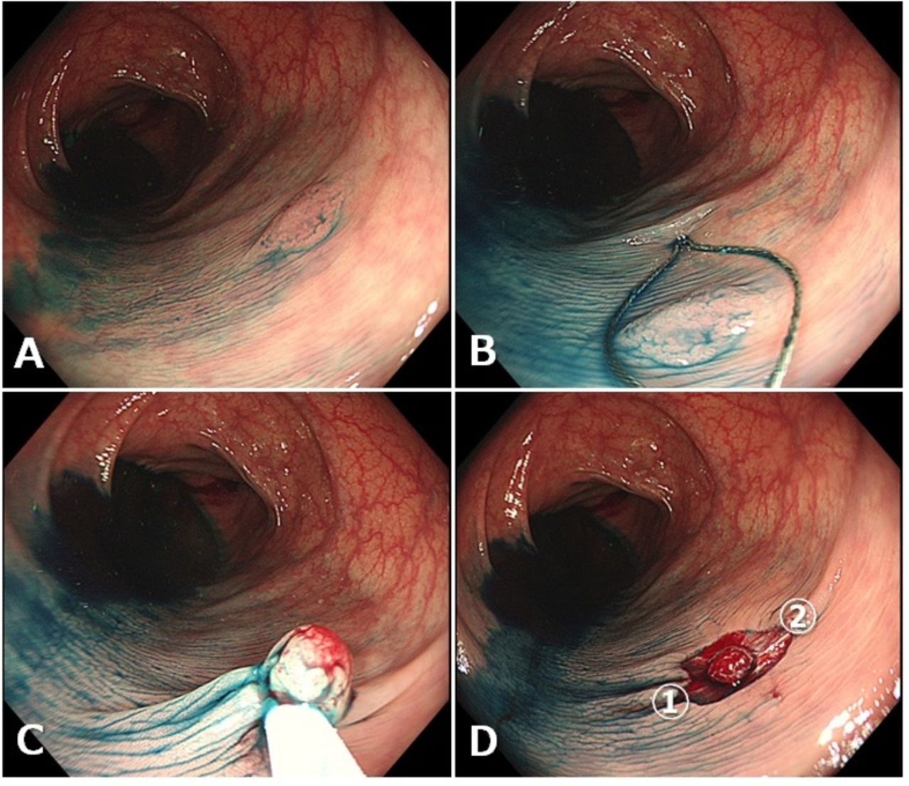
Study procedure for CSP. A flat elevated type polyp detected in the colon (A). The Captivator II polypectomy snare, 10 mm in size, was used for CSP. The size of this polyp was estimated to be 8 mm (B). CSP was performed (C). Biopsies were performed from two marginal sites located symmetrically on the left and right of the mucosal defects to confirm residual polyp tissue (D). CSP, cold snare polypectomy.
Study participants
Patients, ≥20 years of age, who underwent colonoscopy and had provided written informed consent were eligible for enrolment. The exclusion criteria included: (1) inflammatory bowel disease; (2) polyposis of the alimentary tract; (3) use of oral antithrombotic drugs; (4) pregnancy; (5) haemodialysis and (6) an American Society of Anaesthesiologists class III or higher. Only sessile adenomatous colorectal polyps 4–9 mm in size detected during colonoscopy were included in our study. To include only adenomatous polyps in our study, an endoscopic diagnosis of adenoma was made based on macroscopic appearance,15 findings on narrow band imaging (narrow-band imaging international colorectal endoscopic classification type 2)16 or the pit pattern classification (Kudo classification type III–IV).17 The following lesions detected during colonoscopy were ineligible for the study and were treated per usual clinical practice without randomisation even for those 4–9 mm in size: (1) depressed lesions and (2) lesions suspected to be cancerous based on endoscopic appearance. Only three polyps could be included from any one patient, with only one polyp included from each of the following areas: segment from the caecum to the ascending colon, the transverse colon, the descending colon, the sigmoid colon and the rectum. When we detected polyps ≥10 mm or more than one polyp per segment, the ineligible polyps were treated per usual clinical practice.
Interventions
All colonoscopies were performed with appropriate preparation by participating endoscopists who were certified by the Japan Gastroenterological Endoscopy Society or its equivalent. Standard high-definition videocolonoscopes were used, as per usual clinical practice. Magnification and image enhancement functions were not mandatory. A 10 mm round snare (Captivator II, Boston Scientific, Natick, Massachusetts, USA) was used for polypectomy in all cases. Video recording of the procedures was not mandatory.
When a target polyp was identified, eligibility was assessed by its endoscopic findings, with its size estimated using the opening of the snare. The morphology was defined as per the Paris classification.18 Once eligibility of the polyp for the study was confirmed, the polyp was removed by HSP or CSP as randomly determined. Submucosal injection prior to snaring was permitted for HSP but not for CSP. When a polyp could not be removed using the CSP technique, use of a high-frequency electric current was permitted.
After resection, the mucosal defect was washed thoroughly and the marginal mucosa was carefully observed, with magnification and image enhancement used as necessary. When residual polyp tissue was recognised, additional removal using the allocated technique or biopsy forceps was performed. After absence of residual polyp tissue was confirmed by endoscopic inspection, biopsies of the marginal mucosa, from the right and left edges of the postpolypectomy mucosal defect, were performed.
Endoscopic haemostasis was carried out when active haemorrhage continued for ≥30 s, regardless of the treatment group. Preventive haemostasis, defined as prophylactic coagulation of vessels or red spots in the ulcer or clipping of a non-bleeding postpolypectomy mucosal defect, was allowed in the HSP group, but not in the CSP group. Characteristics of the polyp and procedure-related events were input via a web-based case report form.
Outcomes
The primary endpoint of this study was the complete resection rate of adenomatous polyps by endoscopic polypectomy. Complete resection was defined by negative biopsy results from specimens obtained from the resection margin after polypectomy. Secondary endpoints were polyp retrieval rate, number of additional resections (snaring and/or biopsy), rate of difficult/impossible resection by CSP, time required for resection, incidence of immediate and delayed bleeding and other adverse events. Difficult CSP resection was defined as a resection procedure that required ≥5 s after snaring. An impossible CSP resection was defined as a resection procedure that needed high-frequency electric current. Time required for resection was defined as the time between the insertion of the snare into working channel to the end of polyp resection. The time was measured by endoscopists or assistant by using stopwatch that was built in the endoscopic system. When submucosal injection was conducted in the HSP group, the time required for resection was measured from the insertion of the injection needle into the working channel until the end of polyp resection. Immediate bleeding was defined as continuous haemorrhage for ≥30 s immediately after polypectomy. Patients were asked to contact the study institution in the event of a bloody stool or abdominal pain. Delayed bleeding was defined as haemorrhage after colonoscopy requiring endoscopic haemostasis. Before starting this study, we held a meeting for consensus to ensure uniformity in data collection.
Sample size calculation
A recurrence rate of 3.4% has been reported for mucosal or submucosal cancerous polyps <10 mm in size, treated with HSP in Japan.19 By comparison, a residual rate of 6.8% has been reported for these polyps treated with HSP in the USA.20 Assuming a residual rate of 3.4% for both groups in our study, the non-inferiority margins for CSP compared with HSP were defined at 3.4% in absolute risk difference, which would ensure that the CSP results would not exceed the residual rate of 6.8%. For an α-error level of 0.05 (one sided) and a β-error level of 0.20, 352 polyps in each group were required, for a total of 704 polyps. Assuming that approximately 10% of polyps may be excluded from the analysis set, the target sample size was set at 780 polyps in total.
Randomisation and monitoring
As it was not possible to establish if a target polyp would be identified in patients prior to endoscopy, randomisation to the CSP or HSP group was performed at the time of polyp resection, with an assistant opening the randomisation envelope to identify which technique was to be used for the resection. For group allocation, a stratified permuted block randomisation method was employed, using the institution as a stratification factor. The statistical advisor (IY) generated the random allocation sequence, and the contents of envelope were concealed until the intervention group was assigned at the time of polyp resection. Endoscopists were not blinded during the procedures.
After data accumulation, patient data were randomly extracted from the database and provided to a doctor in each institution, who was not part of the research team, to verify consistency of reporting as trial monitoring. The doctors who verified the report signed a prescribed form and faxed it to the data centre. Central reading was not adopted as for endoscopic and pathological images.
Statistics
All efficacy analyses were performed on the full analysis set, which included all randomised polyps. Excluded from the full analysis set were cases in which the allocated intervention was not used, any eligibility criteria were not met, as well as cases in which two biopsies could not be successfully obtained. As for the primary outcome, a lower 90% CI bound of more than −3.4% of the risk difference for complete resection between the two treatment groups was required to confirm the non-inferiority of CSP relative to HSP. Generalised estimating equations were used to control for within-patient correlations because the statistical hypothesis test was valid regardless of strength of correlation.21 Preplanned subgroup analyses based on the size of polyp were conducted based on the above method. Additional post hoc subgroup analyses for the method of observation of the postpolypectomy ulcer were also conducted. The analyses for secondary endpoints were performed using a two-sided 5% significance level.
All statistical analyses were conducted using SAS statistical software V.9.4 (SAS Institute).
Results
Recruitment and participant flow
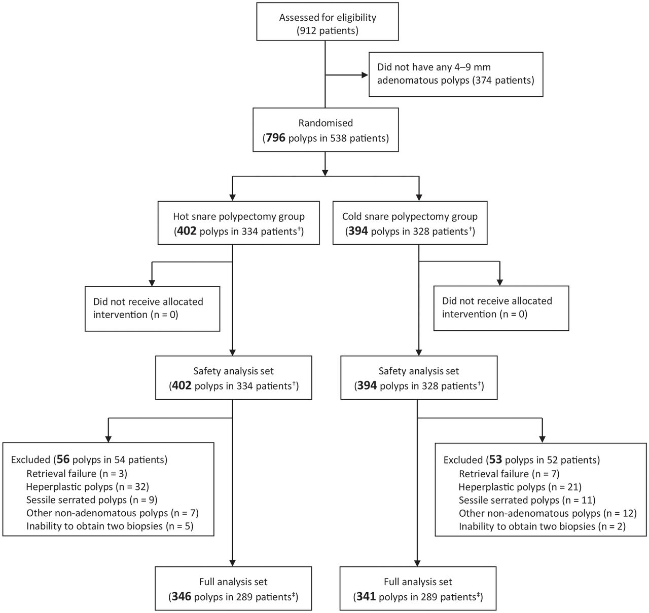
A total of 912 patients were recruited into the study and underwent colonoscopy, with 796 eligible polyps detected in 538 patients between September 2015 and August 2016 (figure 2). Recruitment ended when the number of polyps had reached the predetermined number. With regard to group allocation, 394 polyps were assigned to CSP and 402 to HSP. Of these, 109 polyps (53 in the CSP group and 56 in the HSP group) were excluded for the following reasons: retrieval failure (n=10), hyperplastic polyps (n=53), other non-adenomatous polyps (n=39) and inability to obtain two biopsies (n=7). Finally, 341 polyps from the CSP group and 346 polyps from the HSP were included in the analysis.

Study flow.†Including hot/cold snare polypectomy overlap cases (n=124).‡Including hot/cold snare polypectomy overlap cases (n=102).
Baseline data
Background characteristics of the allocated polyps (location, morphology and size) were comparable between the CSP and HSP groups (table 1). With regard to morphology, overall, 70% of polyps were classified as 0-I and 30% as 0-II types, with a mean size of 5.4 mm.
Characteristics of the polyps
As multiple target polyps in one patient were independently allocated to either the CSP or HSP group, patients were classified into three groups: HSP, CSP and HSP/CSP. Patients’ background characteristics (age, sex, indication, preparation status, endoscope used, number of all neoplastic lesions detected, number of target polyps and distribution of institutions) were similar among the three groups (see online supplementary table 1). Since the Japanese national screening programme of colorectal cancer uses only annual faecal immunochemistry test (FIT) and does not use colonoscopy, as for indication of colonoscopy, numbers of screening examinees were smaller than that of positive FIT and surveillance after polypectomy.
Supplementary file 1
Procedural information
Submucosal injection and prophylactic haemostasis were performed in 152 (43.9%) and 94 (27.2%) of the 346 polyps resected by HSP, respectively (see online supplementary table 2). The non-magnifying white light method for observation of postpolypectomy mucosal defect was used in 70.4% of lesions in the CSP group and 64.2% of lesions in the HSP group. Image-enhanced endoscopy (narrow band imaging or chromoendoscopy), with or without magnification, was used for observation of the remaining polyps.
Study outcomes
The complete resection rate for CSP was 98.2% and 97.4% for HSP. The non-inferiority of CSP for complete resection compared with HSP was confirmed by the +0.8% (90% CI −1.0 to 2.7) in complete resection rate (non-inferiority p<0.0001; table 2). Among 152 polyps with submucosal injection and 194 polyps without injection in the HSP group, an incomplete resection was identified in 5 (3.3%) and 4 (2.1%) polyps, respectively (p=0.505). When we compared the rate of incomplete resection based on patient groups, the rate for the HSP, CSP and HSP/CSP overlap groups was 3.7% (7/187), 2.7% (5/187) and 2.9% (3/102), respectively (p=0.831). In the HSP/CSP overlap group (n=102), incomplete resection was observed for two polyps removed by HSP and one polyp by CSP.
Primary endpoint
The proportion of additional removal for residual polyps was similar between the HSP and CSP groups (table 3). The proportion of difficult and impossible resection by CSP was 5.3% and 0.9% , respectively. The time required for resection was significantly shorter for CSP (60 s) than for HSP (83 s; p<0.001). However, when the HSP group was subdivided into groups with or without submucosal injection, the time required for resection was comparable for the HSP without submucosal injection (65 s) and CSP groups, with the time required being significantly longer for HSP with submucosal injection group (116 s). Both groups showed a similarly high polyp retrieval rate, 98% for CSP and 99% for HSP, with most retrieved polyps having an adenoma with low-grade dysplasia histology (table 4). On subgroup analysis according to the size of the polyp (4–5 mm vs 6–9 mm) and method of observation for postpolypectomy ulcers (white light only vs image enhanced endoscopy with/without magnification), the complete resection rate for CSP and HSP was comparable for both subgroups of polyps (see online supplementary table 3).
Secondary endpoints
Polyp retrieval rates and histology
Adverse events
Immediate bleeding was more frequent in the CSP group (7.1%, 28/394 of polyps) than in the HSP group (3.5%, 14/402 of polyps; p=0.022). However, all the immediate bleeding was handled with endoscopic haemostasis. When the rate of immediate bleeding was calculated using the full analysis set, the difference was not statistically significant (CSP vs HSP: 7.0% (24/341) and 3.8% (13/346), respectively; p=0.057). Based on patient groups, immediate bleeding in the HSP, CSP and HSP/CSP overlap groups was observed in 4.8% (9/187), 9.6% (18/187) and 9.8% (10/102), respectively (p=0.152). In HSP/CSP overlap group (n=102), immediate bleeding was observed in four polyps after HSP and six polyps after CSP.
Four patients in the HSP group and none in the CSP group complained of bloody stool after the procedure. Among four patients in the HSP group, two had bleeding from the eligible polyps, requiring endoscopic haemostasis (delayed bleeding as secondary outcome; 0.5%, 2/402 of polyps), but two had bleeding from postpolypectomy ulcer of ineligible polyps.
A patient in the CSP group, who underwent underwater endoscopic mucosal resection (EMR) for a large colonic polyp in another segment of the colon, complained of abdominal pain. CT imaging revealed free air around the region after underwater EMR. Therefore, we judged that the abdominal pain was caused by perforation after underwater EMR. As for eligible polyps, perforation or other adverse events were not identified in either group.
Discussion
Our multicentre randomised trial provides evidence that the complete resection rate of colorectal adenomatous polyps, 4–9 mm in size, was not inferior for CSP compared with that for HSP. The lower limit of the CI of the risk difference was estimated at −1.0%. This result indicates that the rate of residual tissue by CSP should not exceed 1.0% of the rate for HSP. With the safety profile of CSP having been previously established from several previous studies,3 4 we deem that CSP can be used as one of the standard techniques for the resection of 4–9 mm colorectal adenomatous polyps.
The rate of incomplete resection for HSP in clinical practice was reported by Pohl et al.20 They evaluated polyp residue after polypectomy of neoplastic lesion, including sessile serrated adenoma/polyp (SSA/P), using two biopsies from opposite edges of the mucosal defect after polypectomy for 5–9 mm polyps, as in our present study. They reported the rate of incomplete resection polypectomy to be influenced by the size of the polyp, with a 5.8% rate for 5–7 mm polyps and 9.4% for 8–9 mm polyps. On the other hand, Kim et al evaluated residual adenomatous tissue after CSP using an additional EMR technique, and reported a high complete resection rate of 96.6% for adenomatous polyps ≤7 mm in size.12 A recent single-arm study in Japan reported an incomplete resection rate of 3.9% after CSP for subcentimetre adenomas.22 A pilot study comparing HSP, CSP and CFP for diminutive (<6 mm) polyps reported comparable rates of incomplete resection among the three groups.23 Now, we demonstrated that the rate of complete resection of 4–9 mm adenomas with CSP was comparable to that for HSP in our multicentre, randomised controlled study. Although submucosal injection prior to snaring was permitted only in the HSP group in this study, rates of incomplete resection were almost the same in the subgroups with or without submucosal injection (3.3% vs 2.1%). Therefore, submucosal injection would not be related to the primary endpoint.
The incomplete resection rates reported by Pohl et al are different from ours, with our rate of polyp residue being almost half of the rates they reported.20 Although the reasons for this difference in the rate of incomplete resection are not entirely clear, it is possible that we were more careful to examine for polyp residue at the time of polypectomy in our prospective randomised trial than examination performed in usual practice. Moreover, we provided a comparable high-quality examination for polyp residue for both CSP and HSP groups.
As we described above, the efficacy of CSP and HSP was evaluated in terms of residual polyp tissue as determined using additional EMR or biopsy specimen from the resection margin after polypectomy. Although the additional EMR technique would be more appropriate for evaluation of residual tissue, it is complicated and difficult to apply in a large-scale randomised controlled trial as EMR after HSP increases the potential risk of adverse events, such as post-EMR bleeding and perforation.22 Therefore, we adopted biopsies from the resection margins as the method to determine residual polyp tissue in our multicentre trial. Biopsy of tissue from the bottom margin of the defect after polypectomy was not adopted in the present study. However, a previous study reported that residual tissue with CSP was present in the lateral margins of the defect and not in the bottom margin.22 Therefore, the use of biopsy specimens obtained from opposing resection margins was appropriate.
In our trial, we used an electrocautery snare (Captivator II) in both allocated groups, which allowed us to compare the hot and cold technique specifically rather than the different polypectomy snare. Moreover, the completion rate of resection has been reported to be higher when using a dedicated snare than when using a non-dedicated snare.7 Therefore, our results of a comparable rate of complete resection for CSP and HSP would be valid, whether a dedicated or non-dedicated snare is used.
Delayed bleeding occurred for two polyps in the HSP group, with no occurrence in the CSP group. This supports previous reports of CSP as a safe procedure. Repici et al reported absence of delayed bleeding for a series of 1015 resections of polyps<10 mm, among 823 patients.4 Furthermore, Horiuchi et al reported a low incidence rate of delayed bleeding after CSP even for patients on warfarin treatment.3 In our trial, preventive haemostasis was permitted only in the HSP group and not in the CSP group. However, the actual delayed bleeding rate was similar for both groups (0.5% in the HSP vs 0% in the CSP).
The procedure time for the CSP group (60 s) was significantly shorter than for the HSP group (83 s). However, the procedure time for the subgroup without submucosal injection in the HSP group (65 s) was comparable to that of the CSP group. Horiuchi et al reported that overall procedure time of CSP was significantly shorter than that of HSP, even without submucosal injection.3 We cannot compare overall procedure time among patient groups in the present study because we treated not only eligible polyps but also other ineligible polyps per usual clinical practice. Further study would be needed to evaluate whether CSP is really less time consuming that HSP to perform.
In this study, SSA/Ps were excluded because we specifically wanted to assess the technical aspect of CSP. Endoscopically, the margin of SSA/P is sometimes unclear, and even in cases with a clear pathology, SSA/P is sometimes difficult to distinguish from hyperplastic polyps. Therefore, we concluded that SSA/P was not suitable for assessment of a polypectomy technique. Generally, SSA/P has been reported as one of the characteristic lesions that are associated with incomplete resection.20 Therefore, special attention should be paid when using both CSP and HSP for resection of SSA/P.
This study has several limitations that need to be acknowledged. First, we only evaluated residual tissue just after polypectomy but not long-term outcomes. As the burning effect causes a necrosis of tissues surrounding the defect after a while, biopsy results obtained just after HSP may not reflect the true recurrence rate of removed polyp tissue. In this regard, long-term outcomes should be evaluated in future studies. Second, polyp size may be inaccurately measured due to the inherent difficulty of precise measurement using an open snare.24 However, as we used the same snare for both HSP and CSP, we are confident that measurements can be reliably compared between the groups. Third, we did not systemically monitor patients after the procedures. Patients were asked to contact the hospital in case of bleeding which they believed required treatment. Therefore, delayed bleeding after the procedures may be underestimated in our study. Fourth, endoscopists could not be blinded in this trial. Of course, method of polypectomy could have been blinded in our results if we would have used another endoscopist to perform the biopsy after polypectomies. However, a change in endoscopist during the procedure would have been too complicated. Moreover, it would have been easy for any endoscopist to distinguish postpolypectomy mucosal defects created with or without electrocautery, even with blinding. However, the high rate of complete resection in both groups, which are comparable with previously reported rates, are indicative of the appropriateness of our outcomes.
Despite these limitations, our study provides important information regarding the usefulness of CSP technique for small colorectal polyps. In conclusion, the complete resection rate for CSP is not inferior to that for HSP. CSP can be one of the standard techniques used of the resection of 4–9 mm colorectal adenomatous polyps.
Acknowledgments
We wish to thank Ms Satomi Sakabayashi of the Kyoto Prefectural University of Medicine for her contribution in data analysis. We also wish to thank the staff of each hospital for their cooperation.
References
Footnotes
Contributors TK, YT and NU planned the study. IY and ST were statistical advisors. SA, EA, MK, TA, KT, YK, MI, KK, MN, DW, AY, NF, MS, MO, KF, YS, HK, SH and HI had leadership in each institute and collected data. KT organised the study group. TK wrote the manuscript, and all the authors approved the final version of this manuscript.
Funding This study was funded by the Investigator Sponsored Research Program of Boston Scientific (ISREND00006).
Competing interests This study was funded by the Investigator Sponsored Research Program of Boston Scientific. The funder paid for statistical analysis, construction of electronic data collecting system and English proofreading service. However, the funder was not involved in the study design, recruitment, analysis plan or interpretation of data.
Ethics approval Institutional Review Board of each participating centre.
Provenance and peer review Not commissioned; externally peer reviewed.