Article Text

Abstract
Background The duodenum has become a metabolic treatment target through bariatric surgery learnings and the specific observation that bypassing, excluding or altering duodenal nutrient exposure elicits favourable metabolic changes. Duodenal mucosal resurfacing (DMR) is a novel endoscopic procedure that has been shown to improve glycaemic control in people with type 2 diabetes mellitus (T2D) irrespective of body mass index (BMI) changes. DMR involves catheter-based circumferential mucosal lifting followed by hydrothermal ablation of duodenal mucosa. This multicentre study evaluates safety and feasibility of DMR and its effect on glycaemia at 24 weeks and 12 months.
Methods International multicentre, open-label study. Patients (BMI 24–40) with T2D (HbA1c 59–86 mmol/mol (7.5%–10.0%)) on stable oral glucose-lowering medication underwent DMR. Glucose-lowering medication was kept stable for at least 24 weeks post DMR. During follow-up, HbA1c, fasting plasma glucose (FPG), weight, hepatic transaminases, Homeostatic Model Assessment for Insulin Resistance (HOMA-IR), adverse events (AEs) and treatment satisfaction were determined and analysed using repeated measures analysis of variance with Bonferroni correction.
Results Forty-six patients were included of whom 37 (80%) underwent complete DMR and 36 were finally analysed; in remaining patients, mainly technical issues were observed. Twenty-four patients had at least one AE (52%) related to DMR. Of these, 81% were mild. One SAE and no unanticipated AEs were reported. Twenty-four weeks post DMR (n=36), HbA1c (−10±2 mmol/mol (−0.9%±0.2%), p<0.001), FPG (−1.7±0.5 mmol/L, p<0.001) and HOMA-IR improved (−2.9±1.1, p<0.001), weight was modestly reduced (−2.5±0.6 kg, p<0.001) and hepatic transaminase levels decreased. Effects were sustained at 12 months. Change in HbA1c did not correlate with modest weight loss. Diabetes treatment satisfaction scores improved significantly.
Conclusions In this multicentre study, DMR was found to be a feasible and safe endoscopic procedure that elicited durable glycaemic improvement in suboptimally controlled T2D patients using oral glucose-lowering medication irrespective of weight loss. Effects on the liver are examined further.
Trial registration number NCT02413567
- diabetes mellitus
- therapeutic endoscopy
- endoscopic procedures
- glucose metabolism
- duodenal mucosa
This is an open access article distributed in accordance with the Creative Commons Attribution Non Commercial (CC BY-NC 4.0) license, which permits others to distribute, remix, adapt, build upon this work non-commercially, and license their derivative works on different terms, provided the original work is properly cited, appropriate credit is given, any changes made indicated, and the use is non-commercial. See: http://creativecommons.org/licenses/by-nc/4.0/.
Statistics from Altmetric.com
Significance of this study
What is already known on this subject?
Studies suggest a critical physiological and pathophysiological role of the small bowel in metabolic homeostasis.
Bypassing, excluding or altering the presentation of nutrients to the duodenum results in a weight-independent improvement in glycaemia in people with type 2 diabetes mellitus (T2D), implicating a key role for the duodenum in glucose regulation.
Duodenal mucosal resurfacing (DMR) is a single, minimally invasive endoscopic procedure that involves circumferential hydrothermal ablation of the duodenal mucosa with subsequent regeneration of the mucosa. DMR potentially mimics some of the mechanisms of action of bariatric surgery in a minimally invasive manner.
A first-in-human study showed significant improvements in glycaemia in T2D patients up to 24 weeks after DMR.
Significance of this study
What are the new findings?
In this study, endoscopic DMR was found to be feasible, safe and effective in patients with suboptimally controlled T2D using oral glucose-lowering medication. DMR was completed successfully in the majority (80%) of the patients.
Fifty-two percent of the patients experienced one or more adverse event related to DMR of which 81% was classified as mild. Patients underwent the procedure with minimal GI symptoms post procedure. No unanticipated SAEs were reported and a single DMR-related SAE was reported.
DMR elicited a substantial and clinically significant improvement in glycaemic control and measures of insulin resistance up to 12 months post procedure. Patient-reported treatment satisfaction also improved.
How might it impact on clinical practice in the foreseeable future?
DMR can elicit clinically relevant improvement in glycaemic control without the anatomical disruption seen in bariatric surgery. Absolute mean HbA1c at 24 weeks and 12 months was 10 mmol/mol (0.9%) lower compared with baseline. This improvement is comparable with that seen in studies adding additional pharmacological agents at this stage of diabetes management. In the contemporary diabetes treatment spectrum, DMR may have a role as adjuvant or alternative approach to pharmacological treatment. Our study also adds to the growing body of evidence that the GI tract, and particularly the duodenum, is an important target for interventions to treat T2D and other concomitant metabolic diseases.
Introduction
Type 2 diabetes mellitus (T2D) is increasing at a disturbing rate throughout the world with an estimated global prevalence of 552 million by 2030.1 2 The therapeutic goal of a glycated haemoglobin (HbA1c) level of ≤53 mmol/mol3 is achieved by less than half of the patients with T2D4 despite lifestyle interventions and an increasing number of medical treatment options. Bariatric surgery has proven to be successful in patients with class I, II and III obesity.5–7 In moderately obese patients with T2D, bariatric surgery is superior to intensive medical therapy alone.8 However, bariatric surgery is not a scalable solution for the growing T2D pandemic as the majority of bariatric surgery procedures are invasive, irreversible and associated with some morbidity.9 It appears that excluding or altering the presentation of nutrients to the duodenum contributes to the immediate improvements in glycaemic regulation after bariatric surgery, which do not appear to be due to malabsorption or the substantial weight loss often observed later post surgery.10–12 Studies suggest a critical physiological and pathophysiological role of the small bowel in metabolic homeostasis. The easy endoscopic accessibility of the duodenum makes it a potential target for disease-modifying intervention.13
The duodenal mucosal resurfacing procedure is performed using specially designed catheters (Fractyl Laboratories) which are advanced over a guidewire next to the endoscope. Duodenal mucosal resurfacing (DMR) is a single, minimally invasive endoscopic procedure that involves circumferential hydrothermal ablation of the duodenal mucosa resulting in subsequent regeneration of the mucosa. Before ablation, the mucosa is lifted with saline to protect the outer layers of the duodenum. A first-in-human study showed significant improvements in glycaemia in T2D patients after DMR with a suggestion of a positive relationship between the length of the ablated segment and efficacy.14 This demonstrated the therapeutic potential of DMR but the length of treated duodenum was variable in this initial clinical study and background oral glucose-lowering medication was adjusted during follow-up at the discretion of the investigator, thus confounding the impact of the procedure on ambient glycaemia. In view of these study limitations, we conducted an international multicentre, prospective, open-label study in which patients with T2D using stable oral glucose-lowering medication underwent a standardised DMR procedure to further evaluate efficacy and safety.
Materials and methods
Study design
We conducted an international multicentre, prospective, open-label study to establish the safety and feasibility of the DMR procedure and to evaluate the effect of DMR on glycaemia. The seven study sites were the Academic Medical Centre, Amsterdam, the Netherlands; Erasme University Hospital, Brussels, Belgium; Policlinico Gemelli, Catholic University of Rome, Rome, Italy; University College London Hospital, London, UK; CCO Clinical Centre for Diabetes, Obesity and Reflux, Santiago, Chile; King’s College Hospital, London, UK and University Hospital Leuven, Leuven, Belgium. These centres are tertiary endoscopic intervention centres with tertiary care for T2D. The study protocol was approved by the independent ethics committee of each centre. The study was conducted in accordance with ICH Good Clinical Practice Guidelines and the Declaration of Helsinki. An independent data safety monitoring committee established criteria for stopping the study before enrolment of the first patient and reviewed all adverse events (AEs) that occurred over the course of the study. The study is registered under ClinicalTrials.gov.
Patients
Eligible participants were people with T2D, aged 28–75 years, with body mass index of 24–40 kg/m2 and an HbA1c of 59–86 mmol/mol (7.5%–10.0%) who were on stable diabetes treatment comprising at least one oral glucose-lowering drug for at least 3 months. Exclusion criteria were type 1 diabetes (clinical diagnosis and/or positive GAD antibodies), a history of ketoacidosis, low endogenous insulin production (fasting C-peptide <0.333 nmol/L), use of injectable glucose-lowering medication, hypoglycaemia unawareness or a history of severe hypoglycaemia, known autoimmune disease, previous GI surgery that could affect the ability to treat the duodenum, a history of chronic or acute pancreatitis, active hepatitis or active liver disease, symptomatic gallstones or kidney stones, history of duodenal inflammatory diseases including Crohn’s disease and Celiac disease, upper GI bleeding conditions, use of anticoagulation therapy, P2Y12 inhibitors and/or nonsteroidal anti-inflammatory drugs which could not be discontinued around the DMR-procedure, taking corticosteroids or drugs known to affect GI motility, using weight loss medications, an estimated glomerular filtration rate or modification of diet in renal disesase (MDRD) <30 mL/min/1.73 m2, persistent anaemia (Hb <10 mg/dL), active systemic infection, active malignancy within the last 5 years, not potential candidate for surgery, active illicit substance abuse or alcoholism, pregnancy or expecting to become pregnant and participation in another clinical trial. Written informed consent was obtained from all patients.
Study procedure
The DMR procedure was performed under either general anaesthesia or deep sedation with propofol by a single endoscopist at each site with extensive experience in therapeutic upper GI endoscopy and guidewire management. A screening gastro-duodenoscopy was conducted first to ensure there were no conditions that would preclude the DMR procedure. Subsequently, the location of the papilla of Vater was marked on the contralateral duodenal wall using either argon plasma coagulation or placement of an endoscopic clip. Then, a guidewire was inserted past the ligament of Treitz. DMR catheters were advanced over this guidewire. DMR consisted of submucosal expansion (to provide a protective layer of saline between the mucosa/submucosa and duodenal proper muscle layer) and subsequent stepwise circumferential hydrothermal ablation at 90ᴼC for 10 s over 9–10 cm of the postpapillary duodenum. The first submucosal expansion and duodenal ablation was performed at a position just distal to the papilla of Vater and progressively distal duodenal areas were then ablated. Fluoroscopy was used during the procedure to verify the positioning of the guidewire and catheters. A complete DMR was defined as a duodenal ablation zone of 9–10 cm. If necessary, intravenous paracetamol was administered post procedure. Patients were discharged the same day or after an overnight stay, following the local hospital’s guidelines.
Dietary management
Patients were instructed to follow a 2-week diet post DMR in which clear liquids were gradually transitioned to solid foods. At follow-up visits, patients received per protocol dietary counselling based on standard clinical practice guidelines to educate them on the importance of diet in relation to blood glucose control. During the 2-week post-procedure phase and then out to the full 12 months of follow-up, there was no concerted effort for patients to adhere to a specific hypocaloric regimen beyond standard dietary counselling.
Management of glucose-lowering medication
In patients who met the eligibility criteria, sulfonylureas and meglitinides were discontinued at initial screening to mitigate the risk of potential hypoglycaemia after DMR; other oral diabetes medications were continued unchanged. Participants then entered a 4-week run-in phase with monitoring of medication usage, compliance and blood glucose levels. Patients were instructed to complete a standardised blood glucose diary and record any symptoms related to hypoglycaemia. Patients with ≥3 hyperglycaemic events confirmed by a laboratory blood test (defined as blood glucose level >15 mmol/L fasting or >20 mmol/L non-fasting) or a hypoglycaemic event with a plasma glucose level <3.1 mmol/L or the need for third-party assistance in the run-in phase were excluded. At the subsequent baseline visit, patients were excluded if HbA1c was <59 mmol/mol (7.5%) or >86 mmol/mol (10.0%). Following DMR, glucose-lowering medication was kept stable for at least 24 weeks unless patients experienced persistent hyperglycaemia (three confirmed fasting glucose measurements >15 mmol/L) in which case medication could be increased at the discretion of the investigator. Following the 24-week follow-up visit, glucose-lowering medication was adjusted based on HbA1c measurements; an HbA1c measurement >58 mmol/mol (7.5%) induced a study protocol-based increase in glucose-lowering medication starting with the stepwise addition of sulphonylurea, followed by glucagon-like peptide 1 (GLP-1) and finally insulin, if necessary.
Assessments and outcome measures
At screening, baseline and follow-up visits, physical examination (including anthropometric measurements, systolic and diastolic blood pressure) and laboratory assessment (fasting blood glucose (FPG), glycosylated haemoglobin (HbA1c), fasting insulin, C-peptide, haematology, serum biochemistry and urine microalbumin) were performed alongside recording of medication use and any AEs. At each visit, the local investigator asked for the occurrence of self-measured hypoglycaemia (glucose level <3.1 mmol/L or <56 mg/dL) and the occurrence of any other symptoms or AEs. The number of hypoglycaemic events is reported, but not as a primary outcome. Sulphonylurea derivatives were discontinued at screening to mitigate the risk of hypoglycaemia during study follow-up, which makes reporting hypoglycaemia as a primary outcome irrelevant. AEs were graded in terms of mild (discomfort but no disruption of daily activity), moderate (discomfort sufficient to affect daily activity) and severe (inability to perform daily activity), and the relationship to the device and to the procedure was assessed in terms of not, possibly, probably and definitely based on the temporal association with DMR and the possibility of other aetiologies. Unanticipated adverse device effect was defined as any serious adverse effect (SAE) on health or safety or any life-threatening problem or death caused by or associated with the device if that effect, problem or death was not previously identified in nature, severity or degree of incidence in the investigational plan, or any other unanticipated serious problem associated with the device that relates to the rights, safety or welfare of patients.
Baseline measurements were used for further comparison. The baseline visit was scheduled 4–6 weeks after screening and DMR took place within 14 days after the baseline visit. During DMR, the number of ablations was recorded, as well as procedure time and procedure details in case of an incomplete DMR procedure. Post-DMR follow-up was planned for 2 years with visits scheduled at 4, 12, 18, 24 and 36 weeks and at 12, 18 and 24 months. The primary efficacy endpoint was HbA1c at 24 weeks and we report this data plus follow-up to 12 months.
Patient-reported outcome measures
Patients were also asked to complete Diabetes Treatment Satisfaction Questionnaires (DTSQ) throughout the study. We used the status version (DTSQs)15 and the change (DTSQc)16 version. The DTSQs evaluates absolute treatment satisfaction and was assessed at baseline and at 4, 12 and 24 weeks. The DTSQc measures relative change in treatment satisfaction from previous therapy and was assessed at 24 weeks.
Statistical analysis
The number of patients mentioned in our first protocol was originally based on medical and procedural considerations (n=60). Enrolment was stopped when the DMR procedure had matured to a level ready for initiating a sham-controlled randomised controlled trial. The study closed at 49 patients. Analysis revealed that the current number of patients was sufficient to detect a significant difference at 24 weeks compared with baseline. Statistical analyses were performed using SAS V.9.4. Missing interim data were imputed using multiple imputations (17/276 (6.2%) missing values for HbA1c) (online supplemental methodology multiple imputations). Baseline characteristics are expressed as mean ±SD, change from baseline is presented with SE, and follow-up measurements are presented as mean with SE The intention-to-treat population consisted of all patients who underwent the screening endoscopy. The per-protocol population was defined as all patients who received the complete DMR procedure (defined as 9–10 cm of circumferentially ablated duodenal mucosa). Effect of DMR on glycaemia was evaluated in the per-protocol population analysis. The primary endpoint was the change from baseline in HbA1c at 24 weeks. Secondary efficacy endpoints were the change in HbA1c at 12 months and change in FPG, weight and insulin resistance (as estimated by the homeostatic model assessment index for insulin resistance (HOMA-IR)) at 24 weeks and 12 months post DMR. Efficacy at 12 months was analysed separately in two groups based on glucose-lowering medication use in the 24 weeks to 12 months follow-up interval (stable and increased glucose-lowering medication groups) and compared with baseline. For the primary endpoint (change in HbA1c at 24 weeks compared with baseline), a paired t-test was used. We used ANOVA for repeated measurements with Bonferroni correction for the analysis of multiple measurements of HbA1c, FPG, HOMA-IR and weight after DMR (five multiple tests for the endpoints up to 12 months after DMR, one for each visit assessing the significance of the change from baseline, where the Bonferroni-adjusted p value <0.05 was considered statistically significant). Paired t-tests were used to evaluate DTSQs results at 24 weeks and 12 months compared with baseline. P values <0.05 were considered to be statistically significant. We used Pearson’s correlation to assess the correlation between initial weight loss and improvement in glycaemia.
Supplemental material
Results
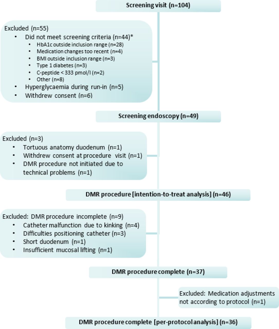
Of the 104 people with T2D screened, 46 patients fulfilled the study criteria at screening, baseline and the screening endoscopy (intention-to-treat population). Thirty-seven patients received a complete DMR procedure. Medication adjustments were not in line with the protocol in a single patient, since this patient intermittently used insulin post DMR. This resulted in a per-protocol population of 36 patients (figure 1). Table 1 shows the screening and baseline characteristics of the intention-to-treat population. At screening, 17 participants (37%) were on sulphonylurea and 2 (4%) were on meglitinide; these drugs were discontinued as per protocol and replaced with a DPP-4 inhibitor if deemed necessary at the investigator’s discretion.

Enrolment flow diagram. *Four subjects were excluded based on two criteria. BMI, body mass index; DMR, duodenal mucosal resurfacing; HbA1c, glycated haemoglobin.
Clinical characteristics at screening and baseline
Procedure feasibility information
The DMR procedure was complete in 37 out of 46 patients (80%). Mean (±SD) procedure time in the per-protocol population (n=37) was 82±28 min. Following the local hospital’s guidelines, general anaesthesia was used for 35 patients and deep sedation with propofol was used for 11 patients. Causes of an incomplete DMR procedure were catheter failure (n=4, 9%), a difficult procedure in terms of tracking and positioning the catheter (n=3, 7%), duodenal tortuosity (n=1, 2%) or inadequate lifting (n=1, 2%) (figure 1). Mean (±SD) duration of hospitalisation after DMR was 0.78±0.87 days.
Safety and tolerability
AEs were evaluated in the intention-to-treat population (n=46). No unanticipated adverse device events were reported. Six SAEs were reported during follow-up of which one SAE was reported to be procedure related. This concerned a patient with general malaise, mild fever (38°C), and increased c-reactive protein (CRP) level on the first day after DMR. The mild fever resolved within 24 hours and CRP level normalised within 3 days. The other SAEs were considered unrelated to the treatment (online supplementary table Serious Adverse Events).
Supplemental material
In total, 54 procedure-related AEs were reported during the first year of follow-up in 24 patients in whom DMR was initiated (24/46, 52%). Of the 54 AEs, 30 (56%) were assessed as possibly procedure related, 16 (30%) as probably procedure related and 8 (15%) as definitely procedure related. Twenty-two (41%) were treated with medication. Details of these AEs are reported in table 2. In 22 patients, no procedure-related AEs were reported.
Summary of adverse events during study 12 months follow-up period (intention-to-treat population, n=46)
Three patients recorded biochemical hypoglycaemia during follow-up (range 2.2 to 3.6 mmol/L). Two of these recorded single episodes of glucose of 3.3 and 3.6 mmol/L at 4 and 30 days post DMR, respectively, and the third experienced four episodes (range 2.2 to 3.1 mmol/L) between days 39 and 55 post DMR. The hypoglycaemic event at day 4 post procedure was assessed as probably procedure related. The other events were considered as not procedure related by the local investigator. No changes to oral diabetes medication were initiated in response to these events. No patients experienced severe hypoglycaemia requiring third-party assistance.
Efficacy
Glucose-lowering medication was stable in the complete per-protocol population up to 24 weeks post DMR; protocol-based medication adjustments due to hyperglycaemia were not necessary in this timeframe. Indices of glycaemia improved significantly after DMR. HbA1c was reduced by 10±2 mmol/mol (0.9%±0.2%) (mean ±SE) at 24 weeks (p<0.001) compared with baseline with preservation of this effect up to 12 months (figure 2A and B). FPG was reduced by 1.7±0.5 mmol/L (p<0.001) and 1.8±0.5 mmol/L (p<0.001) at 24 weeks and 12 months post DMR, respectively, compared with baseline (figure 3A). HOMA-IR continued to improve after DMR (figure 3B). HOMA-IR was reduced by 2.9±1.1 at 24 weeks and by 3.3±0.9 at 12 months post DMR compared with baseline (p<0.001). A modest weight reduction was observed (figure 3C): −2.5±0.6 kg (p<0.001) at 24 weeks and −2.4±0.7 kg (p<0.001) at 12 months.

Change in HbA1c after DMR over 12 months follow-up. (A) Primary endpoint: mean difference ±SE in HbA1c at 24 weeks and 12 months when compared with baseline after a single endoscopic DMR procedure. Analysis with paired t-test. (B) Mean ±SE HbA1c during follow-up up to 12 months after single DMR. ANOVA repeated measurements analysis with Bonferroni correction to apply a more rigorous data analysis. n=36. ‡P<0.0001 when compared with baseline (paired t-test). * P< 0.01 when compared with baseline (ANOVA repeated measurements, Bonferroni-adjusted p value). DMR, duodenal mucosal resurfacing; HbA1C, glycated haemoglobin.
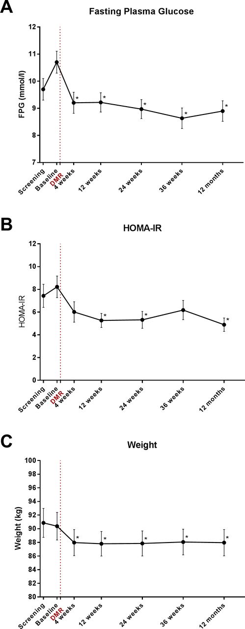
Changes in FPG, insulin sensitivity and weight after DMR over 12 months follow-up. Data represent mean ±SE changes after a single endoscopic DMR procedure in (A) FPG, (B) HOMA-IR and (C) weight. n=36. Analysis with ANOVA for repeated measurements with Bonferroni correction. *Indicates a significant difference (Bonferroni adjusted p value < 0.01) when compared with baseline values. ANOVA, analysis of variance; DMR, duodenal mucosal resurfacing ; FPG, fasting plasma glucose; HOMA-IR, Homeostatic Model Assessment for Insulin Resistance.
Weight loss was observed at 4 weeks post procedure after which weight stabilised. This initial weight loss did not correlate significantly with change in HbA1c at 24 weeks (Pearson’s correlation 0.29, p=0.14) and 12 months (Pearson’s correlation 0.26, p=0.078).
In nine patients (25%) from the per-protocol population, additional glucose-lowering medication was prescribed in the 24 weeks to 12 months window follow-up per study protocol (increased medication group). Six of these patients (67%) had used a sulfonylurea (n=4) or meglitinide (n=2) prior to screening when it was discontinued. No extra glucose-lowering medications were prescribed in 27 of the 36 per-protocol population patients (75%) during 12 months follow-up (stable medication group). This group included one patient whose metformin dose was reduced and replaced by low-dose gliclazide and a second patient whose metformin was replaced by empagliflozin. C-peptide levels before DMR (baseline levels known in 28 patients) did not differ between stable medication (1.1±0.1 nmol/L) and increased medication groups (0.8±0.1 nmol/L) in our study population, neither did fasting plasma insulin levels (stable medication: 94±11 pmol/L vs increased medication: 102±26 pmol/L).
Alanine transaminase (ALT) levels decreased from 40±4 U/L at baseline to 31±2 U/L at 24 weeks (p=0.016) and to 30±3 U/L at 12 months follow-up (p<0.001) (figure 4).

Change in ALT levels after DMR over 12 months follow-up. Post-DMR mean ±SE change in ALT levels. n=36. Analysis with ANOVA for repeated measurements with Bonferroni correction. *Indicates a significant difference (Bonferroni-adjusted p value <0.01) when compared with baseline values. ALT, alanine transaminase; ANOVA, analysis of variance; DMR, duodenal mucosal resurfacing.
Perceived diabetes treatment satisfaction
Mean (SE) baseline treatment satisfaction score was 27.2 (1.1) on the DTSQs. At 24 weeks and 12 months after DMR, treatment satisfaction scores were 30.5 (1.0) and 31.1 (0.9), respectively (p=0.015 and p=0.002 compared with baseline). Mean perceived hyperglycaemia and mean perceived hypoglycaemia at 12 months did not change significantly after DMR. Based on the DTSQc, treatment satisfaction score was +11.8 (1.2) at 24 weeks and +12.7 (0.8) at 12 months, indicating a large and clinically relevant increase in treatment satisfaction.
Discussion
In this first international multicentre, prospective open-label study, the endoscopic DMR procedure was found to be feasible, safe and effective in patients with suboptimally controlled T2D using oral glucose-lowering medication. DMR elicited a substantial improvement in parameters of glycaemia as well as a decrease in liver transaminase levels at 24 weeks which was sustained at 12 months post procedure. These findings were also associated with an improvement in patients’ diabetes treatment satisfaction.
The DMR procedure was completed in the large majority (80%) of the patients, and the observed tolerability and safety profile of DMR was reassuring. Most incomplete DMR procedures could be attributed to the novelty of the procedure for endoscopists and the DMR technology being under development. Patients underwent the procedure with minimal intolerance or GI symptoms post procedure and there were no unanticipated SAEs reported. No devices are left in situ in the GI tract after DMR, so there are no additional risks of long-term device implantation such as device migration or the development of hepatic abscess. Local hospital guidelines were decisive in selecting the type of anaesthesia. General anaesthesia was used in 35 patients and in 11 patients propofol was used for sedation. Since propofol has several advantages over general anaesthesia in terms of rapid induction and recovery and minimal residual effects, propofol could be the preferred type of anaesthesia in centres with experienced usage in future DMR procedures.
As early as 4 weeks post DMR, HbA1c, FPG and HOMA-IR levels decreased and this effect was sustained out to 12 months. Mean HbA1c was 10 mmol/mol (0.9%) lower at 12 months post DMR compared with baseline. Notably, this effect is in the same order of magnitude as adding a second or third oral drug to background therapy17 but without the need to take extra daily medication, and without medication side effects. In the previous first-in-human study of DMR,14 some erosion of the glycaemic effect was observed between 12 and 24 weeks of follow-up, although this observation was confounded by altered background antidiabetic medication use. In this multicentre study, parameters of glycaemia were sustained to 12 months post DMR. This is possibly due to the use of a more standardised duodenal mucosal ablation length and a more stable application of background oral glucose-lowering medication. It should also be noted that the overall glycaemic effect observed occurred despite a significant proportion of patients (approximately one-third) stopping insulin secretagogue therapy before the run-in phase of 4–6 weeks. This might also be a limitation of the understanding of efficacy. HbA1c may not have been at equilibrium during the baseline visit as not enough time had been allotted for HbA1c to re-equilibrate after stopping insulin secretagogue therapy. As such, the HbA1c reduction may in fact be underestimated in our study.
Body weight was reduced at 4 weeks post procedure, most likely related to the 2 weeks post-procedural diet that the patients were asked to follow after DMR. Some weight regain occurred by 12 weeks and thereafter weight stabilised. While the post-procedural diet was not intended to be hypocaloric, it cannot be excluded that the initial weight reduction was at least partially due to some caloric restriction. However, the initial weight loss at 4 weeks did not correlate with change in HbA1c at 24 weeks or 12 months. In a recently published systematic review examining the relationship between body weight change and effects on glycaemia, 1 kg of weight loss was calculated to account for an approximate 1 mmol/mol (or ~0.1%) lowering of HbA1c.18 Thus, logically, the weight loss of ~2.3 kilograms observed in this study cannot fully explain the substantial HbA1c decrease of 10 mmol/mol (~1%) over the 12 months of trial observation.
Medication was kept stable up to 24 weeks post DMR, and no additional rescue medication was prescribed in the per-protocol population. While the majority of patients showed a durable glycaemic response over 12 months, a minority exhibited less benefit from DMR and required additional glucose-lowering medication at 24 weeks. Of note, approximately two-thirds of the patients who required addition of antidiabetic medication in the latter phase of study had undergone insulin secretagogue medication withdrawal at screening. For future study, it may not be necessary to discontinue these medications before DMR, and this will allow an even more precise measure of DMR effect.
We speculated that patients with greater beta-cell reserve (high insulin and C-peptide baseline levels) might benefit more from DMR than patients with lower beta-cell reserve since insulin sensitivity improves after DMR. However, we did not find a clear association between baseline C-peptide or insulin levels and later glycaemic outcome. This is probably due to the selection criteria (orally treated T2D patients with C-peptide ≥0.333 nmol/L), the relative small sample size and lack of power to demonstrate a difference in this parameter. Future research is necessary to identify patient groups in which the endoscopic DMR procedure will be most effective.
A consistent and durable decrease in ALT was also observed. It is well recognised that T2D and non-alcoholic fatty liver disease (NAFLD) are two metabolic conditions where insulin resistance is thought to be a common pathological driver and that the two conditions often co-exist in patients.19 The lowering of ALT observed with DMR therefore suggests an additional beneficial effect of DMR on concomitant NAFLD measures. However, ALT measurements as the only biomarker of NAFLD is relatively aspecific. The mean pre-procedure ALT measurement was normal (40 U/L), so the clinical relevance of a decrease to more normal level can be questioned. A Fibroscan or liver Magnetic Resonance Proton Density Fat Fraction would have added value here. Therefore, effects of DMR on the liver need to be examined further.
Our data indicate a positive and durable effect of DMR on HOMA-IR suggesting DMR elicits an apparent insulin sensitising effect. However, at this point, the precise mechanism underlying the effects of DMR, whether antidiabetic or hepatic or both, is not well understood: changes in gut microbiome,20 bile acid composition21 or gut permeability22 are possible mechanisms of action since these components are already proven to be altered in patients with T2D and are again modified by bariatric surgery. Further mechanistic studies to unravel the potential mechanisms underlying the effect of DMR on glycaemia and the liver bed are eagerly awaited.
The open-label uncontrolled nature of this phase II clinical study is an important limitation. The results of this multicentre study need to be confirmed in a proper controlled study. Nevertheless, this study forms the requisite solid foundation for further research, and controlled studies are currently under way. A clinical study in which DMR is combined with GLP-1 and lifestyle intervention is also under way. Possibly, this treatment combination has a synergistic effect where DMR improves insulin sensitivity, GLP-1 stimulates endogenous insulin production and lifestyle intervention improves the underlying factors of an unhealthy diet and limited exercise.
In conclusion, this study confirms and extends the finding of the first-in-human study, demonstrating that DMR, a single point in time endoscopic intervention, can be implemented safely and is able to exert clinically relevant and durable improvement in glycaemic control over 12 months in patients with T2D.
Acknowledgments
The authors wish to thank Professor Rachel Batterham (University College Hospital, London) and Professor Geltrude Mingrone (Catholic University of Rome) for valuable input during the preparation of this manuscript. The authors thank all study participants.
References
Footnotes
Contributors AvB participated in the design of the study and data analysis, saw patients at the site for screening and follow-ups, and wrote, edited, reviewed and approved the manuscript. FH participated in the design of the study and data analysis, saw patients at the site for screening and follow-ups, provided critical review of the manuscript and edited and approved the manuscript. AM, DH, LC, PV and CG saw patients at the site for screening and follow-ups, and reviewed, edited and approved the manuscript. RH, LRG, MGN, BH, RB, GC and JD performed the study procedures, and reviewed, edited and approved the manuscript. CM and DZ saw patients at the site for screening and follow-ups, provided critical review of the manuscript and edited and approved the manuscript. JT participated in the design of the study and data analysis, provided critical review of the manuscript and edited and approved the manuscript. MN participated in the design of the study, provided critical review of the manuscript and edited and approved the manuscript. JJGHMB participated in the design of the study, performed the study procedures, provided critical review of the manuscript, edited and approved the manuscript. JJGHMB is the guarantor of this work and, as such, had full access to all the data in the study, takes responsibility for the integrity of the data and the accuracy of the data analysis and had final responsibility for the decision to submit for publication.
Funding This study was funded by Fractyl Laboratories.
Competing interests FH reports speaker fees from Sanofi, Bioton and Astra Zeneca. DH reports consultancy for Novo Nordisk, Sanofi and Roche and speaker fees from Novo Nordisk, Sanofi, Roche, Astra Zeneca, Boerhinger, Napp, Medtronic, Sunovion and Fractyl Laboratories. LRG reports consultancy for Fractyl Laboratories. MGN reports consultancy for Fractyl Laboratories, GI Dynamics, GI Windows, Ethicon EndoSurgery, Meditronics, Apollo EndoSurgery, Consultant and Scientific Advisory Board member for GI Dynamics and Faculty in training courses for Ethicon EndoSurgery and Meditronics.
Provenance and peer review Not commissioned; externally peer reviewed.
Patient consent for publication Not required.