Article Text

Abstract
Background Endoscopy is a common clinical practice to evaluate gastrointestinal diseases. Although endoscopy assesses gastrointestinal mucosal surface, it cannot evaluate the shape and volume of gastrointestinal organs and localization of the lesion. The accurate localization of a malignant lesion within the global view of the stomach is crucial for gastric surgeons to make a clinical decision of the operative procedure. Further, the shape of gastrointestinal organs possibly associates with some abdominal symptoms or disorders. Structure from Motion (SfM) is a method to recover 3D scene structure and camera motion from multiple images. SfM may be applied to endoscopy in order to reconstruct the shape of gastrointestinal organs. We aimed to reconstruct the 3D model of the whole stomach from standard endoscopic video image using SfM.
Methods Seven participants underwent gastroscopy under sedation for screening upper gastrointestinal diseases. The endoscope video was captured using a standard endoscope system. The video data was saved as an AVI format in 30 frames per second with full HD resolution. All endoscopic video was converted to RGB frames. The red channel of the extracted RGB frames were put to the SfM, where feature extraction, matching, pose estimation, and feature points triangulation were performed to generate a sparse 3D point cloud. Poisson surface reconstructed was performed to construct a 3D mesh model of the stomach. Finally, the texture of the endoscopic images was applied to the generated 3D mesh model to add more visual detail.
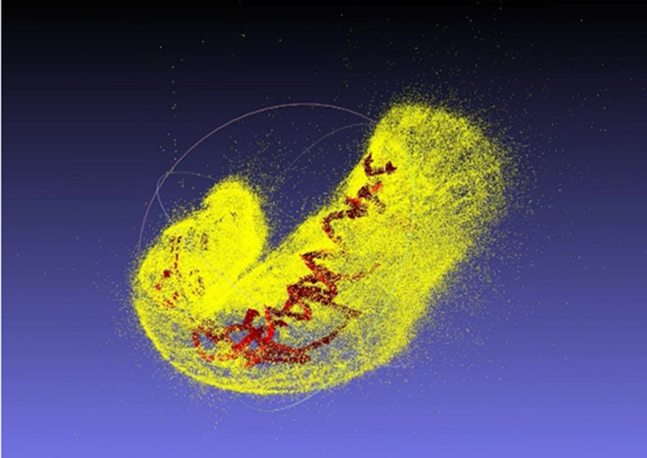
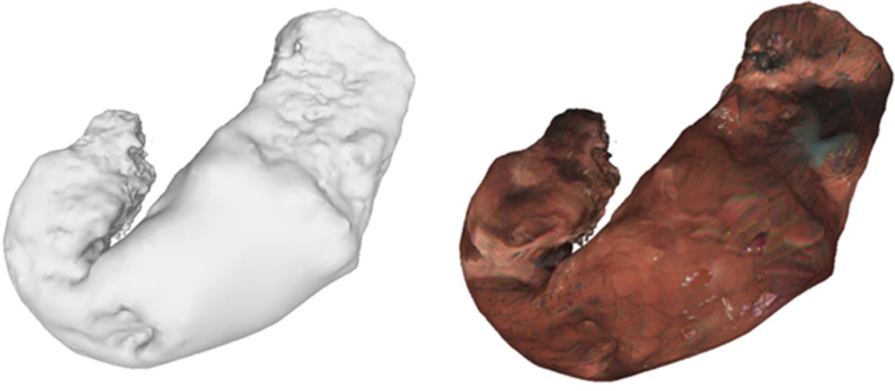
Results Most 3D point clouds were extracted from the red channel of the endoscopic images with the indigo carmine dye (figure 1). 3D meshes and texture representation resembling the whole shape of a stomach were generated from the cloud model (figure 2). Gastric ulcer lesion was clearly localized and reconstructed in one subject.
Conclusions Our study found that 3D reconstruction of the whole stomach can be achieved from a standard endoscopic video image using SfM. Furthermore, gastric ulcer lesion was also localized and reconstructed in 3D reconstruction model. Our future work will be focused on the clinical significance of our proposed method. We will try to evaluate the clinical usefulness of this method for the patients undergone surgery for early gastric cancers.