Article Text

Statistics from Altmetric.com
Introduction
A healthy 41-year-old female was referred for the endoscopic management of a large rectal laterally spreading lesion (LSL). On index colonoscopy, performed for the evaluation of rectal bleeding, multiple proximal sessile serrated polyps were identified meeting diagnostic criteria for serrated polyposis syndrome. Family history was notable for a sister with a history of sessile serrated polyps.
Using an Olympus 190 series high-definition colonoscope (Olympus, Tokyo, Japan), the lesion was evaluated under white-light (figure 1A), narrow-band imaging (NBI) (figure 1B) and near-focus (figure 1C,D).
Question
What is the predicted histopathology of this lesion?
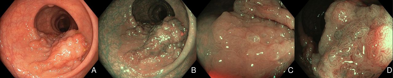
:Optical evaluation including high-definition white-light. (A), narrow-band imaging (B) and near-focus (C, D).
Answer
Using established imaging criteria,1 two distinct surface patterns were identified consistent with serrated (pale colour, lacey vessels, absent surface pattern; NBI International Colorectal Endoscpic (NICE) I; figure 1C) and adenomatous histopathology (brown colour, brown vessels contrasting surrounding white surface pattern; NICE II; figure 1D), respectively. After successful endoscopic submucosal dissection (ESD) (figure 2A), histopathology confirmed a mixed traditional serrated adenoma and tubulovillous adenoma with high-grade dysplasia (figure 2B). At 6-month surveillance colonoscopy, no recurrence was identified.

Resection defect after endoscopic submucosal dissection. (A) and histopathology evaluation (B)
Optical evaluation is critical for managing large LSLs as it empowers the endoscopist to select the appropriate resection technique based on the lesion’s predicted histopathology and the risk for submucosal invasive cancer (SMIC). Moreover, in bulky lesions, location and gross morphology can be used to quantify the risk of invisible or covert SMIC2 ; thereby identifying ideal candidates for ESD and optimising oncological outcomes through the implementation of a selective resection algorithm.3
While serrated-class lesions are an established precursor to colorectal cancer,4 5 to our knowledge this is the first description of the optical features of a mixed lesion containing a traditional serrated adenoma and tubulovillous adenoma; which is believed to be precipitated through KRAS mutation. Moreover, this case highlights the ability to identify these lesions preresection. Until more is known about their malignant potential, a selective resection algorithm similar to that employed for adenomatous lesions, may be most appropriate.
Ethics statements
Footnotes
Contributors Drafting of the article: NS. Critical revision of the article for important intellectual content: WAvH, SV, MJB. Final approval of the article: MJB.
Funding The authors have not declared a specific grant for this research from any funding agency in the public, commercial or not-for-profit sectors.
Competing interests None declared.
Provenance and peer review Not commissioned; externally peer reviewed.