Article Text

Statistics from Altmetric.com
A very recent publication in Gut highlights that faecal microbiota transplantation (FMT) from alginate oligosaccharide (AOS)-dosed animals improves mouse sperm quality and spermatogenesis after busulfan treatment.1 The results suggest the potential of FMT for the improvement of infertility,1 since worldwide 10%–15% of couples are infertile and many of them have failed spermatogenesis.1 2 In addition, many investigations have found that gut microbiota may affect male or female reproduction.3 4 Although the improvement of male infertility is an emerging novel area of interest and many investigations have attempted to ameliorate spermatogenesis by various methods, little progress has been achieved.5 6 In the study done by Zhang et al,1 FMT from AOS-dosed animals increased spermatozoa quality and the process of spermatogenesis; however, that gut microbiota from AOS-dosed animals can actually increase fertility rate is as yet unknown.
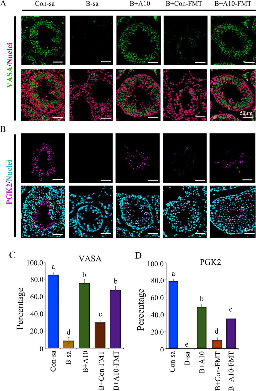
To confirm the beneficial advantages of FMT from AOS dosed animals, we set out to explore the fertility rate (pregnancy rate and number of live pups/litter) following FMT from AOS-dosed animals to busulfan-treated mice (online supplemental file 1 and online supplemental figure 1). We found that B+A10 FMT (busulfan plus gut microbiota from AOS 10 mg/kg mice) significantly increased pregnancy rate (10-fold) and number of live pups/litter (twofold) compared with busulfan (B-sa; figure 1A,B). Notably, the number of live pups/litter was almost the same for B+A10 FMT and control (Con-sa; blank control) which suggested that A10-FMT had a strong potential for rescuing male fertility. However, B+Con FMT (busulfan plus gut microbiota from control mice) did not significantly increase the pregnancy rate or number of live pups/litter compared with busulfan (figure 1A,B). At the same time, we compared the beneficial advantages of AOS 10 mg/kg (A10) and A10-FMT after busulfan treatment. A10 and A10-FMT produced a similar improvement on the pregnancy rate and number of live pups/litter (figure 1A,B). In our earlier studies,1 5 we discovered that AOS 10 mg/kg improves the gut microbiota to, in turn, improve spermatogenesis and semen quality. Furthermore, A10-FMT similarly benefited gut microbiota1 through an increase in the ‘beneficial’ bacteria7 Bacteroidales, Bifidobacteria, Sphingomonadales and Campylobacterales which have beneficial effects such as protecting the intestinal barrier,8 production of antioxidant compounds9 and the possession of reduction enzymes.10 It is also interesting that the microbes from A10-FMT showed a good correlation with sperm concentration/motility, blood metabolome and testis metabolome.1 It is even more profoundly important that the microbiota from A10-dosed mice and A10-FMT-treated mice were well correlated.1 5 Moreover, A10, A10-FMT and Con-FMT did not affect the fertility rate of control mice (without busulfan; figure 1C,D) which indicated that these treatments did not pose a disadvantage for male animal reproduction. In the current investigation, spermatogenesis was significantly improved by A10-FMT as shown by the germ cell marker VASA (figure 2). There were almost no VASA-positive cells in the busulfan group (B-sa) and a very small number in B+Con FMT group; however, a significant number of VASA-positive cells were found in the B+A10 FMT and B+A10 groups (figure 2), which suggested that spermatogenesis was improved by A10-FMT, since busulfan mainly disrupted germ cells.2 5 6 At the same time, protein levels of the sperm cell marker PGK2 were determined by immunofluorescence staining.1 2 5 There were almost no PGK2-positive cells in the B+Sa and B+Con FMT groups (figure 2). However, a similar number of PGK2-positive cells were found in the B+A10, B+A10 FMT and control (Con-sa) groups (figure 2). The data further revealed that A10-FMT rescued busulfan disrupted spermatogenesis, while Con-FMT did not. The data in this investigation confirmed that gut microbiota from AOS-dosed mice had the potential to improve spermatogenesis and then to increase male fertility rate.
Supplemental material
Supplemental material
Supplemental material

Mouse pregnancy rate and the number of live pups/litter. (A) Mouse pregnancy rate (number of pregnant mice/total mice in each group: Con-sa (experiment II), B-sa (experiment II), B+A10 (experiment III), B+A10 FMT (experiment II) and B+Con FMT (experiment II); n=60/group). a,b,c Means not sharing a common superscript are different (p<0.05). (B) The number of live pups/litter. The average number of live pups/litter in each group: Con-sa (experiment II), B-sa (experiment II), B+A10 (experiment III), B+A10 FMT (experiment II) and B+Con FMT (experiment II); (n=60/group). a,b,c Means not sharing a common superscript are different (p<0.05). (C) Mouse pregnancy rate (number of pregnant mice/total mice in each group: Con (experiment II), A10 (experiment III), A10-FMT (experiment II) and Con-FMT (experiment II); n=60/group). a,b,c Means not sharing a common superscript are different (p<0.05). (D) The number of live pups/litter. The average number of live pups/litter in each group: Con (experiment II), A10 (experiment III), A10-FMT (experiment II) and Con-FMT (experiment II); (n=60/group). a,b,c Means not sharing a common superscript are different (p<0.05). Note: (1) Con-sa (dosed with saline); (2) B-sa (busulfan (a single injection 20 mg/kg body weight of busulfan)1 4 plus saline); (3) B+A10 (busulfan plus AOS 10 mg/kg); (4) B+Con FMT (busulfan plus gut microbiota from regular mice); (5) A10-FMT (busulfan plus gut microbiota from AOS 10 mg/kg dosed mice); (6) Con (dosed with saline); (7) A10 (dosed with AOS 10 mg/kg); (8) Con-FMT (dosed with gut microbiota from regular mice); (9) A10-FMT (dosed with gut microbiota from AOS 10 mg/kg dosed mice). See more detailed information in online supplemental file 1). AOS, alginate oligosaccharide; FMT, faecal microbiota transplantation.

VASA and PGK2 staining of mouse testicular samples. (A) Germ cell marker VASA staining of mouse testicular samples. (B) Sperm cell marker PGK2 staining of mouse testicular samples. (C) Quantification data for VASA staining. (D) Quantification data for PGK2 staining. Note: (1) Con-sa (dosed with saline); (2) B-sa (busulfan (a single injection 20 mg/kg body weight of busulfan)1 4 plus saline); (3) B+A10 (busulfan plus AOS 10 mg/kg); (4) B+Con FMT (busulfan plus gut microbiota from regular mice); (5) A10-FMT (busulfan plus gut microbiota from AOS 10 mg/kg dosed mice). For more details, see information in online supplemental file 1). The letters a, b, c, d and e indicate a significant difference among different treatments (p<0.05). AOS, alginate oligosaccharide; FMT, faecal microbiota transplantation.
Ethics statements
Patient consent for publication
Acknowledgments
We thank the investigators and staff of The Beijing Genomics Institute (BGI) for technical support.
Supplementary materials
Supplementary Data
This web only file has been produced by the BMJ Publishing Group from an electronic file supplied by the author(s) and has not been edited for content.
Footnotes
CZ, BX and LC contributed equally.
Contributors CZ, BX, LC, WG, SY and YF performed the experiments and analysed the data. HZ, WS and YZ designed and supervised the study. ZS, QS, HZ, WS and YZ wrote the manuscript. All the authors edited the manuscript and approved the final manuscript.
Funding This study was supported by the National Natural Science Foundation of China (31772408 to YZ; 31672428 to HZ).
Competing interests None declared.
Provenance and peer review Not commissioned; externally peer reviewed.
Supplemental material This content has been supplied by the author(s). It has not been vetted by BMJ Publishing Group Limited (BMJ) and may not have been peer-reviewed. Any opinions or recommendations discussed are solely those of the author(s) and are not endorsed by BMJ. BMJ disclaims all liability and responsibility arising from any reliance placed on the content. Where the content includes any translated material, BMJ does not warrant the accuracy and reliability of the translations (including but not limited to local regulations, clinical guidelines, terminology, drug names and drug dosages), and is not responsible for any error and/or omissions arising from translation and adaptation or otherwise.