Article Text

Abstract
Objectives Alterations in the intestinal microbiota are linked with a wide range of autoimmune and inflammatory conditions, including inflammatory bowel diseases (IBD), where pathobionts penetrate the intestinal barrier and promote inflammatory reactions. In patients with IBD, the ability of intestinal macrophages to efficiently clear invading pathogens is compromised resulting in increased bacterial translocation and excessive immune reactions. Here, we investigated how an IBD-associated loss-of-function variant in the protein tyrosine phosphatase non-receptor type 2 (PTPN2) gene, or loss of PTPN2 expression affected the ability of macrophages to respond to invading bacteria.
Design IBD patient-derived macrophages with wild-type (WT) PTPN2 or carrying the IBD-associated PTPN2 SNP, peritoneal macrophages from WT and constitutive PTPN2-knockout mice, as well as mice specifically lacking PTPN2 in macrophages were infected with non-invasive K12 Escherichia coli, the human adherent-invasive E. coli (AIEC) LF82, or a novel mouse AIEC (mAIEC) strain.
Results Loss of PTPN2 severely compromises the ability of macrophages to clear invading bacteria. Specifically, loss of functional PTPN2 promoted pathobiont invasion/uptake into macrophages and intracellular survival/proliferation by three distinct mechanisms: Increased bacterial uptake was mediated by enhanced expression of carcinoembryonic antigen cellular adhesion molecule (CEACAM)1 and CEACAM6 in PTPN2-deficient cells, while reduced bacterial clearance resulted from defects in autophagy coupled with compromised lysosomal acidification. In vivo, mice lacking PTPN2 in macrophages were more susceptible to mAIEC infection and mAIEC-induced disease.
Conclusions Our findings reveal a tripartite regulatory mechanism by which PTPN2 preserves macrophage antibacterial function, thus crucially contributing to host defence against invading bacteria.
- bacterial infection
- cellular immunity
- E. Coli
- macrophages
- mucosal immunology
This is an open access article distributed in accordance with the Creative Commons Attribution 4.0 Unported (CC BY 4.0) license, which permits others to copy, redistribute, remix, transform and build upon this work for any purpose, provided the original work is properly cited, a link to the licence is given, and indication of whether changes were made. See: https://creativecommons.org/licenses/by/4.0/.
Statistics from Altmetric.com
Significance of this study
What is already known on this subject?
Alterations in microbial composition are associated with several inflammatory disorders, including inflammatory bowel disease (IBD).
Increased presence of adherent-invasive Escherichia coli (AIEC) occurs in IBD patients.
Variants in protein tyrosine phosphatase non-receptor type 2 (PTPN2) are associated with increased risk to develop IBD and loss of PTPN2 affects immune responses.
What are the new findings?
PTPN2 crucially affects the ability of macrophages to clear invading bacteria.
Increased invasion and intracellular replication of AIEC occurs in PTPN2-deficient macrophages.
PTPN2 restricts bacterial growth and invasion of macrophages by three distinct mechanisms in vivo: PTPN2 restricts expression of carcinoembryonic antigen cellular adhesion molecule proteins that promote bacterial entry; PTPN2 maintains autophagy of invading bacteria; PTPN2 preserves lysosomal acidification that is required for bacterial clearance.
How might it impact on clinical practice in the foreseeable future?
Provides a mechanistic explanation for the altered microbial composition in patients ith IBD carrying PTPN2 variants.
Our results indicate PTPN2 as a crucial factor for anti-bacterial defence, and thus, position PTPN2 as a potential therapeutic target in patients with AIEC overgrowth.
Introduction
The human intestine is populated with over 1000 different bacterial species, which form a complex ecosystem that significantly impacts health.1 The vast majority of these bacteria are harmless or even beneficial commensals and do not pose danger to the host.2 Certain bacterial species, however, can cause diseases or contribute to the emergence of chronic inflammatory conditions, including inflammatory bowel disease (IBD),3 which comprises ulcerative colitis (UC) and Crohn’s disease (CD). UC and CD are associated with intestinal dysbiosis3 4—a term describing reduced overall bacterial diversity and overgrowth of pathogenic strains, including adherent-invasive Escherichia coli (AIEC) species.4–6 In contrast to their harmless, non-invasive counterparts, AIEC can penetrate the mucus layer, adhere to intestinal epithelial cells (IEC), and invade and survive in intestinal macrophages.7 8
In healthy individuals, overgrowth and invasion of pathogenic microbes is restricted by a thick mucus layer, a tight epithelial cell barrier,9 and immune cells in the lamina propria, and especially macrophages residing directly beneath IEC efficiently eliminate invading pathogens.10 11 These macrophages display an anti-inflammatory, highly phagocytic phenotype12 and remove cell debris, dying cells and harmless bacteria in an immune-silent manner.13 Nevertheless, in response to barrier defects and/or infiltration of invading pathogens, intestinal macrophages acquire an inflammatory phenotype, secrete large amounts of inflammatory cytokines and initiate full-blown immune responses.11–13 Once an infection is cleared, the anti-inflammatory setting is restored.11 12 However, when clearance of invading pathogens is disturbed, due to overgrowth of pathogenic bacteria or genetic factors that compromise the return to an immune silent state, exacerbated immune responses cause chronic inflammation, and drive the development of IBD and other intestinal disorders.14
Besides microbial/immunological factors, variants in over 200 genes have been associated with an enhanced risk to develop IBD.15–17 Among these, single nucleotide polymorphism (SNP) rs1893217 in the gene locus encoding protein tyrosine phosphatase non-receptor type 2 (PTPN2), not only contributes to enhanced risk of IBD, but also promotes other inflammatory diseases including rheumatoid arthritis, type 1 diabetes, and metabolic syndrome—diseases which are associated with alterations in the intestinal microbiota.18 19 PTPN2 (also known as T cell protein tyrosine phosphatase) loss, or presence of SNP rs1893217, results in aberrant T cell activation/differentiation,20 hyper-responsiveness to interferon (IFN)-γ and EGF,21 and defective autophagy—a process important for intracellular bacteria handling.22 In mice, full-body Ptpn2-knockout leads to severe systemic inflammation and death 3–5 weeks after birth.23 T cell-specific deletion promotes susceptibility to intestinal inflammation and alters the intestinal microbiome (depletion of potentially beneficial microbes, decreased bacterial richness, reduced levels of butyrate producers, increase of potential pathobionts20) similar to that observed in IBD patients carrying PTPN2 SNP rs1893217.24 This demonstrates the importance of PTPN2 and its potential role in controlling bacterial invasion. However, it is unclear how loss of PTPN2 affects the ability of macrophages to prevent bacterial infections.
We previously found that full-body Ptpn2 knockout mice exhibit a significant expansion of a novel mouse AIEC (mAIEC).25 The aim of this study was to identify whether loss of PTPN2 activity compromises macrophage-mediated host defences against AIEC and to assess the mechanisms by which loss of PTPN2, or the presence of SNP rs1893217 SNP, compromises the ability of macrophages to clear AIEC infections.
Methods
Reagents
Sources of material and reagents used in this study are listed in online supplemental table S1.
Supplemental material
Patient-derived peripheral blood mononuclear cells
Whole blood samples were obtained from previously genotyped, sex and age-matched patients from the Swiss IBD cohort and healthy volunteers. All patients presented with quiescent disease at the time of sample collection. A list of patient characteristics and medication at the time of blood collection can be found in online supplemental table S2. All patients and healthy controls signed informed consent before study inclusion and the local ethics commission approved the study (Cantonal Ethics Commision Zurich, Switzerland; Approval number EK-1755).
Mice
Heterozygous Ptpn2 knockout (Ptpn2-Het) mice on a Balb/c background were obtained from Michel L. Tremblay at McGill University. Ptpn2-Het mice were bred with each other to obtain wild-type (WT), Ptpn2-Het and Ptpn2-KO littermates and cells obtained at an age of 3 weeks. Mice with a loxP flanked Ptpn2 gene expressing Cre under the Lysozyme2 promoter (Ptpn2-LysMCre mice26) were bred with Ptpn2fl/fl mice to obtain Ptpn2fl/fl and Ptpn2-LysMCre littermates. Eight to ten weeks old littermates were used. For in vivo autophagy activation, mice were injected intraperitoneally daily with 1 mg/kg rapamycin in vehicle (4% ethanol, 5% polyethylene glycol, 5% tween 80) starting 1 day prior to the first bacterial gavage. Vehicle treated mice served as controls. For in vivo carcinoembryonic antigen cellular adhesion molecule (CEACAM)1 inhibition, mice were injected intraperitoneally daily with 10 mg/kg anti-CEACAM1 antibody (clone CC1) starting 1 day prior to the first bacterial gavage.
Bacterial strains and infections
For experiments, K12 (ATCC), LF828 and mouse AIEC (mAIEC)27 were cultured in Luria-Bertani (LB) medium overnight from frozen stocks, subcultured into fresh LB, harvested in the log-phase, washed in PBS, and macrophages infected at an MOI of 20. After 2 hours, macrophages were washed twice with Gentamycin (100 µg/mL)-containing culture medium and incubated in gentamycin (20 ug/mL)-containing culture medium. Unless otherwise stated, incubation times are given as the time of incubation after the Gentamycin washes. mAIEC expressing a plasmid containing the mCherry fluorescent protein were used for localisation studies.
Macrophages
Macrophages were generated from human peripheral blood mononuclear cells, mouse bone marrow and THP-1 monocytes as detailed in online supplemental methods. Purity and differentiation was checked by flow cytometry and >96% of the cells were identified as macrophages. THP-1 cells with stable PTPN2 knockdown using shRNA and transfected with WT PTPN2, variant PTPN2 or an empty vector (EV) were obtained from Prof. Scharl at the University Hospital Zurich, University of Zurich, Zurich, Switzerland. Due to partially retained endogenous PTPN2 expression, variant expressing cells functionally resemble cells from heterozygous variant carriers.
Flow cytometry
For flow cytometry, bone marrow derived macrophages (BMDM) or peritoneal macrophages were infected with mAIEC expressing a mCherry expressing plasmid or pHRhodo-K12 particles, detached from the culture plate using 2 mM ethylenediaminetetraacetic acid (EDTA) in PBS, washed twice and directly analysed on an LSRII flow cytometer. For measurement of reactive oxygen species (ROS), the cells were incubated with CellROX reagent (500 nM, Thermo Fisher Scientific) for 30 min at 37°C, washed in PBS and analysed immediately on an LSRII flow cytometer. For measurement of lysosomal acidification, the cells were incubated with LysoTracker green (25 nM, Thermo Fischer Scientific) for 1 hour, washed with PBS and analysed immediately on an LSRII flow cytometer.
Immunofluorescent imaging, confocal microscopy
For immunofluorescence imaging, macrophages were seeded on cover slides after PMA pulsing and infected 24 hours later with mAIEC-mCherry or pHRhodo-K12 particles as described above. Staining for lysosome-associated membrane protein (LAMP)-1 and LC3B was performed according to standard procedures and as detailed in online supplemental methods.
CEACAM1 and CEACAM6 inhibition in vitro
For blocking of CEACAMs, cells were incubated for 1 hour with 10 µg/mL antimouse CEACAM1 or antihuman CEACAM6 antibody prior to infection with K12, LF82 or mAIEC.
siRNA treatment
STAT1 was silenced in peritoneal macrophages using a predesigned pool of 3 small-interfering RNAs (siRNA) from Dharmafect and Lipofectamine transfection reagent according to the manufacturer’s instructions, medium was replaced 16 hours after the transfection. Bacterial infections were carried out 48 hours later.
Protein and RNA extraction, Western Blotting and quantitative PCR was performed according to standard procedures and as described in online supplemental methods.
PTPN2 activity assay
PTPN2 phosphatase activity was assessed as described previously28 using a phosphatase activity assay (Thermo Fisher Scientific). In brief, PTPN2 was immunoprecipitated from the lysates and samples dissolved in phosphatase-assay buffer, DiFMUP substrate added, and phosphatase activity measured every 10 min for 120 min to reliably determine phosphatase activity.28
In vivo AIEC infection
Mice aged 8–10 weeks old were infected on four consecutive days with 109 K12, LF82 or mAIEC/day. Disease activity was evaluated using the following parameters: weight loss, reduced grooming, reduced activity, stool consistency, overall appearance. Bacterial load was determined by homogenisation of faeces or tissues in 0.5 mL PBS and plating on LB agar.
Quantification and statistical analysis
Data are represented as mean of n biological repetitions±SE of the mean. Data followed a Gaussian distribution and variation was similar between groups for conditions analysed together. Significant differences were determined using GraphPad Prism V.9 software using analysis of variance. P values below 0.05 were considered significant. Mice were randomly distributed into experimental groups after matching for age and gender. Numbers of replicates are given in the figure legends. Investigators were blinded for weight recording, evaluation of disease activity, and sample collection. No data points were excluded from statistical analysis.
Results
Loss of PTPN2 promotes AIEC uptake and survival in macrophages
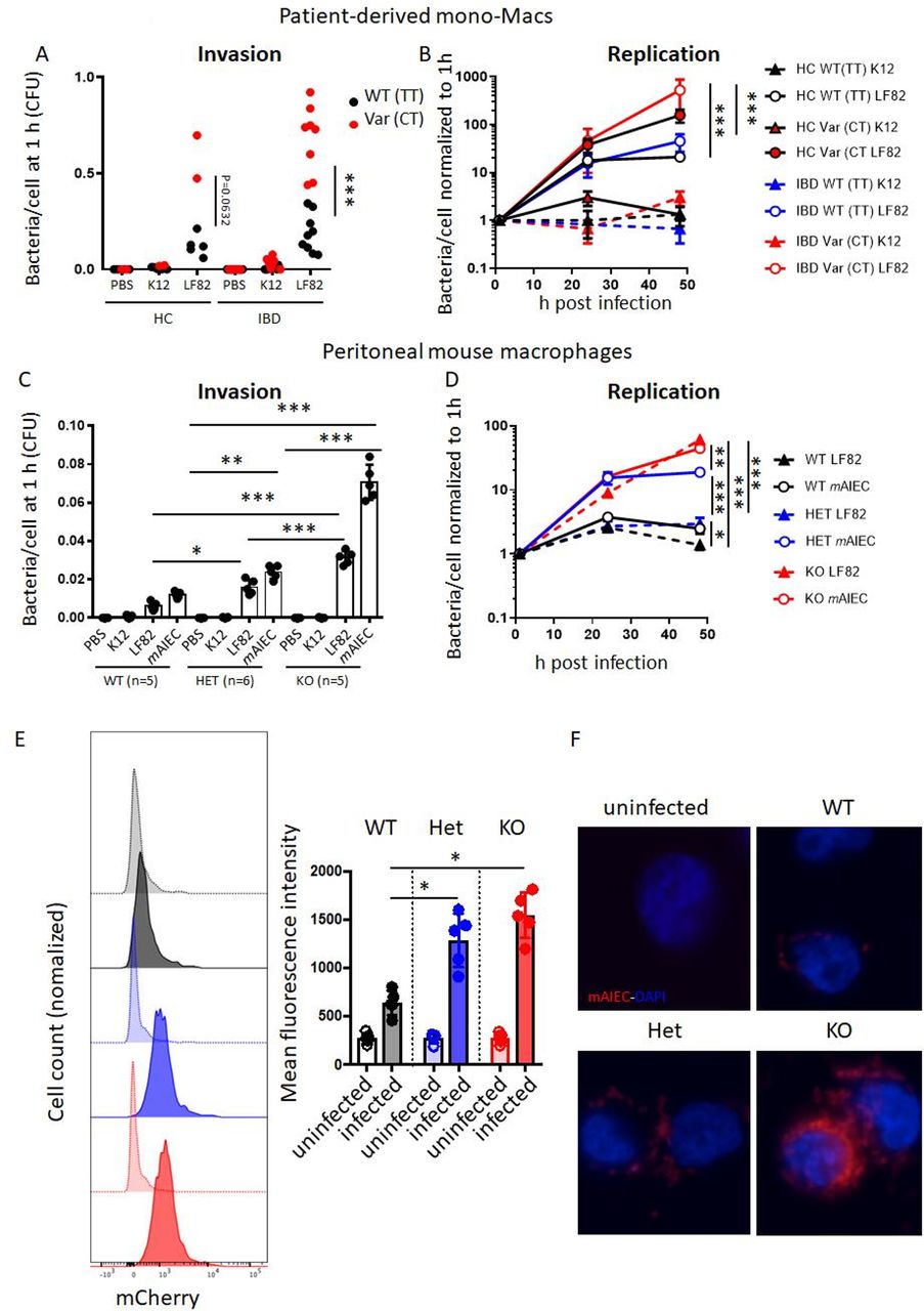
First, we assessed how loss of PTPN2 or presence of the disease-associated variant in SNP rs1892317 in the gene locus encoding PTPN2 affects bacterial uptake and intracellular replication in macrophages. Therefore, we employed PTPN2-silenced THP-1 monocytes that were stably transfected with lentiviral constructs containing normal (WT) PTPN2, an EV or the disease-associated PTPN2 variant (Var; online supplemental figure S1A).29 These cells were exposed to non-invasive E. coli K12, the human LF82 AIEC, or a novel mAIEC recently discovered in our lab.27 EV-transfected (PTPN2-deficient) macrophages were highly susceptible to LF82 and mAIEC uptake, an effect mirrored in macrophages expressing the PTPN2 loss-of-function variant (online supplemental figure 1A,B). Bacterial replication was increased in PTPN2-deficient and PTPN2-variant macrophages when compared with macrophages expressing the WT variant (online supplemental figure 1C). To confirm the effect of PTPN2 SNP rs1893217, we used monocyte-derived macrophages from IBD patients from the Swiss IBD cohort previously genotyped as WT (TT) or heterozygous (CT) for SNP rs1892317. While uptake and replication in macrophages from PTPN2 WT healthy control and IBD patients was similar, there was highly increased bacterial uptake and increased bacterial proliferation in macrophages from PTPN2-variant patients (figure 1A,B). Consistent with previous reports,29 presence of SNP rs1893217 reduced PTPN2 phosphatase activity, while protein expression levels were not affected (online supplemental figure 1D–G). We did not observe differences between macrophages from UC versus CD patients (data not shown).

Loss of PTPN2 promotes uptake and replication of AIEC in macrophages. (A, B) Macrophages from healthy controls (HC), or patients with IBD homozygous for the wild-type (=C allele; WT (CC), HC: n=5, IBD: n=9) or heterozygous for the disease-associated (=T allele) variant (Var (CT), HC: n=2, IBD: n=8) in PTPN2 SNP rs1893217 were infected with non-invasive K12 E. coli or the AIEC strain LF82 for 2 hour, washed with PBS and incubated with gentamycin and macrophage uptake after 1 hour (A) and intracellular replication over time (B) analysed. (C, D) Peritoneal macrophages were isolated from WT, Ptpn2-Het (HET) or Ptpn2-KO (KO) mice and infected with K12, LF82 or a mouse AIEC (mAIEC) strain as described in A, B and analysed for bacterial uptake (C) and replication (D). (E, F) Bone marrow-derived macrophages were generated from WT, HET and KO mice, infected with mCherry-tagged mAIEC as in B and mCherry fluorescence measured after 3 hours by flow cytometry (E) and fluorescent imaging (F). Scale bar: 50 µm. Asterisks denote statistical significances (*P<0.05, **p<0.01, ***p<0.001, ANOVA with Bonferroni correction for multiple testing). See also online supplemental figure 1. AIEC, adherent-invasive Escherichia coli; ANOVA, analysis of variance; IBD, inflammatory bowel disease.
In peritoneal macrophages from Ptpn2-het mice, uptake of LF82 and mAIEC was clearly increased, and further enhanced in homozygous Ptpn2-KO macrophages (figure 1C). Moreover, bacterial replication was elevated in Ptpn2-KO macrophages, an effect also partially visible in macrophages from Ptpn2-het mice (figure 1D). Increased bacterial uptake on loss of Ptpn2 was confirmed by flow cytometry (figure 1E) and immunofluorescent imaging (figure 1F) in BMDM infected with mCherry-tagged mAIEC. Conversely, K12 uptake was very low (not shown). The inability of PTPN2-defective macrophages to clear intracellular bacteria was not limited to AIEC, but was also observed when macrophages were infected with the mouse enteropathogen C. rodentium (online supplemental figure 2). Taken together, this indicates that Ptpn2-deficient macrophages take up more bacteria than Ptpn2-competent macrophages, and that loss of Ptpn2 compromises intracellular bacterial handling and elimination.
Increased bacterial uptake is mediated via enhanced CEACAM6/CEACAM1 expression in PTPN2-deficient/defective macrophages
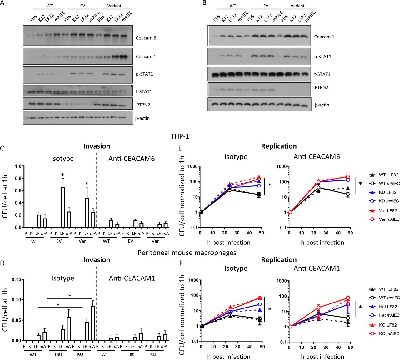
LF82 uses CEACAM6 to adhere to and invade human IEC.30 To identify whether altered CEACAM expression contributes to increased AIEC uptake/invasion in PTPN2-deficient macrophages, we determined expression levels of CEACAM6 (human cells) and CEACAM1 (mouse cells; CEACAM6 is not expressed in mice).31 PTPN2-deficient and PTPN2-variant human THP-1 cells expressed elevated basal levels of CEACAM6 and CEACAM1, and AIEC further increased CEACAM1/6 expression (figure 2A). In addition, we observed enhanced levels of Signal-transducer and activator of transcription (STAT1) phosphorylation, which has been reported to promote CEACAM1, and to a lesser extent CEACAM6, expression32 (figure 2A). In mouse macrophages, we detected increased levels of CEACAM1 in Ptpn2-het and Ptpn2-KO macrophages and bacteria-induced STAT1 phosphorylation was increased (figure 2B). CEACAM1 (mouse) and CEACAM6 (human) inhibition with specific antibodies reduced bacterial invasion (figure 2C,D), indicating that AIEC entered the cells at least partially by attaching to CEACAM1/CEACAM6. In contrast, CEACAM inhibition did not affect intracellular bacterial replication, and even after CEACAM1/CEACAM6 inhibition, PTPN2-deficient and PTPN2-knockdown/variant THP-1 macrophages showed a faster increase in intracellular bacterial load (figure 2E,F), indicating that the increased replication was not due to increased bacterial uptake.

Loss of PTPN2 promotes CEACAM1 and CEACAM6 expression in macrophages. (A) PTPN2 was silenced in THP-1 macrophages prior to transfection with the major (WT) allele in SNP rs1893217, an empty vector (EV), or the minor (Var) allele in PTPN2. After selection of stable clones, cells were infected with K12, LF82 or mAIEC for 1 hour. Depicted are representative Western blot images of the indicated proteins. (B) Peritoneal macrophages were isolated from WT, Ptpn2-Het (HET) or Ptpn2-KO (KO) mice and infected with K12, LF82 or mAIEC for 1 hour. Depicted are representative Western blot images of the indicated proteins. (C–F) WT, EV and Var THP-1 cells (C, E) and peritoneal macrophages from WT, Het and KO mice (D, F) were infected with K12, LF82 or mAIEC for 2 hours, incubated with gentamycin and bacterial load (C, D) and replication (E, F) determined at the indicated time points. Asterisks denote statistical significances (*p<0.05, **p<0.01, ***p<0.001, ANOVA with Bonferroni correction for multiple testing). ANOVA, analysis of variance; mAIEC, mouse adherent-invasive Escherichia coli; PTPN2, protein tyrosine phosphatase non-receptor type 2; WT, wild-type.
Given the increased levels of STAT1 phosphorylation and previous reports that IFN-γ-driven STAT1 activation promotes CEACAM1 and CEACAM6 expression,32 we assessed whether knockdown of STAT1 affected bacterial load. STAT1 siRNA-treatment decreased bacterial uptake (figure 3A) but did not affect bacterial replication (figure 3B). STAT1 silencing inhibited bacteria-induced CEACAM1 mRNA and protein induction (figure 3C,D), indicating that STAT1 mediates increased CEACAM1 expression in Ptpn2-KO macrophages.
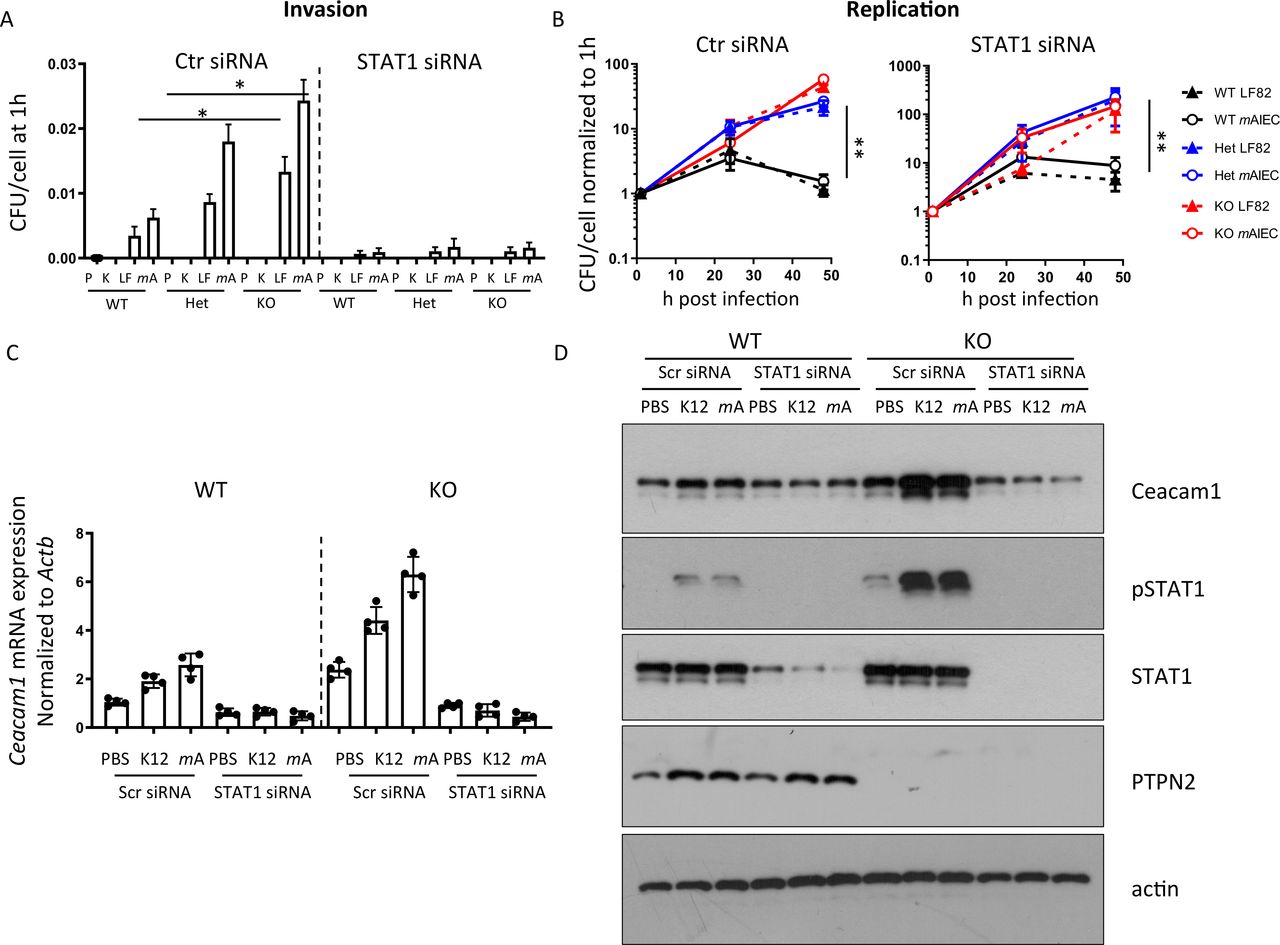
STAT1 inhibition normalises CEACAM expression and inhibits bacterial uptake. Peritoneal macrophages were isolated from WT, Ptpn2-Het (HET) or Ptpn2-KO (KO) mice, STAT1 silenced using siRNA and cells infected 24 hours later with K12, LF82 or mAIEC for 2 hours, incubated with gentamycin containing medium and (A) bacterial uptake and (B) replication analysed at the indicated time points; (C) mRNA expression of CEACAM1 normalised to untreated control and Actb after 24 hours; or (D) for the indicated proteins by Western blot after 30 min. Asterisks denote statistical significances (*p<0.05, **p<0.01, ANOVA with Bonferroni correction for multiple testing). ANOVA, analysis of variance; CEACAM, carcinoembryonic antigen cellular adhesion molecule; mAIEC, mouse adherent-invasive Escherichia coli; STAT1, signal-transducer and activator of transcription 1; WT, wild-type.
Increased bacterial survival is partially due to defects in autophagy
Loss of PTPN2 compromises autophagy in IEC and THP-1 monocytes.29 Since autophagy is an important factor for bacterial handling in the intestine,33 we investigated whether increased bacterial replication in PTPN2-deficient/variant macrophages might be due to defects in autophagy. In WT macrophages, infection with K12, LF82, or mAIEC resulted in enhanced conversion of LC3B into its lipidated form (LC3B-II) and a decrease of p62, indicating autophagy activation, while levels of ATG16L1 were not affected. PTPN2-deficient or variant THP-1 cells, however, failed to induce autophagy (figure 4A). Likewise, autophagy induction following bacterial infection was reduced in macrophages from Ptpn2-Het and completely absent in those from Ptpn2-KO mice (figure 4B). In WT and Ptpn2-Het macrophages, mAIEC colocalised with autophagosomes (visible as LC3B bright punctae that colocalise with mCherry-tagged mAIEC) although to a higher extent in WT than in Ptpn2-Het cells. In Ptpn2-KO cells, which exhibited a higher bacterial burden than WT cells, however, very few LC3B bright punctae were visible and they did not colocalise with mCherry-tagged mAIEC (figure 4C). This clearly indicates that Ptpn2-deficient and PTPN2-variant cells are defective in autophagy induction on bacterial infection. Autophagy induction via rapamycin induced autophagy in Ptpn2-Het and Ptpn2-KO macrophages (figure 4D), but had no effect on bacterial uptake (figure 4E), and only partially reduced bacterial replication (figure 4F). Conversely, autophagy inhibition using 3-methyladenine (3-MA) resulted in enhanced bacterial proliferation in WT macrophages, but had no effect on bacterial proliferation in PTPN2-deficient macrophages (online supplemental figure 3A). This indicates that loss of (functional) Ptpn2 results in defective autophagy but the effect on bacterial replication seems only partially due to deficient autophagy. While autophagy was clearly reduced in PTPN2-deficient/variant macrophages, loss of (functional) PTPN2 had no effect on bacteria-induced ROS production (online supplemental figure 3B).
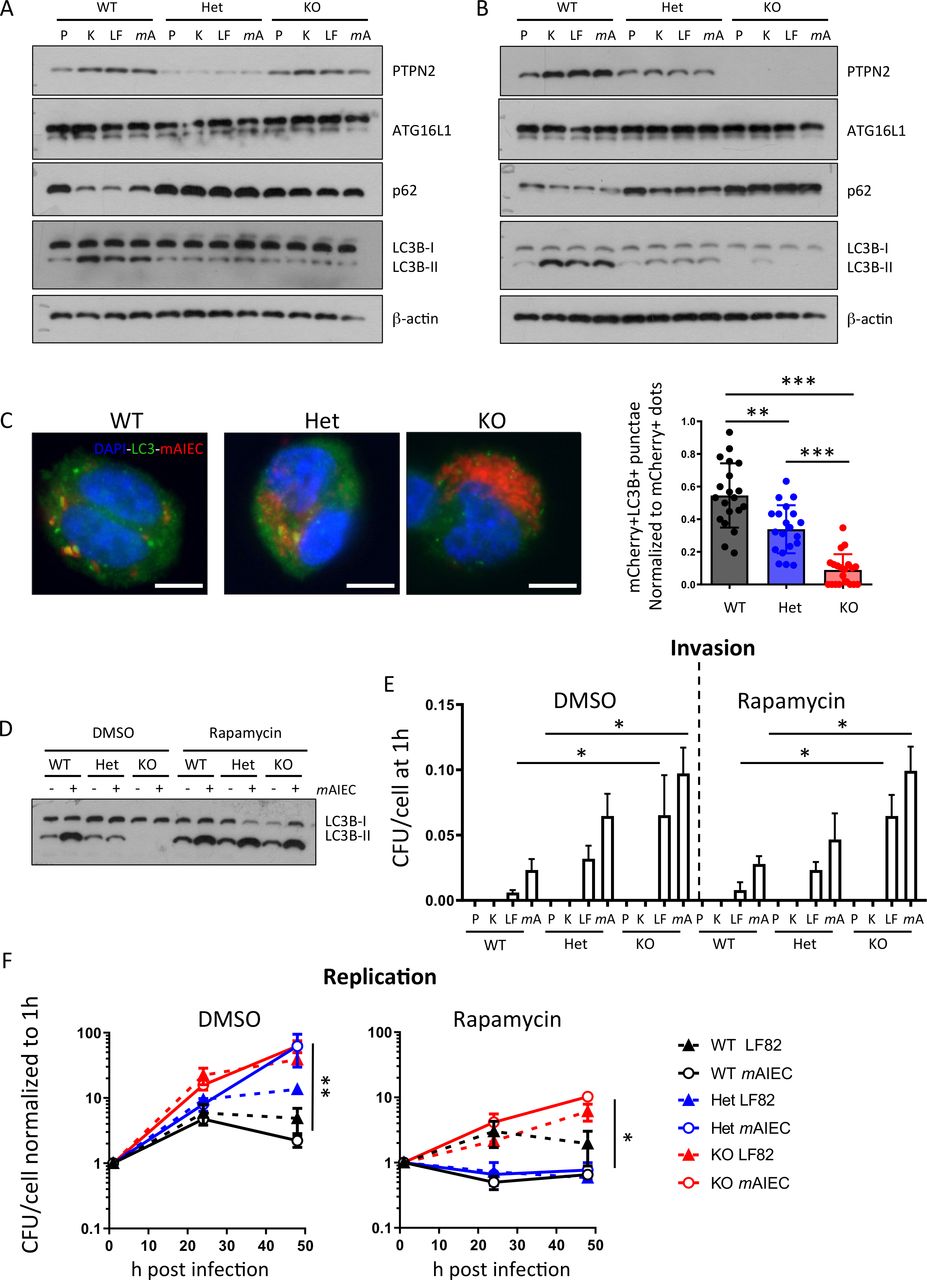
Defective autophagy in PTPN2-deficient cells contributes to enhanced bacterial replication. (A) PTPN2 was silenced in THP-1 macrophages prior to transfection with WT PTPN2, an empty vector (EV), or Var PTPN2 as in figure 1 and infected with K12, LF82 or B2 for 24 hours. Depicted are representative Western blots for the indicated proteins. (B) Peritoneal macrophages were isolated from WT, Ptpn2-Het (HET) or Ptpn2-KO (KO) mice and infected with K12, LF82 or mAIEC for 24 hours. Depicted are representative Western blot pictures for the indicated proteins. (C) Peritoneal macrophages were infected with mCherry expressing mAIEC for 4 hours and stained for LC3B. Bacteria colocalising with autophagosomes versus total bacteria were counted in 20 cells/condition. Scale bar: 10 µm. (D–F) Peritoneal macrophages were infected with K12, LF82 or mAIEC and incubated in the presence of rapamycin for the indicated time. (D) Representative Western blot picture for LC3B, (E) bacterial uptake after 1 hour, and (F) bacterial replication. (*P<0.05, **p<0.01, ***p<0.001). ANOVA, analysis of variance; mAIEC, mouse adherent-invasive Escherichia coli; PTPN2, protein tyrosine phosphatase non-receptor type 2; WT, wild-type.
Ptpn2-deficiency results in defective lysosomal acidification
Macrophages usually degrade phagocytozed dead cells and bacteria in lysosomes, a process promoted by autophagy and dependent on acidification of lysosomes.34 35 Since Ptpn2-defective macrophages were not able to degrade bacteria efficiently even when autophagy was restored, we next assessed whether this might be due to defective lysosomal acidification. For this aim, we used pHRhodamine-coupled bacterial particles, which show an increase in fluorescence in acidic environments. In WT macrophages, fluorescence was clearly visible indicating that the bacterial particles were transported into lysosomes with low pH (figure 5A). In Ptpn2-Het macrophages, the Rhodamine signal was reduced, but still detectable, while in Ptpn2-KO cells, no Rhodamine bright bacteria were visible, indicating that there was either a defect in bacterial transport to the lysosomes or a defect in lysosomal acidification (figure 5B). Consistent with this, staining with lysoTracker, which stains acidic lysosomes, was significantly reduced in Ptpn2-Het and Ptpn2-KO macrophages (figure 5C). To further investigate the mechanism leading to defective lysosomal acidification in Ptpn2-KO macrophages, we assessed the mRNA expression of proteins involved in transporting protein cargo from the endoplasmatic reticulum to lysosomes, or in maintaining the proton gradient across the lysosomal membrane, including cation-independent and cation-dependent mannose-6-phosphate receptor (CI-M6pr and CD-M6pr, respectively), glucocerebrosidase (Gba), the chloride channel Clc7, LAMP1 and LAMP2, and lysosomal integral membrane protein 2 (encoded by Scarb2). We found significantly reduced expression of CI-M6pr in Ptpn2-Het and Ptpn2-KO macrophages, consistent with defects in trafficking of mannose-6-phosphate tagged enzymes required for lysosomal acidification (figure 5D).36 Additionally, LAMP1 staining demonstrated that bacteria did not colocalise with lysosomes in Ptpn2-KO macrophages (figure 5E), indicating defective bacterial handling and compromised transport to lysosomes.

Disturbed lysosomal function in PTPN2-defective cells. (A, B) Peritoneal macrophages from WT, Ptpn2-Het (HET) or Ptpn2-KO (KO) mice were infected with K12-pHRhodamine particles that are fluorescent in acidic environment. Depicted are (A) representative images and (B) flow cytometry measurements of rhodamine fluorescence. (C) Peritoneal macrophages were infected with mAIEC and stained with LysoTracker and analysed by flow cytometry. (D, E) Peritoneal macrophages were infected with mCherry-expressing mAIEC. The graphs show (D) mRNA expression of the indicated genes and representative Western blot for cation-independent mannose-6-phosphate receptor protein after 24 hours, and (E) representative images from LAMP1 immunofluorescence. Scale bars: 10 µm. *p<0.05, **p<0.01, ANOVA. ANOVA, analysis of variance; LAMP1, lysosome-associated membrane protein; mAIEC, mouse adherent-invasive Escherichia coli; M6pr, mannose-6-phosphate receptor; PTPN2, protein tyrosine phosphatase non-receptor type 2; WT, wild-type.
Defects in bacterial transport to lysosomes together with defects of lysosomal acidification in Ptpn2-KO macrophages
Since autophagy is essential for intracellular transport of invading bacteria into lysosomes,37 we next addressed whether autophagy activation by rapamycin restores localisation of bacteria into lysosomes in Ptpn2-KO cells. In rapamycin-treated Ptpn2-KO macrophages, localisation of bacteria into lysosomes was indeed restored (figure 6A, colocalisation of mAIEC with LAMP1, subpanel viii), but there were still no pHRhodamine bright spots detectable on infection with pHRhodo particles, indicating that autophagy activation can restore the ability to transport bacteria into lysosomes in Ptpn2-KO cells, but that a defect in lysosomal acidification persists (figure 6A, subpanel iii). Treatment with IFN-γ and activation of STAT1 decreases expression of CI-M6PR.38 Thus, we hypothesised that the elevated levels of STAT1 observed in Ptpn2-KO macrophages might contribute to the reduced CI-M6pr protein expression and subsequent defect in lysosomal acidification in these cells. To test this hypothesis, we silenced STAT1 and subsequently activated autophagy by rapamycin treatment after infection with mAIEC or pHRhodo particles. In this setting, we clearly observed bacteria in LAMP1-positive lysosomes and pHRhodamine-bright spots (figure 6A, subpanels v and x). In addition to restoring lysosomal acidification in Ptpn2-KO macrophages, STAT1 silencing also restored CI-6Mpr expression (online supplemental figure 4). Concomitant STAT1 silencing and autophagy induction completely normalised the increased bacterial uptake and replication (figure 6B,C). Similar effects on bacterial replication were observed in rapamycin-treated PTPN2-deficient THP-1 cells that overexpressed CI-M6pr (figure 6D,E, online supplemental figure 4C). Taken together, this indicates that the defect in bacterial handling observed in Ptpn2-deficient cells results from a defect in both autophagy and lysosomal acidification, while restoration of both processes re-established the ability to clear intracellular bacteria in Ptpn2-KO cells.
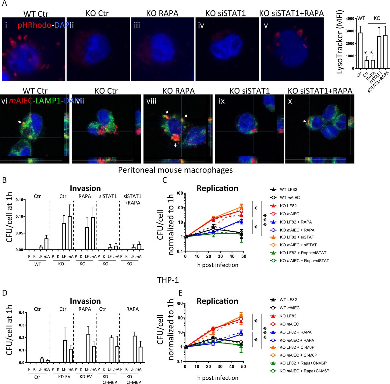
Autophagy activation combined with STAT1 silencing normalises bacterial handling in PTPN2-defective cells. (A) Peritoneal macrophages from WT, Ptpn2-Het (HET), or Ptpn2-KO (KO) mice were infected with pHRhodamine linked K12 (A, images in upper panel) or with mCherry-expressing mAIEC and autophagy induced using rapamycin and/or STAT1 silenced using siRNA. Depicted are representative fluorescent images and mean fluorescent intensity for LysoTracker stained cells. Scale bars: 10 µm. (B, C) Peritoneal macrophages were infected with K12 (K), LF82 (LF) or mAIEC (mA) and treated with rapamycin and/or STAT1 siRNA and analysed for (B) bacterial invasion after 1 hour, and (C) bacterial replication at the indicated time points. (D, E) THP-1 cells expressing PTPN2-specific shRNA (KD) were transfected with an empty vector (EV) or a CI-M6P overexpressing vector and infected with K12 (K), LF82 (LF) or mAIEC (mA) and treated with rapamycin. The graphs show (B) bacterial uptake after 1 hour, and (C) bacterial replication at the indicated time points. *P<0.05, **p<0.01, ***p<0.001, ANOVA. ANOVA, analysis of variance; mAIEC, mouse adherent-invasive Escherichia coli; M6P, mannose-6-phosphate; PTPN2, protein tyrosine phosphatase non-receptor type 2; STAT1, signal-transducer and activator of transcription.
Mice lacking Ptpn2 in macrophages are more susceptible to mAIEC infection
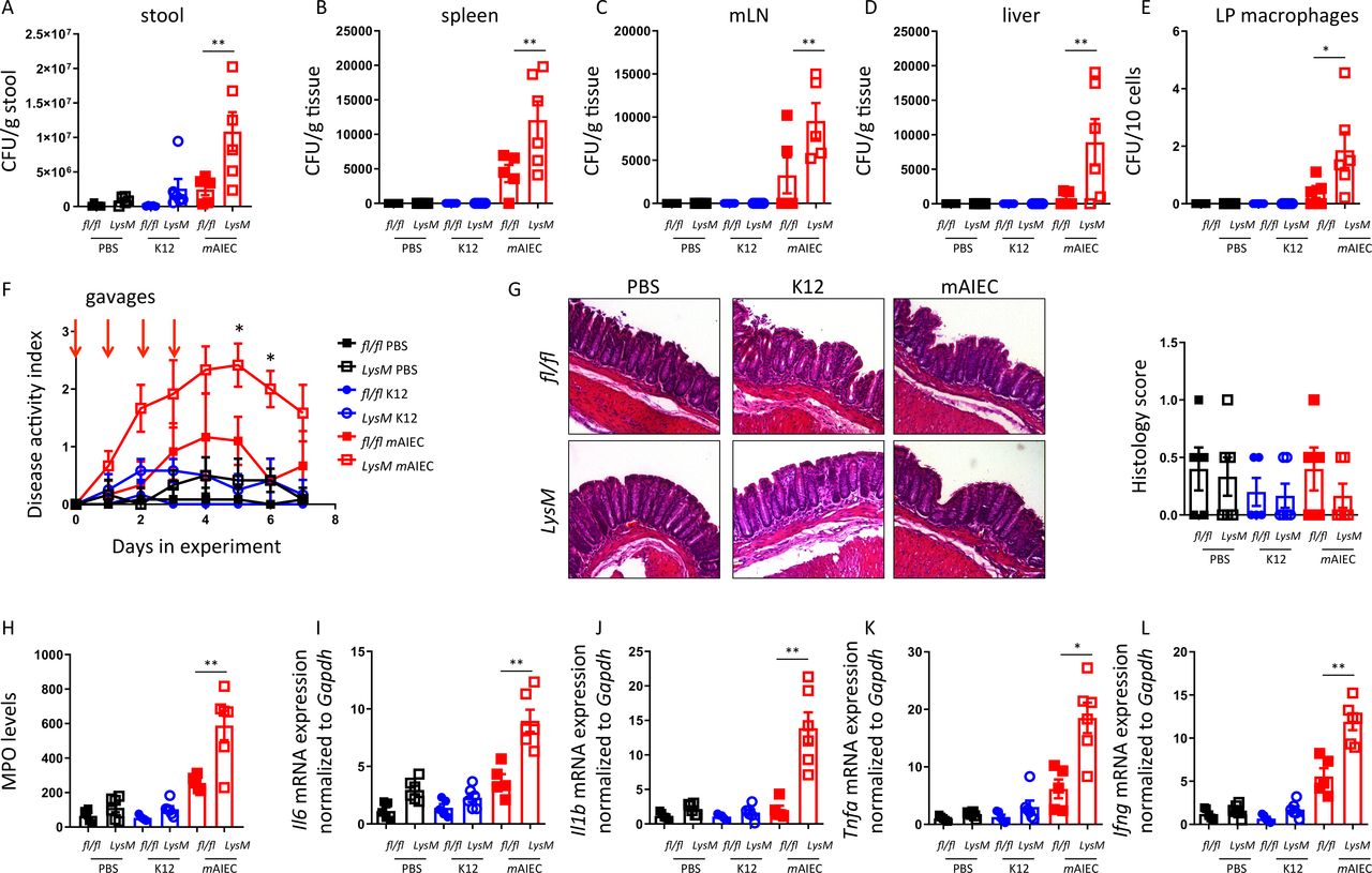
To test the in vivo consequences of defective bacterial handling in macrophages with PTPN2-loss, mice lacking PTPN2 in myeloid cells (Ptpn2-LysMCre mice; predominant PTPN2-loss in macrophages26) and Ptpn2fl/fl controls were orally gavaged on four consecutive days with 109 mAIEC or non-invasive K12 E. coli. Consistent with an in vitro defect in bacterial handling, we observed increased mAIEC load in the stool of Ptpn2-LysMCre mice and enhanced bacterial translocation to the spleen, mesenteric lymph nodes and liver, and elevated bacterial counts in lamina propria macrophages (figure 7A–D). mAIEC induced mild disease only in Ptpn2-LysMCre but not Ptpn2fl/fl littermates (figure 7E,F), but there were no overt differences detectable in histological scores from infected vs non-infected and Ptpn2fl/fl or Ptpn2-LysMCre mice (figure 7G), while inflammatory markers, such as myeloperoxidase and mRNA levels of inflammatory cytokines were elevated in AIEC-infected Ptpn2-LysMCre mice (figure 7H–L). Consistent with our in vitro observations, activation of autophagy using rapamycin, or inhibition of CEACAM1 using an inhibitory anti-CEACAM1 antibody, partially prevented increased bacterial translocation/uptake in macrophages and normalised disease activity in Ptpn2-LysMCre mice (online supplemental figure 5). These data clearly indicate that loss of Ptpn2 in macrophages hampers the clearance of potentially pathogenic bacteria and promotes susceptibility to AIEC-induced disease in vivo.
Mice lacking PTPN2 in macrophages are more susceptible to mAIEC infection.Ptpn2fl/fl and Ptpn2-LysMCre littermates were orally infected for 4 days with 109 LF82 or mAIEC and Escherichia coli load in (A) the stool at day 5, (B) in the spleen, (C) mesenteric lymph nodes, (D) the liver and (E) lamina propria macrophages determined by plating on LB agar plates. (F) Disease activity index (DAI) over the course of the experiment. (G) Representative histological pictures and histopathology scoring. (H) Myeloperoxidase (MPO) levels in colon pieces. (I–L) mRNA expression levels of the indicated genes in the colon normalised to Gapdh and the median of untreated controls. *P<0.05, **p<0.01, ANOVA. ANOVA, analysis of variance; mAIEC, mouse adherent-invasive Escherichia coli; PTPN2, protein tyrosine phosphatase non-receptor type 2.
Discussion
We demonstrate that normal PTPN2 expression and function in intestinal macrophages/myeloid cells is crucial to clear intestinal adherent-invasive bacteria and that PTPN2-deficiency severely compromises effective macrophage-mediated elimination of invading bacteria, thus promoting AIEC-induced intestinal disease. Macrophages prevent systemic dissemination of bacteria that breach the epithelial barrier39 and macrophage-induced immune reactions towards luminal and adherent bacteria restrict overgrowth of pathogens and pathobionts,40 thus shaping the intestinal microbiota composition.41–43 The microbiome of patients with IBD carrying PTPN2-SNP rs1893217 differs from the microbiome of patients without the variant.24 44 In line with this, presence of SNP rs1893217 or loss of Ptpn2 resulted in increased translocation of invasive bacteria, coupled with defective clearance, effects that might contribute to the changes in bacterial composition observed in PTPN2 variant carrier IBD patients. The defective response to AIEC in macrophages with compromised PTPN2 function is of clear clinical importance given the elevated levels of AIEC found in intestinal biopsies from IBD patients.5 In addition to explaining the alteration in microbial composition in PTPN2 variant carriers, our findings identify additional mechanisms how PTPN2 variants contribute to an increased risk of developing IBD.
Ptpn2-LysMCre mice lacking Ptpn2 primarily in macrophages/monocytes26 showed increased adherence and invasion of AIEC on challenge, and furthermore show extra-intestinal translocation and AIEC-induced disease. Hence our data indicate that the defects in autophagy and lysosomal acidification observed in Ptpn2-deficient macrophages have a clear in vivo relevance.
Bacteria are actively taken up and subsequently degraded in macrophages by two distinct mechanisms: (1) uptake via endocytosis/phagocytosis and delivery into lysosomes,45 and (2) autophagy/xenophagy of bacteria, which targets bacteria that adhere to the membrane and enter the cytosol and/or escape the endosome-lysosome pathway.33 Intracellular vesicles are highly dynamic and interconnected, and autophagy vesicles promote the fusion of late endosomes with lysosomes.34 35 Thus, defects in autophagy, as observed in PTPN2-deficient/defective cells, crucially hamper the ability to combat invasive bacteria. This aligns with our observations and previously published in vitro studies showing defective autophagy in IEC and monocytes lacking PTPN2.22 Genetic studies point towards an important contribution of defective autophagy to the development of IBD,46 and variants in the autophagy-inducing receptor NOD2 and the autophagy-initiator molecule ATG16L1 were among the first genes associated with IBD.47 48 Bacterial products induce recruitment of autophagosomes to the site of bacterial entrance.37 It has been described that AIEC can subvert autophagy by blocking autophagosome-lysosome fusion, resulting in apoptosis of infected neutrophils and increased overall AIEC burden.49 These findings show that defects in autophagy hamper clearance of AIEC and ultimately promote survival of invading pathogens. Notably, we observed autophagy-activation not only in AIEC-infected cells, but also in the presence of non-invasive K12 E. coli, which was again abrogated in PTPN2-deficient macrophages. This indicates that loss of PTPN2 not only affects the response to pathogens/pathobionts, but also compromises physiological host reactions to benign commensals.
One important observation with regards to mouse models of IBD and studying the relevance of AIEC infections in those models is that the most widely used AIEC model strain, LF82, enters enterocytes and macrophages via binding to CEACAM6.30 However, mouse cells do not express CEACAM6 and are thus not very susceptible to infection with human AIEC strains.30 This issue is overcome in some studies by utilising transgenic mice that express human CEACAM3, 5, 6 and 7.50 The expression pattern of CEACAMs in IECs of these mice corresponds to that observed in humans, however, it also resulted in crypt hyperplasia and aberrant crypt morphology.50 Therefore, this model might not be optimally suited to study AIEC-host interactions. In contrast, our recently identified mAIEC27 was effective in invading mouse macrophages since it binds to and enters host cells by using CEACAM1, which is highly expressed in mouse enterocytes and intestinal macrophages.51 Thus, this novel mAIEC strain might represent a more appropriate strain for studying the effect of AIEC colonisation in mouse models of IBD.
AIEC manipulate the host response to infection, that is, it has been reported that the AIEC strain LF82 promotes its survival in macrophages via suppression of nuclear factor (NF)-κB signalling,52 but more recent studies showed that intracellular AIEC survival on early infection depends on NF-κB activation,53 while AIEC attenuate the very same signalling pathway later on.54 Furthermore, AIEC are more resistant to lysosomal superoxides than non-invasive E. coli strains.54 AIEC are also more resistant to the acidic pH in lysosomes, allowing them to survive within macrophages without the need to escape phagosomes/endosomes.55 This is of great interest given the reduced lysosomal acidification in PTPN2-deficient macrophages. Hence, loss of functional PTPN2 likely facilitates intracellular survival of AIEC in two ways: (1) compromising transport of bacteria into lysosomes and (2) restricting acidification within lysosomes, thus compromising the cell’s ability to degrade bacteria.
In conclusion, we show that the autoimmune risk gene PTPN2 is involved in handling invasive pathogens and its loss compromises macrophages to clear pathobionts. We not only identified several molecular mechanisms contributing to defects in bacterial handling, but also demonstrate the importance of efficient bacterial clearance by macrophages to prevent intestinal inflammation on AIEC infection. This might not only functionally explain the alterations observed in the microbial composition in patients with IBD carrying PTPN2 SNP rs1893217,24 44 but may also explain, at least in part, why patients carrying this variant are more susceptible to the development of IBD.
Acknowledgments
We thank Prof. Nicole Beauchemin (Goodman Cancer Research Centre, McGill University) for critical reading of the manuscript and for valuable input on CEACAM biology.
References
Supplementary materials
Supplementary Data
This web only file has been produced by the BMJ Publishing Group from an electronic file supplied by the author(s) and has not been edited for content.
Footnotes
Contributors MRS: study and experiment design, data acquisition, analysis and interpretation, writing of the manuscript; ASh, PC, VC, ASa, AS-B: data acquisition and analysis; MS: provided patient samples; JB: support with bacteria isolation and culture, data interpretation and critical intellectual input; DFM: data interpretation, study design, supervision of the experiments, funding, editing of the manuscript; MLT and MS provided mice. All authors reviewed the manuscript.
Funding This study was supported by research grants from the NIH to DFM (2R01DK091281; and 1R01AI153314); a Senior Research Award from the Crohn’s and Colitis Foundation to DFM; and a research stipend from the Swiss National Science Foundation (P300PB_177932) and a grant from the Hartmann Müller Foundation to MRS. This study was supported by an Advanced. Postdoc Fellowship from the Swiss National Science Foundation to MRS (Project Nr. P300PB_177932), by NIH grant 2R01DK091281 and 1R01AI153314 to DFM, and a Crohn’s and Colitis Foundation Senior Research Award to DFM.
Competing interests None declared.
Patient consent for publication Not required.
Provenance and peer review Not commissioned; externally peer reviewed.
Data availability statement All data relevant to the study are included in the article or uploaded as online supplemental information.
Supplemental material This content has been supplied by the author(s). It has not been vetted by BMJ Publishing Group Limited (BMJ) and may not have been peer-reviewed. Any opinions or recommendations discussed are solely those of the author(s) and are not endorsed by BMJ. BMJ disclaims all liability and responsibility arising from any reliance placed on the content. Where the content includes any translated material, BMJ does not warrant the accuracy and reliability of the translations (including but not limited to local regulations, clinical guidelines, terminology, drug names and drug dosages), and is not responsible for any error and/or omissions arising from translation and adaptation or otherwise.