Article Text

Statistics from Altmetric.com
Introduction
A diabetic patient presented with 10 kg weight loss and slightly elevated C reactive protein without diarrhoea, abdominal pain, fever or leucocytosis. Serum lipase, transaminases, bilirubin, carcinoembryonic antigen and CA19-9 were normal. Ultrasound showed prominent perihilar bile ducts and a cystic mass of the pancreas (60×57 mm) with echogenic internal structures, which was identified as the main pancreatic duct on endoscopic ultrasound (EUS), without enlarged peripancreatic lymph nodes (figure 1A,B). At endoscopic retrograde cholangio-pancreatography (ERCP), the compressed common bile duct was secured by a plastic stent. Cannulation of the pancreatic duct resulted in massive putrid secretion and confirmed an enormously dilated main pancreatic duct over its full length (figure 1C,D).
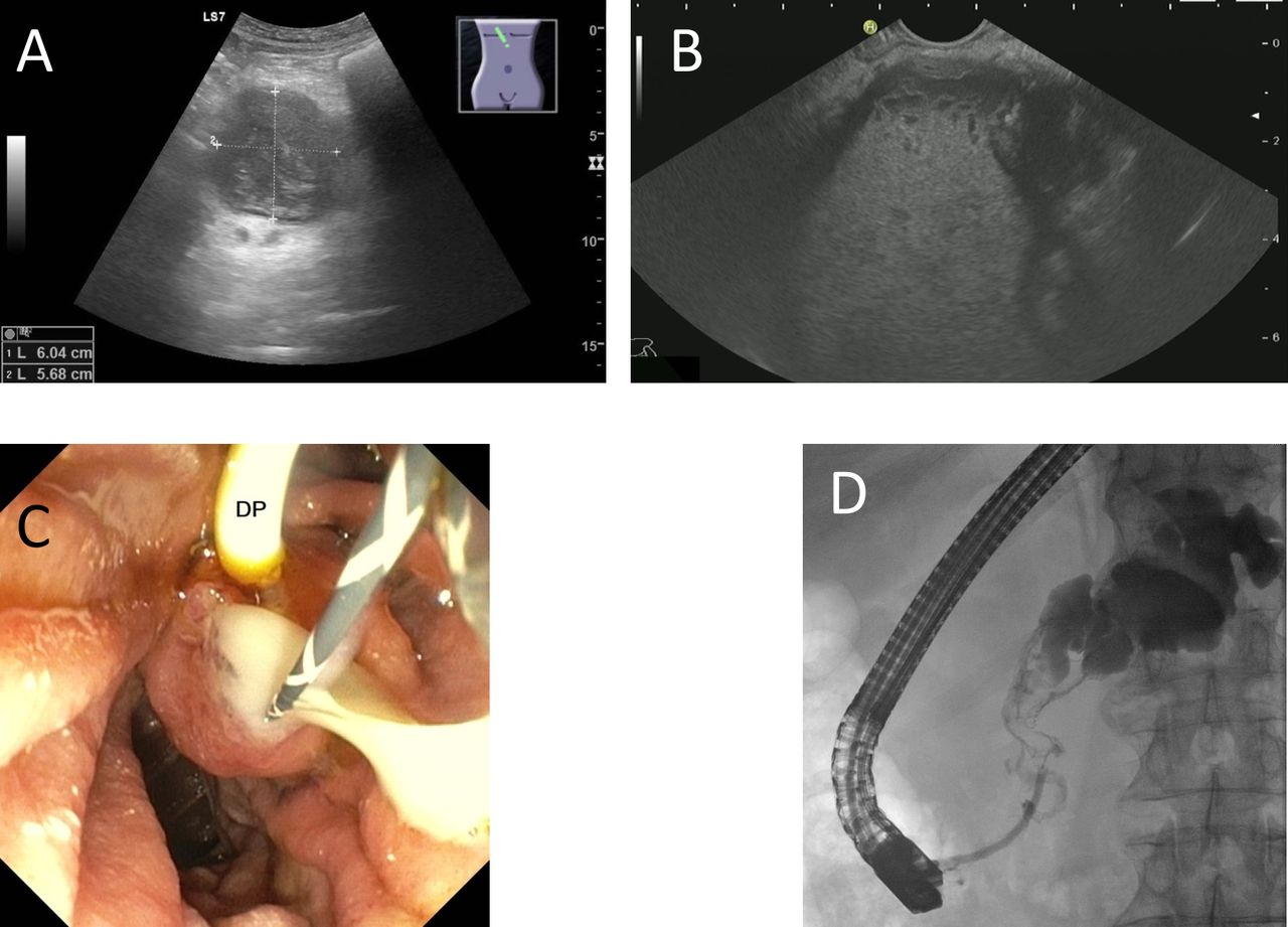
(A) Ultrasound of the pancreas. (B) Endoscopic ultrasound of the pancreas. (C) Endoscopic retrograde cholangio-pancreatography (ERCP) after double pigtail (DP) stent insertion in the common bile duct. Guidewire in pancreatic duct. (D) ERCP shows the dilated pancreatic duct.
Question
What is the most likely diagnosis and what is the next step?
Answer
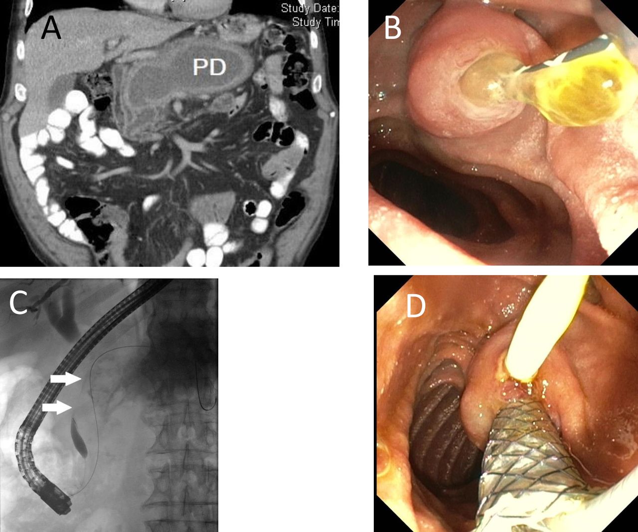
CT scan demonstrated the pancreatic duct dilatation up to 50 mm (figure 2A). ERCP showed contrast dye notches due to very viscous mucus, which could be rinsed out together with massive pus (figure 2B). The congested pancreatic and the compressed common bile duct (figure 2C) were splinted by a fully covered 8×60 mm self-expanding metal stent (fcSEMS) to allow mucus and pus evacuation and an 8F double pigtail, respectively (figure 2D). Aspirates from the pancreatic duct exhibited superinfection with Lactobacillus salivarius and Streptococcus salivarius. CT and ERCP were consistent with the diagnosis of main duct intraductal papillary mucinous neoplasm (MD-IPMN). Due to the high risk of malignancy (main duct diameter ≥10 mm),1 total pancreatoduodenectomy was performed and confirmed MD-IPMN of adenoma type on histopathology with low-grade dysplasia. The postoperative course and follow-up were uneventful. While mucinous dilatation is the hallmark of MD-IPMN, the characteristic fish mouth papilla was obscured in our patient by massive purulent secretion. Superinfection of IPMN is an extremly rare condition in the absence of fistula2 3 and might have been facilitated by the enlarged orifice and the occluding viscous mucus.

(A) CT shows a massively dilated pancreatic duct (PD). (B) Endoscopic retrograde cholangio-pancreatography (ERCP): papilla after cannulation of the pancreatic duct with efflux of highly viscous mucus. (C) ERCP: compression of the common bile duct (arrows) by the dilated pancreatic duct. (D) ERCP after additional insertion of a fcSEMS in the pancreatic duct.
Ethics statements
Patient consent for publication
Ethics approval
We obtained the written consent of the patient to anonymously publish his case.
Acknowledgments
We thank Dr Martin Ruch (Radiology, Rhine-Main-Centre for Diagnostics, Weiterstadt, Germany) for providing the CT image.
Footnotes
Contributors RK: performed EUS/2.ERCP, wrote manuscript. ML: performed EUS/1.ERCP, wrote manuscript, took care of patient in outpatient setting. TH: wrote manuscript, surgical care. ML/TH: outpatient follow-up.
Funding The authors have not declared a specific grant for this research from any funding agency in the public, commercial or not-for-profit sectors.
Competing interests None declared.
Provenance and peer review Not commissioned; externally peer reviewed.