Article Text

Abstract
Crohn's disease is characterised by recurrent and/or chronic inflammation of the gastrointestinal tract leading to cumulative intestinal tissue damage. Treatment tailoring to try to prevent this tissue damage as well as achieve optimal benefit/risk ratio over the whole disease course is becoming an important aspect of Crohn's disease management. For decades, clinical symptoms have been the main trigger for diagnostic procedures and treatment strategy adaptations. However, the correlation between symptoms and intestinal lesions is only weak. Furthermore, preliminary evidence suggests that a state of remission beyond the simple control of clinical symptoms, and including mucosal healing, may be associated with better disease outcome. Therefore monitoring the disease through the use of endoscopy and cross-sectional imaging is proposed. However, the degree of mucosal or bowel wall healing that needs to be reached to improve disease outcome has not been appropriately studied. Furthermore, owing to their invasive nature and cost, endoscopy and cross-sectional imaging are not optimal tools for the patients or the payers. The use of biomarkers as surrogate markers of intestinal and systemic inflammation might help. Two biomarkers have been most broadly assessed in Crohn's disease: C-reactive protein and faecal calprotectin. These markers correlate significantly with endoscopic lesions, with the risk of relapse and with response to therapy. They could be used to help make decisions about diagnostic procedures and treatment. In particular, with the use of appropriate threshold values, they could determine the need for endoscopic or medical imaging procedures to confirm the disease activity state.
- Abdominal Mri
- Crohn's Disease
- Endoscopic Procedures
Statistics from Altmetric.com
Introduction
Crohn's disease (CD) is a chronic inflammatory disorder of the gastrointestinal tract leading to cumulative intestinal tissue damage and complications such as fistulas and strictures requiring surgical resection.1 A classical step-up strategy in which the treatment is adapted according to the clinical activity of the disease does not seem to be able to change the natural history of the disease, particularly to avoid the need for intestinal surgical resection.2 Achieving mucosal healing has been associated with better patient outcome in several population-based studies and clinical trials.3–6 For these reasons and although there is no current proof that optimising therapy in a patient who has achieved clinical remission but not mucosal healing will lead to improved outcome, there is an intuitive trend to aim to achieve such healing, particularly in patients with worse prognoses. Because of the poor correlation between clinical activity of CD and mucosal healing,7 this requires specific monitoring, defined as the systematic use of objective tools able to assess the state of biological activity of the disease, beyond the simple assessment of clinical symptoms. Endoscopy is an invasive and costly procedure, and regular monitoring of the disease by endoscopy may not be realistic. Moreover, it is not adapted for small-bowel location above the terminal ileum, and it does not provide information about the transmural nature of the inflammation. For these locations and aspects, cross-sectional imaging may be more appropriate, but the specific features associated with tissue healing have been less well described.8 Biomarkers may represent an attractive alternative for both colonic and small-bowel disease, as they represent potential surrogate markers of intestinal inflammation and tissue lesions. Among the very large number of biomarkers assessed in CD, the vast majority have not been adequately studied to determine their potential clinical usefulness. For most of these markers, abnormal stool or blood concentrations have been shown in CD, but no correlation has been clearly demonstrated with disease activity, intestinal lesions or disease evolution and risk of complications. The main exceptions are blood C-reactive protein (CRP) and faecal calprotectin, which have been more extensively studied, including assessment of their ability to predict response to treatment, mucosal healing, and risk of relapse and of complications.9 Depending on the clinical situation, different combinations of these imaging and biomarker monitoring tools may be used. The aim of this review is to describe the potential use, advantages and limitations of endoscopy, cross-sectional imaging and biomarkers for the monitoring of CD. We will more specifically tackle these issues through various clinical scenarios, including confirmation of disease activity, response to induction therapy, confirmation of sustained mucosal healing, and prevention of relapse or recurrence. We will not address the use of these tools for the diagnosis of CD or when the clinical presentation leads to the suspicion of a complication of the disease.
Endoscopy
Preliminary mainly retrospective data or post hoc analyses indicate that achieving endoscopic mucosal healing may improve outcome of the disease.3–6 However, endoscopy suffers from a series of significant drawbacks: it is not that well accepted by the patients,10 although it is usually safe,11 it is relatively expensive, it does not give information on the deep layers of the intestine and the extraintestinal signs of inflammation,12 and finally the significance of different types of lesion and the degree of endoscopic healing that should be achieved is not well established. Illustrations of unhealed, partly healed and fully healed mucosa are shown in figure 1. Advantages and drawbacks of endoscopy as a monitoring tool are summarised in table 1. The potential role of endoscopy in monitoring patients with clinically active disease or in remission is developed in the following paragraphs and summarised in tables 4 and 5.
Monitoring of Crohn's disease with ileocolonoscopy

Examples of various degrees of endoscopic healing in Crohn's disease. (A) Absence of healing characterised by the persistence of a deep ulcer in the caecum; (B) absence of healing characterised by the persistence of extensive longitudinal and transversal ulcers in the sigmoid colon; (C) absence of healing characterised by the persistence of multiple deep ulcers in the left colon; (D) partial healing characterised by the presence of small pseudo-polyps and the persistence of focal erythema and tiny superficial ulcers in the right colon; (E) partial healing characterised by the presence of small pseudo-polyps, healed ulcers with modification of the vascular pattern, and the persistence of small superficial ulcers in the sigmoid colon; (F) partial healing characterised by the persistence of tiny aphthous lesions throughout the colon; (G) full mucosal healing characterised by the presence of longitudinal healed whitish areas; (H) full mucosal healing characterised by the presence of whitish healed areas and mucosal bridges; (I) full mucosal healing characterised by the presence of mucosal bridges converging to a healed stellar-shaped area.
Confirming disease activity and severity of lesions
The correlation between clinical activity of CD and the severity of endoscopic lesions is only weak.7 Of patients with clinically active disease, a significant proportion will have no significant endoscopic lesions. These patients do not respond optimally to the most effective treatment of CD, such as immunosuppressant and anti-tumour necrosis factor (TNF) combination therapy.13 Preliminary data also indicate that deep colonic ulcers covering more than 10% of a colonic segment are associated with increased risk of colectomy over the next 8 years.14 Stricturing lesions have been associated with lower response rate to medical therapies and a greater need for surgery.15 To be fully informative, an endoscopic procedure for CD should thus lead to a very precise description of the type, location and extent of the lesions. Although endoscopic indexes of disease severity such as the CD Endoscopic Index of Severity (CDEIS)16 and Simplified Endoscopic Score of CD (SESCD)17 have been used in several studies, and specific thresholds of these scores have been associated with CD outcome, these thresholds have not been broadly validated. Hence, therapeutic decisions still seem to be best based on the precise description of the lesions, instead of a specific quantitative or semiquantitative threshold.
Confirming mucosal healing
Achieving endoscopic healing after medical therapy has been associated with a better disease outcome. In a population-based study from Norway, the presence of mucosal healing 1 year after the diagnosis of CD tended to be associated with less need for surgical resection, although this difference did not reach statistical significance.3 In the Accent 1 trial, patients achieving mucosal healing at weeks 12 and 54 experienced fewer relapses and hospitalisations.18 In the experience of a tertiary referral centre in Belgium, patients achieving at least partial healing subsequently underwent fewer surgical resections.4 Very interestingly and intriguingly, the patients with partial healing had no more surgery than those with complete healing, emphasising the relevance of the question about the degree of healing required to improve disease outcome in CD. The long-term follow-up of the so-called ‘step-up top-down trial’ also revealed that patients with complete mucosal healing 2 years after the beginning of the trial (whatever the type of treatment they received) experienced fewer flares and were more often in remission without steroids and without anti-TNF over the next 2 years.19 In a controlled trial showing the superiority of adalimumab maintenance over placebo to achieve early and sustained mucosal healing in CD, the patients with mucosal healing after 12 weeks had fewer relapses and hospitalisations over the 1-year follow-up.6 On the basis of these preliminary data, endoscopic monitoring after induction or during maintenance therapy is now advocated by many experts. However, there are currently no data to clarify the timing of this monitoring, the degree of healing that needs to be reached, and, above all, the management of insufficiently healed patients. No prospective or even retrospective study can report on an improved outcome in these unhealed patients after a change in therapy. This is even more troubling because the only available data in this field, dating back to the time of corticosteroid therapy, do not support such treatment optimisation. Indeed, in the 1990s, a GETAID Study showed that prolonging steroid treatment in patients in clinical remission but with unhealed mucosa after 6 weeks of full steroid induction did not improve the relapse rate over 1 year, despite a slight increase in the proportion of patients whose mucosa was healed.20 Because of all these unsettled issues, the use of endoscopic monitoring to confirm tissue healing in CD can only be empirical. An endoscopy should only be discussed if a clear disease management plan can be proposed to the patient depending on the results of this endoscopy. In our view, this could be particularly adapted when there is concern about disease progression and its consequences. In some situations, such as extensive small-bowel disease, previous multiple intestinal resections, deep and extensive colonic (particularly sigmoid or rectal) ulcers, due to the consequences of uncontrolled disease, endoscopic monitoring and treatment optimisation in the case of persisting significant lesions may be proposed.
Assessing the patient before therapy de-escalation
In order to achieve optimal benefit/cost and benefit/risk ratio for the patient, treatment de-escalation in patients who have reached sustained remission may be as important as treatment optimisation in those who have not reached this target. Whether treatment de-escalation is possible in a subset of patients and what the criteria should be is not precisely known. However, preliminary results are provided by a prospective cohort study from the GETAID. In this study, full endoscopic healing was associated with a lower risk of relapse after infliximab withdrawal in patients treated with immunosuppressant/infliximab combination therapy for more than 1 year and in steroid-free remission for more than 6 months.21 In patients without mucosal healing, defined by a CDEIS >0, the relapse rate over 1 year was above 60%. Therefore endoscopic exploration could be advocated in these patients before a decision is made on such drug withdrawal. However, mucosal healing is not the best individual predictor, and the prediction is significantly improved if some demographic characteristics, blood tests and faecal calprotectin are integrated.
Predicting postoperative recurrence
The postoperative setting is a situation where the use of endoscopic monitoring has been particularly widely used in routine practice. The disease is usually clinically quiescent, but the recurrence rate is known to be very high. This recurrence has been well described in the seminal paper by Rutgeerts et al.22 Over 8 years after an ileocolonic resection, ∼90% experienced endoscopic recurrence, 60% clinical recurrence and 30% surgical recurrence. This study also showed that the clinical recurrence rate was strongly associated with endoscopic recurrence within 1 year of ileocolonic resection. Diffuse ileitis, strictures and large or deep ulcers (Rutgeerts scores i3 and i4) were associated with almost 90% of clinical relapse over 8 years, while no lesion or a few aphthoid ulcers (Rutgeerts scores i0 and i1) were associated with a clinical relapse rate of ∼10%. Following these results, it has become common practice to perform ileocolonoscopic surveillance once, between 6–12 months after ileocolonic resection in order to adapt treatment according to the results.
Potential role for small-bowel capsule endoscopy
Small-bowel capsule endoscopy may have much better acceptance than classical endoscopic explorations. It can also visualise the whole small bowel, and its diagnostic yield in small-bowel lesions of CD has been well established.23 It could thus be of help in assessing disease activity or mucosal healing in the small bowel. However, it is hampered by a relatively high retention rate in CD, above 10%.23 Although this can usually be avoided by a test with a patency capsule, obstructive accidents may still occur. Furthermore, for this disease location, it is in competition with cross-sectional imaging techniques (see below), which may also offer information on transmural inflammation and assess stricturing and fistulising complications.8 Some lesions missed at cross-sectional imaging may be diagnosed by capsule endoscopy, particularly in the proximal small bowel, but their clinical significance is still unclear.24 Several small-bowel capsule endoscopy indexes of severity have been proposed but not yet broadly validated.23
Small-bowel capsule endoscopy has been specifically assessed in the postoperative recurrence setting. Its correlation with ileocolonoscopy appears to be good, and it may thus represent an alternative in this situation.23
Cross-sectional imaging techniques for the monitoring of CD
Although it may currently be considered the gold standard, the monitoring of CD activity and mucosal healing by ileocolonoscopy or small-bowel capsule endoscopy is hampered by a series of drawbacks highlighted above. Therefore the use of cross-sectional imaging techniques to monitor the disease is rapidly increasing.8 These techniques, including ultrasonography (US), CT and MRI, show both parietal and extraparietal changes caused by the disease, and allow evaluation of small-intestinal regions inaccessible to ileocolonoscopy, enabling the identification of a whole spectrum of lesions with good resolution.8
MRI is considered the standard imaging technique for assessment in patients with CD who require many follow-up examinations and are usually a young population.25 The absence of ionising radiation, along with very high soft-tissue contrast, multiplanar images, low incidence of adverse events related to the intravenous contrast, and high diagnostic accuracy in the evaluation of luminal and extraluminal abnormalities, justify its application.26 CT has a similar accuracy to MRI for assessing bowel damage in CD,27 but the risk of radiation exposure should limit its use as a monitoring tool. US is another non-ionising alternative,28 but has some drawbacks such as the difficulty of visualising deep bowel segments, high interobserver variability, and often incomplete exploration due to gas interposition.29
An illustration of monitoring of multifocal small-bowel CD with MR enterography is shown in figure 2. Advantages and drawbacks of cross-sectional imaging as a monitoring tool are summarised in table 2. The potential role of cross-sectional imaging in monitoring patients with clinically active disease or in remission is developed in the following paragraphs and summarised in tables 4 and 5.
Monitoring of CD with cross-sectional imaging

Example of monitoring of small-bowel Crohn's disease (CD) with MR enterography. This patient had been operated on three times for recurring stricturing and occlusive CD in December 1998, March 2002 and December 2005. He had been continuously treated with azathioprine between the second and third operations. He was prescribed methotrexate 15 mg/week subcutaneously immediately after the third operation. Six months later, in June 2006, he was in clinical remission and had a normal C-reactive protein (CRP) concentration. Because of the multifocal small-bowel CD, it was decided to monitor him with MR enterography instead of colonoscopy. The MR enterography performed in June 2006 showed thickening of the bowel wall without strong contrast enhancement at the ileocolonic anastomosis (B) and mild thickening and wall enhancement in a jejunal segment (A). In January 2007, the patient was still in clinical remission with a normal CRP concentration, but had mild iron-deficiency anaemia (haemoglobin concentration 11.7 mg/dl). The MR enterography performed at that stage showed a stable lesion at the ileocolonic anastomosis (D) and marked contrast enhancement and thickening of the wall in the jejunal segment, together with hyperaemia of the vasa recta (comb sign) (C). The patient was then treated with infliximab in combination with methotrexate. In September 2007, the patient was still in clinical remission with normal CRP concentration. The anaemia had disappeared. The MR enterography showed a significant decrease in wall thickening and contrast enhancement of the jejunal segment, but a partial small-bowel obstruction due to jejunal disease (E), while the anastomotic stricture remained unchanged (F). Maintenance treatment combining methotrexate 15 mg/week orally and infliximab 5 mg/kg every 8 weeks was prescribed .
Assessment of disease activity and severity and response to treatment
The high accuracy of MR enteroclysis, enterography and enterocolonography has been demonstrated in the assessment of activity and severity in CD, with several parameters related to the degree of activity being identified.30 Globally, enterography has been preferred to enteroclysis because of its better acceptance. Enterocolonography implies a colonic distension by enema and may provide information on the whole gastrointestinal tract in only one procedure. The main MRI findings that correlate with intestinal inflammation and disease activity and severity include wall thickening, bowel wall enhancement, mural oedema (signal hyperintensity on T2-weighted sequences) and presence of ulcers. It is in the terminal ileum and the colon that the ability of MRI to assess disease activity has been best validated. Using the above parameters, Rimola et al30 ,31 proposed and validated a simplified Magnetic Resonance Index of Activity (MaRIA) score to quantify disease activity based on MRI findings in each ileocolonic segment, which strongly correlated with CDEIS. For the detection of active CD in the terminal ileum and the colon, the sensitivity of this score was 87%, specificity 89%, positive predictive value (PPV) 98%, negative predictive value (NPV) 88% and overall accuracy 98%. This score was also able to accurately predict severely ulcerated CD. Therefore, the MaRIA score represents an objective, quantitative and reproducible measure of activity and could categorise disease severity and monitor response to therapeutic interventions in the terminal ileum and the colon.
These results agree with those from other studies identifying MRI signs associated with pathological inflammation mainly in the small bowel, using surgical examination as a reference method.32 ,33 In a systematic review, Panes et al8 reported a sensitivity and specificity of MRI for the assessment of disease activity on a per patient basis of 80% and 82%, respectively. The use of oral and intravenous contrast agent promotes bowel lumen distension and improves detection of these features.34 CT has a similar accuracy to MRI for distinguishing activity in the terminal ileum, with a sensitivity of 81% and specificity of 88%.8 ,35 ,36
US has been established as a reliable imaging technique for the assessment of disease activity in CD. The wall thickness and vascularisation pattern shown by Doppler US are particularly useful for the detection of active disease. The overall sensitivity and specificity of US for detecting disease activity have been found to be ∼85% and ∼91%, respectively,8 although its performance is largely dependent on the operator and the disease location.37 The use of contrast-enhanced US seems to increase its accuracy for the evaluation of activity in CD, with a sensitivity of 93% and specificity of 94%.38 It also seems to better classify disease severity than Doppler US signal and measurement of wall thickening.
Few studies have investigated the capability of MRI for assessing treatment response after a flare of CD. Sempere et al39 evaluated this capability of MRI compared with ileocolonoscopy in patients in the active phase of disease or in remission and in healthy controls. These authors reported that contrast enhancement of the bowel wall decreased significantly from the active to the remission phase. The mean contrast enhancement in active CD was significantly greater than in the control group, although there was no difference between patients in remission and the healthy controls. The same occurred with bowel wall thickness, which was significantly decreased in the remission phase. However, the segments remained thickened in patients with CD compared with healthy controls. This is probably due to a double component: an acute factor (inflammation and oedema) and a chronic factor (fibrosis), which may not reverse despite therapeutic response. Another study has evaluated MRI for monitoring therapeutic responses using the MaRIA score and ileocolonoscopy as a reference standard. In this setting, the MaRIA score predicted endoscopic remission with a sensitivity of 82% and specificity of 85%.40
All these data suggest that cross-sectional imaging could be used for the assessment of disease activity and response to therapy in CD. The use of a CT scanner should be avoided in this setting to minimise irradiation. MRI enterography could be used essentially to visualise small-bowel CD not evaluable by standard endoscopic procedures. MRI enterocolonography, could be used as a single exploration of the whole gastrointestinal tract, particularly in ileocolonic disease, decreasing the need for supplementary colonoscopies. This would be of particular interest for patients with both small-bowel and colonic lesions. US is an attractive alternative, particularly in attempts to confirm active disease. However, it is less powerful than MRI for broadly assessing lesion extent and severity, because of its poorer performance in the colon and inability to systematically see all intestinal segments.
Detection of complications
Chronic intestinal inflammation in CD can result in complications during the course of the disease. Although they may present as acute modifications of the clinical situation leading to emergency work up, they can also develop more silently, and their identification through disease monitoring may reveal disease aggressiveness and influence treatment strategy. The ability of cross-sectional imaging methods to demonstrate extramural changes makes them accurate procedures for detecting complications.
US, CT and MRI have a high sensitivity and specificity for the diagnosis of stenosis affecting the large or small bowel8 identified as an intestinal loop with wall thickening, narrowed lumen and prestenotic dilation. A systematic review of pooled results from seven studies reported that MRI had a sensitivity for detection of stenosis of ∼89% and a specificity of ∼94%.8 This is usually considered to be slightly higher than with CT or US. Distinguishing between inflammatory and fibrotic strictures is important, as it may have a significant effect on response to medical treatments. Making an exclusive distinction between an inflammatory or fibrotic pattern is difficult as they usually coexist, especially in patients with severe disease.41 Nevertheless, here again, MRI may be of help. Collagen deposition in the bowel wall is known to result in late gadolinium enhancement. A decrease in the signal intensity of the thickened wall and a reduction in bowel-wall early contrast enhancement are usually related to intestinal fibrosis.42
The diagnostic accuracy of MRI for intra-abdominal fistulas has been evaluated in multiple studies.8 Pooled results showed a sensitivity of ∼76% and a specificity of ∼96%. CT has a similar accuracy,43 while US is significantly less efficient.8 Likewise, MRI detected intra-abdominal abscesses with a sensitivity ranging from 86% to 100%, and specificity from 93% to 100%.27 With CT, the sensitivity was 84% and specificity 97%.8 The value of US for detection of abscesses reached a sensitivity of 81–100% and specificity of 92–94%.8 However, US accuracy was highly related to disease location,44 and its diagnostic accuracy was slightly lower than that of CT and MRI because of false-positive cases. Combining CT with US did not significantly improve their diagnostic accuracy for detection of abscesses in CD.45
Assessment of postoperative disease recurrence
Although ileocolonoscopy is the gold standard technique for evaluating recurrence of CD after intestinal resection, MRI and US may be valuable alternatives to avoid repeated colonoscopies. An MRI-based score has even been validated for the detection of postoperative recurrence compared with endoscopic Rutgeerts score.46 ,47 Mild bowel-wall thickening and enhancement without stricture were considered to be signs of low-grade recurrence. In contrast, the presence of a clear stricture and increased bowel-wall thickness and enhancement correlated with severe recurrence. The sensitivity and specificity of MRI for detecting moderate to severe recurrence were 100% and 89%, respectively.
Monitoring tissue damage
Tissue damage in CD is characterised by intestinal resections, strictures and fistulising lesions.48 One of the main aims of optimised therapeutic strategies for CD, including early intensive treatment and tight disease control, is to limit, or even suppress, the development of tissue damage. Owing to its ability to visualise and quantify both stricturing and penetrating lesions of CD, MRI currently represents the best candidate for monitoring tissue damage in CD. A tissue damage score is currently under development.48
Biomarkers
CRP is an acute phase reactant produced by the liver.49 In CD, there is also significant CRP production by the mesenterium itself.50 The main trigger for CRP production is interleukin 6.51 In CD, interleukin 6 is produced in the whole intestinal wall and probably the mesenterium, at the site of inflammation, by a broad selection of cell types including lymphocytes, monocytes, granulocytes, fibroblasts, and epithelial and endothelial cells. CRP increases very rapidly during acute inflammation and may remain elevated during chronic inflammation.
Calprotectin is a heterodimer or heterotetramer combining S100A8 and S100A9 proteins.52 It is produced at the site of inflammation mainly by granulocytes, but also monocytes and epithelial cells. Owing to this direct production by intestinal epithelial cells, increased amounts of calprotectin may be found in the stool even in cases of mild mucosal inflammation.53 Production and secretion of calprotectin is activated by stimulating producing cells by inflammatory cytokines such as interleukin 1, activated complement, immunoglobulins through Fc receptor binding, and bacterial products such as lipopolysaccharide.
Monitoring of a patient with CD in remission with blood CRP and faecal calprotectin is illustrated in figure 3. Advantages and drawbacks of blood CRP and faecal calprotectin as monitoring tools are summarised in table 3. Their potential roles in monitoring patients with clinically active CD or in remission is developed in the following paragraphs and summarised in tables 4 and 5. Other blood and faecal biomarkers, such as faecal lactoferrin, show promise as potential biomarkers for CD, but have been studied far less extensively.9 Their place in CD monitoring cannot yet be discussed.
Monitoring of Crohn's disease with blood C-reactive protein and faecal calprotectin
Potential monitoring of a patient with clinically active CD
Potential monitoring of patients with CD in clinical remission
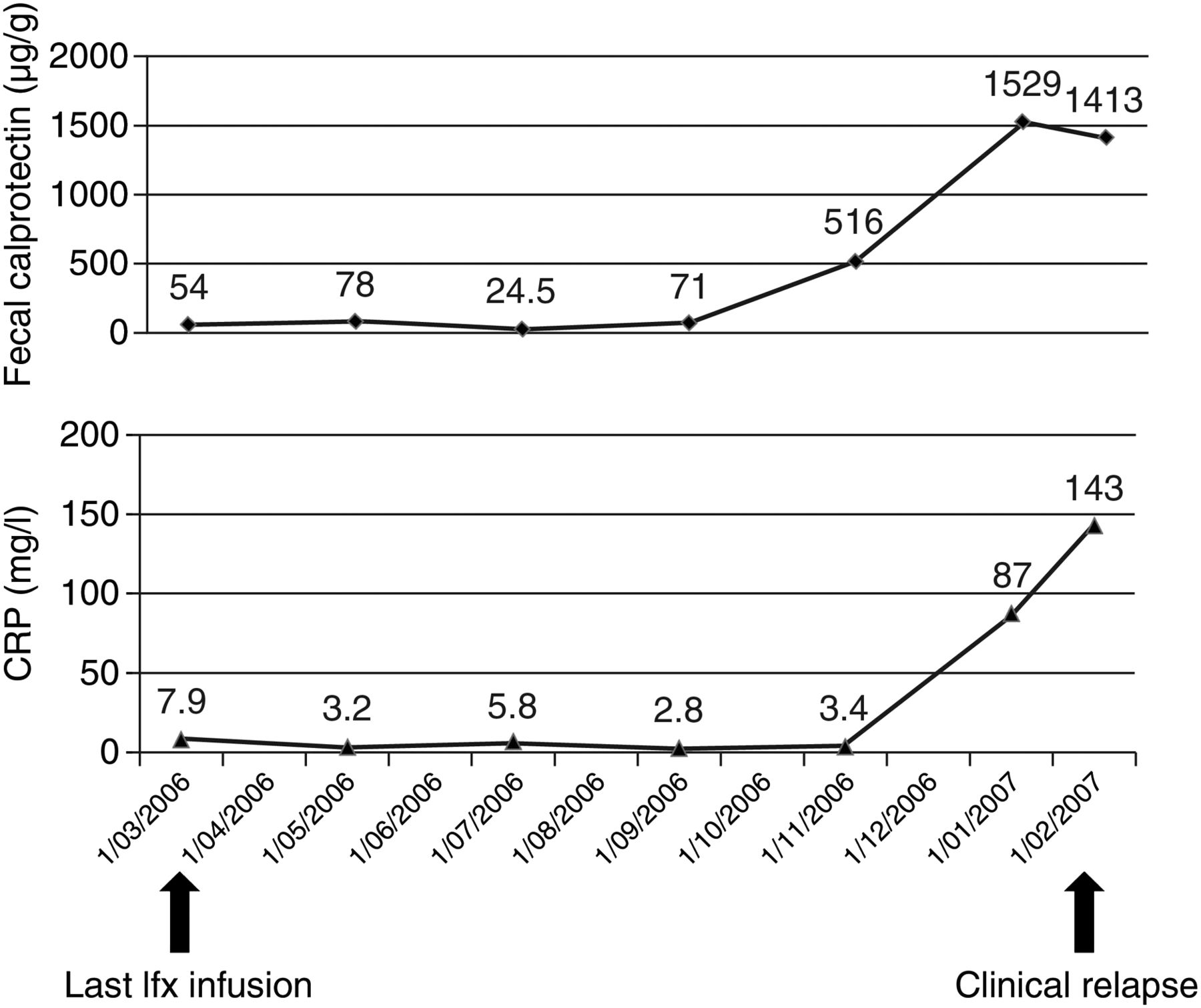
Example of monitoring a patient with Crohn's disease (CD) in clinical remission by using blood C-reactive protein (CRP) and faecal calprotectin. This patient had been treated with azathioprine/infliximab combination therapy since July 2003 for ileocolonic refractory CD. In March 2006, he had been in stable steroid-free remission for more than 1 year. Following a request from the patient, it was decided to withdraw infliximab and to continue with azathioprine monotherapy. The last infliximab (Ifx) infusion was administered on 15 March 2006. At that time, CRP was slightly increased at 7.9 mg/l, but faecal calprotectin was low at 54 μg/g. CRP and faecal calprotectin were measured every 2 months after the last infliximab infusion. While the patient was still in remission in November 2006, a significant increase in faecal calprotectin was noticed (516 μg/g), whereas CRP was still normal (3.4 mg/l). Two months later, faecal calprotectin had further increased to 1529 μg/g, and CRP had also significantly increased to 87 mg/l. Clinical relapse finally occurred 1 month later in February 2007.
Biomarkers to confirm disease activity and assess response to treatment
The importance of confirming inflammatory activity in a patient with clinical symptoms before starting or escalating medical treatment for CD has been highlighted above in the discussion of monitoring by endoscopy and cross-sectional imaging. Blood markers could be of help here to avoid repeating these invasive procedures. Another feature is rapid confirmation of drug efficacy when the treatment has been started. Here again the early repetition of endoscopy and/or cross-sectional imaging is inappropriate, and blood markers could provide important information.
Confirming disease activity before treatment
Although it does not correlate perfectly with endoscopic scores of activity, CRP represents an objective marker of active inflammation. Response to medical therapy for CD, particularly anti-TNF antibodies, has been shown to be better when an increase in CRP was present to confirm disease activity.54 In contrast, in patients with a normal CRP despite clinical activity of the disease, a substantial proportion of patients may have functional disorders or the aftermath of previous flares or surgeries. A prospective study has specifically addressed this point in patients with a CDAI >150 but a CRP <5 mg/l. A colonoscopy was systematically performed; this showed only minor lesions in the majority of the patients, but still one-third of them had a CDEIS >6, confirming clinically significant lesions.55 From this, it seems logical to advocate controlling endoscopy or medical imaging for signs of disease activity in patients with clinically active disease but a normal CRP concentration. Faecal calprotectin correlates better with endoscopic scores of disease severity, with a correlation coefficient of ∼0.70.56 Although it may represent a more efficient marker than CRP in this setting, this correlation is still imperfect. CRP and faecal calprotectin may thus be best used as first-line tools to decide whether or not there is a need for endoscopic or cross-sectional imaging reassessment. In this perspective, an interesting meta-study was recently performed, analysing the data of six studies that included more than 550 patients and provided blood CRP and faecal calprotectin levels together with endoscopic scores of severity.57 In patients with symptoms (CDAI >220), the sensitivity of CRP ≤5 mg/l or calprotectin ≤200 μg/g to anticipate a CDEIS ≤6 was 83% and the specificity 71%. The PPV ranged from 66% to 81% and NPV from 86% to 73% for a prevalence of CDEIS ≤6 between 40% and 60%. It thus means that, out of 100 patients with clinically active disease, if endoscopy was only performed when one of the markers was below the threshold, 38–50 colonoscopies could be avoided. Those with both markers above the threshold would be considered to have active lesions. However, 7–10 of them would have a CDEIS <6 and may thus be overtreated.
Confirming response to treatment
A decrease in CRP has been clearly demonstrated in patients clinically responding to medical treatment. The same has been shown for faecal calprotectin.58 More recently, it was shown that a persisting increase in CRP under anti-TNF therapy was associated with future loss of response to the drug.59 This suggests that biomarkers such as CRP and faecal calprotectin could be used to confirm the response to therapy and that an adequate response should be accompanied by normalisation of CRP and a dramatic decrease in faecal calprotectin. Normalisation of calprotectin (<50 μg/g) is certainly more difficult to achieve than normalisation of CRP, and may not be considered as a therapeutic target. Nevertheless, it may represent a state of deeper remission, probably associated with more profound mucosal healing.21
Biomarkers to confirm tissue healing and predict disease relapse
Correlation coefficients between faecal calprotectin and endoscopic scores of disease activity ranged from 0.42 to 0.73, and the sensitivity and specificity to predict absence of mucosal healing were 70–100% and 44–100%, respectively, depending on the calprotectin concentration threshold used.56 ,60–62 In the largest study so far, a faecal calprotectin concentration >250 μg/g predicted large ulcers with a sensitivity of 60% and a specificity of 80%, and a concentration <250 μg/g predicted mucosal healing (CDEIS <3) with a sensitivity of 94% and specificity of 62%.63 The weaknesses of faecal calprotectin as a biomarker may be a weaker correlation with endoscopic activity in the small bowel,60 the imperfect reflection of the transmural inflammatory process, and finally the unpleasant requirement for the patient to bring a stool sample to the laboratory. While no other marker has been specifically studied for the assessment of small-bowel mucosal inflammation, the transmural process may be better reflected by CRP. Indeed, in a retrospective study assessing correlation between CRP concentration and various semiological features at CT cross-sectional imaging, CRP correlated more closely with transmural and mesenteric signs of inflammation than with purely mucosal signs of inflammation.64 In some studies, the correlation between serum inflammatory markers, including CRP, and endoscopic activity of the disease was close to that of faecal calprotectin, with a correlation coefficient of ∼0.70.65 For the assessment of mucosal healing, a recent subanalysis of the STORI cohort suggested that the combination of faecal calprotectin (at a threshold of 250 μg/g) and CRP (at a threshold of 5 mg/l) may improve the ability to predict such healing by significantly increasing specificity above 70% while sensitivity remained reasonably good, also above 70%.66 As for the prediction of endoscopically active disease, the prediction of mucosal healing with biomarkers is imperfect, with an inaccuracy of 30–40%. It is thus again as first-line tests and in combination with endoscopy and/or cross-sectional imaging that these biomarkers may be best used. The previously mentioned meta-study also addressed this question.57 When patients with inactive disease (CDAI ≤150) are considered, the sensitivity of the association of CRP ≤10 mg/l and calprotectin ≤200 μg/g to predict a CDEIS ≤3 was 78% and the specificity 58%. The PPV ranged from 65% to 88% and NPV from 73% to 40% for a prevalence of CDEIS ≤3 between 50% and 80%. This means that, out of 100 patients in clinical remission, if endoscopy was only performed when both markers were below the thresholds, 30–40 colonoscopies could be avoided. Patients with CRP or calprotectin greater than the threshold would be considered to have active lesions. Of these 60–70 patients, only 11–18 would have a CDEIS ≤3 and would thus risk overtreatment.
CRP and faecal calprotectin are also predictors of CD relapse. Globally, the sensitivity and specificity of faecal calprotectin to predict CD clinical relapse was 43–90% and 43–88%, respectively, again depending on the threshold used.67–69 In various studies, CRP was, overall, a less powerful predictor, although increased CRP was associated with a significant increase in relapse risk, the relative risk ranging between 3 and 58.70–72 The frequency with which these biomarkers should be measured in the follow-up of patients who have achieved clinical remission is not well established. Most studies measured these markers only once and then assessed the time to relapse or the relapse rate over 1 year. However, preliminary data from the GETAID STORI cohort indicate that both CRP and faecal calprotectin start to increase 4–6 months before the relapse, and thus measurement of these markers every 3–4 months should be able to catch this increase and enable the clinician to adapt the therapeutic strategy.73
As emphasised above, in patients with longstanding stable steroid-free remission, treatment de-escalation may be contemplated to try to optimise benefit/risk and benefit/cost ratio. The data from the STORI cohort indicate that biomarkers, particularly CRP and faecal calprotectin, may be complementary to endoscopic assessment in predicting the risk of relapse upon infliximab withdrawal.21 Increased CRP has already been associated with increased risk of relapse after azathioprine withdrawal.74
Biomarkers have been little studied in the postoperative setting to try to predict disease recurrence. Preliminary data indicate that CRP is probably not sensitive enough to be useful in this clinical setting.75 A postoperative follow-up study clearly showed the absence of correlation between endoscopic recurrence and CRP. Inflammation at this stage may be confined to the mucosa in many cases and badly translate into systemic inflammation. Faecal calprotectin seems to be much more promising. A surgical series showed normalisation of faecal calprotectin in all patients after uncomplicated curative surgery within 2 month of the resection.76 This marker remained low in patients without relapse, while it increased significantly in patients experiencing clinical relapse. However, the precise timing of this increase was not specifically assessed in this study and thus the predictive ability of faecal calprotectin in that setting cannot be determined.
Conclusion
Endoscopic techniques, cross-sectional imaging and biomarkers represent a range of potential monitoring tools for CD. They all have theoretical advantages and drawbacks in helping clinicians to optimise treatment strategies. However, none of these tools has really been prospectively validated in a study aimed at improving CD outcome. Therefore their use in the monitoring of patients with CD in routine practice remains empirical. A proposed algorithm of CD monitoring based on the limited available evidence and our experience is shown in figure 4. The standard of care in CD is still to manage patients according to disease evolution based on clinical symptoms. However, on a case by case basis, monitoring can be used to optimise therapeutic strategy, aiming at decreasing cumulative tissue damage and its consequences. This could be particularly adapted in situations where there is concern about disease progression, because of disease location and extent, or history of the patients. In this case, the use of endoscopy, cross-sectional imaging and biomarkers should be proposed, aiming to answer specific questions leading to treatment adaptation. Biomarkers such as blood CRP and faecal calprotectin concentration often represent informative first-line tests to try to predict disease activity, tissue healing or the risk of relapse. By choosing the thresholds of these markers appropriately to optimise sensitivity or specificity, up to half of endoscopic or cross-sectional imaging procedures could be avoided. New biomarkers still in development, as well as blood drug levels, including trough levels of biological agents, may also improve the efficacy of monitoring in the future. Prospective studies to confirm the effect of monitoring-based therapeutic changes on key outcomes of CD are urgently required.

Proposed algorithm for Crohn's disease (CD) monitoring based on the limited available evidence (see also tables 4 and 5) and our experience. It illustrates how CD monitoring can affect disease management. In the presence of only two clinical situations (clinically active or inactive disease), CD monitoring can reveal five very different situations: controlled disease, symptomatic but controlled disease, controlled disease but with increased risk of relapse, silent uncontrolled disease, uncontrolled disease. These five different situations will lead to different treatment strategies and further monitoring. A clear definition and criteria for significant and non-significant endoscopic or cross-sectional imaging lesions as well as of controlled and uncontrolled disease are lacking. Minor persisting endoscopic or cross-sectional imaging lesions and slightly raised faecal calprotectin (calpro) may still be compatible with controlled disease. This will be left to the clinician's discretion, after also taking into account the patient's history (cumulative tissue damage, response to treatment). CRP, C-reactive protein.
Summary Box 1
-
Monitoring of Crohn's disease with endoscopy, cross-sectional imaging and biomarkers aims to achieve tighter disease control to try to prevent tissue damage, with an optimal benefit/risk and benefit/cost ratio.
-
Mucosal healing assessed by ileocolonoscopy is associated with improved disease outcome including fewer relapses, hospitalisations and operations.
-
The degree of mucosal healing required to improve outcome is not completely established.
-
Cross-sectional imaging provides information on the small bowel and the transmural and extramural features of the inflammatory process.
-
MRI score of activity correlates well with endoscopic score of activity.
-
Faecal calprotectin and blood C-reactive protein, can predict mucosal healing and relapse of the disease with an accuracy of 60–70%.
Summary Box 2
-
No prospective or even retrospective study has validated a treatment strategy based on disease monitoring aimed at improving disease outcome.
-
Crohn's disease monitoring with endoscopy, cross-sectional imaging and biomarkers can only be empirical and should be decided on a case by case basis.
-
Patients at high risk of disease progression or having a rapidly aggressive disease are the best candidates for empirical disease monitoring.
-
Preference should be given to non-invasive and cheap first-line tests such as biomarkers.
-
In asymptomatic patients, the decision whether to perform endoscopic or cross-sectional imaging monitoring could be based on disease history and results of the biomarkers.
-
Treatment should be empirically adapted according to monitoring results to optimise benefit/cost and benefit/risk for the patient.
References
Footnotes
-
Contributors J-MB and EL wrote the first draft of the manuscript. M-AM reviewed and amended the part on biomarkers. CR and CVK reviewed and amended the part on endoscopy. PM reviewed and amended the part on cross-sectional imaging. All authors approved the final manuscript.
-
Competing interests None.
-
Provenance and peer review Commissioned; externally peer reviewed.