Article Text

Abstract
Background About 10% of patients with IBS report the start of the syndrome after infectious enteritis. The clinical features of postinfectious IBS (PI-IBS) resemble those of diarrhoea-predominant IBS (IBS-D). While altered faecal microbiota has been identified in other IBS subtypes, composition of the microbiota in patients with PI-IBS remains uncharacterised.
Objective To characterise the microbial composition of patients with PI-IBS, and to examine the associations between the faecal microbiota and a patient's clinical features.
Design Using a phylogenetic microarray and selected qPCR assays, we analysed differences in the faecal microbiota of 57 subjects from five study groups: patients with diagnosed PI-IBS, patients who 6 months after gastroenteritis had either persisting bowel dysfunction or no IBS symptoms, benchmarked against patients with IBS-D and healthy controls. In addition, the associations between the faecal microbiota and health were investigated by correlating the microbial profiles to immunological markers, quality of life indicators and host gene expression in rectal biopsies.
Results Microbiota analysis revealed a bacterial profile of 27 genus-like groups, providing an Index of Microbial Dysbiosis (IMD), which significantly separated patient groups and controls. Within this profile, several members of Bacteroidetes phylum were increased 12-fold in patients, while healthy controls had 35-fold more uncultured Clostridia. We showed correlations between the IMD and expression of several host gene pathways, including amino acid synthesis, cell junction integrity and inflammatory response, suggesting an impaired epithelial barrier function in IBS.
Conclusions The faecal microbiota of patients with PI-IBS differs from that of healthy controls and resembles that of patients with IBS-D, suggesting a common pathophysiology. Moreover, our analysis suggests a variety of host–microbe associations that may underlie intestinal symptoms, initiated by gastroenteritis.
Statistics from Altmetric.com
Significance of this study
What is already known about this subject?
-
Infectious gastroenteritis leads to new IBS symptoms in 3–36% of patients.
-
The faecal microbiota of patients with postinfectious IBS (PI-IBS) has so far not been characterised, whereas several microbial changes have been linked to other subtypes of IBS.
What are the new findings?
-
Twenty-seven discriminant bacterial groups separated the faecal microbiota profile of postinfectious patients from that of healthy controls.
-
We could create an Index of Microbial Dysbiosis, which correlated with pain quality and multiple somatic symptoms but not anxiety or depression.
-
Significant associations between the host mucosal gene expression and histology and faecal microbiota were recognised, indicative of an increased inflammatory response and compromised intestinal barrier in PI-IBS.
How might it impact on clinical practice in the foreseeable future?
-
Profiling of the faecal microbiota represents one of the first objective measures for IBS, and allows further subclassification of this heterogeneous patient group into those with a predominantly peripheral gut abnormality and those with a predominantly central nervous system basis for the syndrome.
-
We identified several host–microbe associations, implying that a compromised intestinal barrier may underlie both immunological and microbiological deviations in PI-IBS, providing hypotheses for further mechanistic research.
Introduction
IBS affects about 10% of the population.1 The patient group is heterogeneous but subtyping according to the predominant bowel habit (diarrhoea, constipation or mixed) is clinically useful. There is no single unifying cause for IBS; prospective studies have indicated several factors that increase the risk of subsequently developing IBS.2 Genetic factors play a role,3 as does prior anxiety and depression,4 but the strongest predictor is a gastroenteritis episode in the previous year.5 Patients with postinfectious IBS (PI-IBS) show increased mucosal lymphocytes and increased intestinal permeability, which has also been noted in IBS of non-infectious origin.6 Several studies have reported deregulation of tight junction formation.7 This may be important in IBS symptoms, as severity of pain has been correlated with gut permeability.8
Acute gastroenteritis is characterised by inflammation, which markedly depletes the colonic microbiota. The success of a pathogen relies on its ability to overcome the colonisation resistance provided by the commensal microbiota—for example, by activating the host inflammatory response.9 Depletion of the commensals with antibiotics has been shown to impair colonic salvage of non-absorbable dietary carbohydrate.10 This may lead to increased stool weight and frequency, all characteristic features of PI-IBS.11 A marked shift in microbiota following acute gastroenteritis could lead to prolonged changes in the host–microbiota interactions. These, so far uncharacterised microbiota changes which could be long lasting might be responsible for the prolonged symptoms of PI-IBS, long after the infectious insult has gone.12
Several studies have investigated the faecal microbiota of patients with IBS.13 Although many have reported differing microbial composition between cases and controls, there is little consensus on the bacteria that differentiate these groups. Most of these studies have examined patients from several IBS subgroups, and the study designs and methodologies have varied between the cohorts making them hard to compare. There is evidence, however, that the faecal microbiota of patients with IBS could be used to stratify them according to whether the predominant abnormality lies in the gut or the central nervous system.14
The goal of this study was to characterise for the first time the microbial composition of patients with PI-IBS, and to examine the associations between the faecal microbiota and a patient's clinical features. As the development of IBS is multifactorial, we hypothesised that the observed symptoms and development of PI-IBS arise from the interplay between the faecal microbiota, host immune response and psychological factors. Hence we performed a detailed characterisation of the faecal microbiota of patients in a different degree of recovery from gastroenteritis and compared the results with those for healthy controls (HC) and patients with diarrhoea-predominant IBS (IBS-D). We correlated GI microbiota composition with measures of host mucosal gene expression, peripheral blood cytokine production and psychological and gastroenterological symptoms in order to find host–microbe associations underlying the development of IBS. This represents a new approach to examining the associations between clinical phenotype, host gene expression and faecal microbiota, and is a step forward in understanding the complex relationships between the host and microbiota in the pathogenesis of IBS and PI-IBS.
Methods
Participants
The participants were a subset of a study previously reported in Gut describing candidate genes related to IBS after a Campylobacter infection.15 The present cohort included 57 subjects (for the inclusion and exclusion criteria see online supplementary material), 11 HC and 46 patients from four groups; subjects who 6 months after gastroenteritis either experienced no bowel dysfunction (PI-nonBD, n=12) or had recurrent bowel dysfunction (PI-BD, n=11), patients with IBS reporting the start of the symptoms after gastroenteritis from 6 months up to 3 years previously (PI-IBS, n=11), and patients with IBS-D (n=12). Both IBS groups fulfilled the Rome II criteria. All postinfectious patients had a prior positive stool culture for Campylobacter jejuni in the Nottingham Public Health laboratory. When applicable, the participants were age and gender matched from the original cohort, although the patients with PI-nonBD were significantly older since older age is protective against acquiring PI-BD.16
Experimental design
Factors related to IBS and gastroenteritis were recorded from three different sources. First, all subjects donated a faecal sample for faecal microbiota composition analysis. Second, objective measures of participants’ intestinal health were collected, including colon permeability assay, rectal mucosal enterochromaffin cells, goblet cells, eosinophils and mast cells. Host gene expression in rectal biopsies and peripheral blood mononuclear cell (PBMC) cytokine production was also studied from a subset of participants (for technical details see online supplementary material). Third, the quality of abdominal pain and subjective measures of health were assessed from the participants using the McGill pain,16 Hospital Anxiety and Depression Scale17 and Patient Health Questionnaire 12 Somatic Symptom Scale (PHQ-12 SS)18 questionnaires. Additionally, the bowel movement frequency, consistency, urgency and mucus in stools were recorded using the validated Talley Bowel Symptom questionnaire.19 We did not attempt to assess or control the participants’ diet. All patients gave written informed consent and the study was approved by the Nottingham research ethics committee (reference Q1030308).
Host gene expression in rectal epithelium
We analysed the host gene expression levels in rectal biopsy samples from 29 subjects (10 healthy, 8 PI-BD, 5 PI-nonBD, 5 PI-IBS, 1 IBS-D) with Affymetrix Human Genome U133 Plus 2.0 Array (for technical details see online supplementary material). The data were normalised with the Robust Multichip Average method20 using custom probe set mappings, V.15.21 The expression levels of 14 potential IBS biomarker genes were measured with TaqMan real-time reverse transcription PCR (RT-PCR) in the rectal biopsy specimen as described before.15 These included Calpain 8 (CAPN8), major histocompatibility complex class II DQA1 (HLA-DQA1), interleukin 1β (IL-1B), interleukin 6 (IL-6), thyrotrophic embryonic factor and tumour necrosis factor (TNF). The full list of genes and primer sequences is shown in the online supplementary material.
Compositional analysis of the faecal microbiota
Stool samples were delivered to the hospital within 2 h for immediate freezing at −70°C. The faecal microbial DNA was extracted with repeated bead-beating method22 and analysed by using a previously described and benchmarked,23–,25 custom-made, phylogenetic microarray, the Human Intestinal Tract Chip (HITChip). The raw signal intensity was normalised as described previously,24 followed by quantile normalisation and background signal subtraction. The probe signals were summarised into 130 genus-like taxonomic groups, referred to with the type species’ name and relatives (hereafter “et rel.”).23
In addition to the phylogenetic microarray, specific bacterial groups were analysed using quantitative PCR (qPCR) with previously described primers and analysis conditions; Archaea26 and Bacteroides–Prevotella–Porphyromonas,27 a species related to Ruminococcus torques28 and species related to Collinsella aerofaciens.28
Statistical analysis
All analyses were carried out with logarithm-transformed data using R, V.2.15.1.29 For the microbial abundance, questionnaire and clinical data the between-group comparisons were analysed using analysis of variance (ANOVA) with Tukey's honest significant differences post hoc analysis. All correlations between two datasets were analysed using Spearman's coefficient (ρ) with pairwise comparisons, with the exception of correlations between RT-PCR and host microbiota abundances, which were calculated using Pearson's coefficient (r). All subsequent p values were adjusted for multiple comparisons with Benjamini–Hochberg false discovery rate correction and values <0.05 were considered significant. The microbial profile separating the study groups was identified with redundancy analysis using bootstrap aggregation (baggedRDA) with 5000 bootstrap datasets to increase the robustness of the estimates. Significance and generalisation error for holdout data was estimated with the bootstrap .632+ method.30 Separation between the patient groups was additionally evaluated with UniFrac distance.31 Significant associations between the microbiota and host gene expression were analysed with gene set analysis (GSA),32 ,33 and visualised with an eye diagram.34 For technical details of the statistical analysis see online supplementary material.
Results
The study cohort contained 57 subjects from five groups including patients with a history of past gastroenteritis and varying degree of recorded symptoms; patients with PI-IBS fulfilling the Rome II criteria, patients with persistent bowel dysfunction after gastroenteritis (PI-BD) and patients whose bowel habit returned to normal (PI-nonBD) (table 1). We aimed to examine the effects of past gastroenteritis and the occurrence of IBS or IBS-like symptoms in relation to the faecal microbiota composition. The findings were compared with those for patients with IBS-D and the HC.
Characteristics of the study groups
Compositional analysis of the faecal microbiota
We examined the microbial differences in the postinfectious patient groups by conducting a comprehensive and deep microbiota analysis with a phylogenetic microarray. Based on multivariate analysis using bootstrap aggregated redundancy analysis (RDA), we identified a microbial profile, instead of individual taxa, that separated the patient groups and HC (the first RDA axis, explained 13% of the total variation in the microbiota; figure 1A). This profile consisted of 27 genus-like bacterial taxa (table 2), and separated the HC from the patients with IBS or IBS-like symptoms (IBS-D, PI-IBS, PI-BD; p<0.05, table 1). The PI-nonBD group did not differ from any of the study groups (figure 1A). However, a separate RDA showed significant difference (p<0.05) between the subjects who considered themselves healthy (HC and PI-nonBD) and those having IBS or IBS-like symptoms (figure 1B,C), indicating a gradual change in the microbiota from healthy to PI-IBS. The microbial changes in the postinfectious groups resembled those detected in patients with IBS-D. When all participants were indexed according to the abundance of bacterial taxa in the discriminant microbial profile, a specific ordering of the study subjects was obtained, reflecting their health status (figure 1D).
Panel of 27 discriminant bacterial genera that jointly separated the patients from HC

Bootstrap aggregated redundancy analysis (RDA) of the microbiota composition in all patient groups and controls. (A) A set of bacteria significantly contributing to separation between the healthy controls (HC) and the patient groups. (B) Separation among the patients with a history of gastroenteritis but not diagnosed with IBS. (C) Separation between the patients with IBS and HC. (D) Patients were transposed on the first axis of the RDA, which explains the majority of the microbial separation, creating an index of microbial dysbiosis. *Subjects who underwent rectal gene expression analysis. IBS-D, diarrhoea-predominant IBS; PI-BD, persistent bowel dysfunction; PI-IBS, postinfectious IBS; PI-non-BD, no bowel dysfunction.
Of the 27 discriminant taxa in the microbial profile, 15 significantly separated the patient groups and HC also when analysed separately with linear models (table 2). The patient groups alone showed no significant differences in the microbial abundances when measured with linear models or RDA using UniFrac distance. The major difference in the IBS-type microbiota was the increase of several members of the Bacteroidetes (6.0 fold, SD 1.14) and decrease of uncultured Clostridiales (16.8-fold, SD 16.52) compared with HC. We confirmed the increased amount of Bacteroidetes with two independent approaches. First, the abundance of the Bacteroides-Prevotella-Porphyromonas group, measured with qPCR, was increased in the patients (3.2-fold, SD 0.8, p<0.05). Second, to rule out the possibility that the observed differences were due to large interindividual differences, characteristic for this bacterial group, we tested the cohort against additional healthy individuals. The analysed 12 Finnish subjects (details in Jalanka-Tuovinen et al24) also possessed a significantly lower amount of Bacteroidetes spp than the patient groups from this cohort (p<0.05, see online supplementary figure S1).
We complemented the community-wide microbial analysis with qPCR. Our focus was on the presence and abundance of microbial groups previously associated with IBS, including methanogenic Archaea, and uncultured phylotypes related to C. aerofaciens and R. torques.28 Methanogens were less prevalent with IBS or IBS-type patients (PI-IBS 17%, IBS-D 8%, PI-BD 9%) than in healthy subjects (HC 33% and PI-nonBD 25%, p<0.05) and their abundance inversely correlated with the frequency of loose stools (ρ=−0.4, p<0.05). Bacteria related to the R. torques-like phylotype were more prevalent in the patients with IBS than HC (85% in PI-IBS, 67% in IBS-D and 42% in HC, p<0.05) and this was confirmed by the phylogenetic microarray data (data not shown). Moreover, their abundance was associated with increased pain sensation, increased amount of mast cells and expression of IL-6 and TNF genes (see online supplementary table S1). The bacteria related to the uncultured C. aerofaciens-like phylotype, previously shown to be decreased in patients with IBS,28 correlated negatively with inflammatory cytokines (IL-1B, IL-6, IL-8, TNFα and IL-10) produced from unstimulated PBMCs (see online supplementary table S1).
Associations between the faecal microbiota and host gene expression
Previous studies have shown that the faecal microbiota composition can be used to stratify patients with IBS further from the current subtyping categories.14 To test if this was applicable also for this cohort, we correlated the first axis of RDA creating a profile from HC to IBS-type microbiota (RDA1, see figure 1D; for a detailed description of the method see online supplementary material) with diagnostic variables (figure 2A). We found positive correlations with the IBS-type microbiota and intestinal complaints including pain (ρ=0.34), bowel movement frequency (ρ=0.37), loose stools (ρ=0.35) and multiple somatic symptoms (PHQ-12 SS, ρ=0.43; all p<0.05). However, correlations with anxiety and depression characteristic of IBS were smaller and not significant, (ρ=0.25 and ρ=0.22, respectively, p>0.05). Altogether, these results indicate that the discriminant microbial profile was more closely associated with bowel rather than psychological symptoms of the disease phenotype, and may be a useful objective measure of disturbed bowel function in IBS. Hence, we refer to the profile as an Index of Microbial Dysbiosis (IMD).

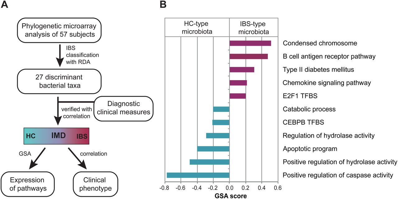
(A) Definition and implementation of the Index of Microbial Dysbiosis (IMD). The flow chart shows the generation of the IMD, an objective measure of IBS and its relation to host gene expression and clinical phenotype. (B) Associations between IMD and host expression pathways, expressed in terms of gene set analysis (GSA) scores. Pathways correlating with IBS-type microbiota are indicated with purple and pathways correlating with healthy control (HC)-like microbiota are indicated with blue; RDA, redundancy analysis; TFBS, transcription factor binding site.
To further study the IMD, we investigated its associations with the patients’ clinical phenotype, including the expression of selected marker genes, immunological and histological variables. There was a positive correlation between the IBS-type microbiota and inflammatory markers, including eotaxin (CLL11) and the number of mast cells (ρ=0.38 and ρ=0.39 respectively, p<0.05). In addition, the amount of goblet cells (ρ=0.35, p<0.05) and the expression of serotonin transporter (SCL6A4) were associated with the IBS-type microbiota (ρ=0.45, p<0.05). On the other hand, the IMD correlated negatively with the number of enterochromaffin cells (ρ=0.55, p<0.05) and the expression levels of an orphan G-coupled receptor (GPR161) (ρ=−0.31, p<0.05).
We then analysed the potential associations of the discriminant microbiota on host gene expression by mapping the expression measurements from rectal biopsies to known biological functions using GSA.32 Pathway analysis seeks for coordinated changes in sets of functionally related genes, allowing to step up from the expression levels of single genes into detecting activated/silenced cellular functions. GSA, unlike many other pathway analysis methods, can use continuous data such as the bacterial abundances or the IMD value. The method determines a GSA score, which reflects the correlation between the expression of genes in a given pathway and the IMD or bacterial abundance. The IMD was associated with altered expression of 11 cellular pathways (figure 2B). The IBS-type microbiota was positively correlated with increased expression of chemokine production and B cell antigen receptor signalling. Moreover, several pathways for apoptosis were negatively associated with the IBS-like microbiota.
Association of discriminative microbial taxa with host gene expression patterns
To further investigate the linkages between the microbiota and host gene expression GSA was conducted separately for each of the 27 taxa of the IMD, examining the postinfectious host–microbe cross-talk network. The abundances of the discriminant taxa were significantly associated with 279 differentially expressed pathways (see online supplementary table S2). To avoid false-positive results, we concentrated our analysis on the 40 pathways associated with at least two bacterial taxa, and a previously described function (figure 3, see online supplementary table S3). The largest group of concordant correlations, comprising a total of eight genus-like groups, involved expressional changes in the metabolism of glycine, serine and threonine. These amino acids are important for gut integrity, as serine and threonine are the backbone amino acids of mucin.35 Seven out of the eight taxa were Bacteroides spp (see online supplementary figure S2) and the only genus positively associated with this pathway was HC-type related Collinsella spp. In addition, GSA identified seven other pathways linked to amino acid metabolism to which the IBS-type microbiota was negatively, and HC-type microbiota positively, correlated (figure 3, see online supplementary table S3).

Host–microbiota associations. Differences in the expression levels of host gene expression pathways were associated with the IBS-type and healthy controls (HC)-type microbiota using the gene set analysis. The genera increased in the patients with IBS and the positive/negative associations with the host pathways are indicated with IBS+ and IBS−, respectively. The genera increased in the HC and their positive and negative associations with host pathways are indicated with Healthy+ and Healthy−, respectively. The width of the line indicates the number of genera with significant associations to the given pathway. NER, nucleotide excision repair; TFBS, transcription factor binding site.
The abundance of IBS-related bacteria was also associated with pathways regulating cell junctions (see online supplementary figure S3). The HC-type microbiota was positively correlated with increased expression levels of basolateral membrane and cell junction pathways. Bacteroides spp correlated negatively with the expression of apical cell junction complex and cell junction gene pathways. In addition, IBS-related bacteria associated positively with the abundance of genes known to be activated with a transcription factor tyrosine kinase oncogene (ELK1). These genes include the myosin light chain kinase, which promotes opening of tight junctions.36
Finally, to link the microbiota to host responses previously associated with IBS, we correlated all bacterial abundances with the RT-PCR data and with other clinical measurements. Seven of these genes and two of the clinical variables significantly correlated with microbial abundances (table 3).
Statistically significant correlations between faecal microbiota abundance and host mRNA expression (for gene names, see ‘Methods’)
Bacteria related to Lactobacillus gasseri negatively correlated (r=−0.52, p<0.05) with measured total cytokine production from PBMCs. The expression of IL-6 correlated positively with several Proteobacteria and Bacilli, and negatively with bacteria related to Prevotella melaninogenica and Butyrovibrio crossotus. A single group of Proteobacteria related to Enterobacter aerogenes also correlated positively with the expression of HLA-DQA1 as well as measures of increased colon permeability (r=0.566, p<0.05).
Discussion
Postinfective IBS is known to be associated with faster colonic transit,37 mucosal immune activation7 and now, as our study shows, disturbances in the faecal microbiota. PI-IBS and IBS-D have many similar clinical features, and our data suggest similarities also in the faecal microbiota. The microbiota analysis revealed a gradient of abnormality that coincides with the disease phenotype, ranging from diagnosed IBS, to HC through patients with past gastroenteritis experiencing recurrent bowel dysfunction to non-symptomatic subjects (figure 1D). The patients with IBS-like symptoms (PI-BD, PI-IBS and IBS-D) had similar microbial profiles. This IMD correlated with the severity of symptoms, pain, increased stool frequency and looseness, features associated with acceleration of gut transit. Additionally, we found that immunomarkers, including mast cells, were associated with the IMD value, but not when the patient groups were analysed separately. However, we did not find any significant correlations between IMD and psychological symptoms. This is in line with previous findings where IBS-type microbiota was associated with intestinal symptoms, and patients with IBS with a microbiota resembling that of the HC had significantly more psychological symptoms.14 These data suggest that the differences in the microbiota, as shown by the IMD, could be used as a method to identify a more homogeneous subgroup of patients with IBS and stratify those with a predominantly gut, rather than central nervous system, basis for the syndrome.
The discriminant bacteria between the patients and HC consisted of 27 genus-like phylogenetic groups. Though some of these taxa did not individually show significant differences between the disease groups, their joint abundances constituted a bacterial signature that was characteristic for all postinfective patients and patients with IBS. The most prominent change was the increased amount of Bacteroides spp in the patients together with the decrease of uncharacterised and uncultured Clostridia. It is plausible to hypothesise that the IBS-related bacteria might harbour some common metabolic features or cooperate in a trophic chain. Unfortunately, little is known about the interactions among these particular intestinal commensals, and the potential mechanisms of the diagnostic profile cannot yet be defined. Hence, our interpretations are based on the findings of the implicated bacteria individually.
Alterations in the microbiota may arise from several sources, including differences in the host physiology or immunology. Rapid intestinal transit is a typical feature of patients with IBS with diarrhoea (PI-IBS and IBS-D) and it is known to modify the colonic milieu and presumably the microbiota. We observed that Bacteroides spp were over-represented, and methanogens under-represented, in patient groups with IBS symptoms. A recent study in mice showed that chemically accelerated transit increased the amount of Bacteroides spp,38 while carriage of methanogenic Archaea is associated with delayed transit,39 indicating that an altered transit rate itself shapes the microbiota. It is not yet known whether the microbiota change is causing IBS or secondary to IBS pathophysiology.
The role of Bacteroides spp in IBS aetiology or pathophysiology is unclear. Previous studies have reported opposing results for this genus; an increased amount of Bacteroides spp has been reported in patients with IBS28 ,40 ,41 and in related functional bowel disorders,42 but also in HC.14 ,43 ,44 The discrepancy in the microbiota findings may suggest that rather than a single pathobiont, a microbial dysbiosis is important in the pathophysiology of IBS. However, these differences may also rise from heterogeneous patient materials, high individuality of the faecal microbiota, and therefore inadequate sample sizes, or different methodological approaches. Here, we attempted to minimise the technical variation and avoid false-positive correlations by concentrating on a more homogeneous IBS group with a known disease aetiology, and by applying analysis methods such as bootstrapping, which give robust estimates even with small sample sizes. In addition, we used p value adjustments for multiple comparisons and concentrated on host effects that were significant across several bacterial taxa.
One classic symptom in IBS is abdominal pain. Several intestinal bacteria have been linked to increased sensation of pain.14 ,24 ,45 Our study verified that an IBS-related, uncultured R. torques-like phylotype28 was more prevalent in the patients with IBS of this cohort and was associated with increased pain sensation and immune response. On the other hand, the health-associated uncultured relative of C. aerofaciens negatively correlated with PBMC-produced proinflammatory cytokines. Interestingly, the genus Collinsella was also among the 27 discriminant taxa and associated with several gene expression pathways. This highlights the importance of further characterisation of the implicated uncultured phylotypes as possible biomarkers for IBS and PI-IBS.
In addition to the microbial signature characteristic for subjects with past gastroenteritis, our study provides unique insights into associations between the faecal microbiota and host gene expression. We showed that the IMD correlated with increased inflammation, particularly PBMC IL-6 secretion, and with a decreased level of apoptosis. Changes in apoptosis may be important in the immune activation documented in IBS-D.46 ,47 Crohn's disease is associated with proinflammatory cytokine secretion, which inhibits apoptosis leading to increased mucosal lymphocytes48; a similar process may be activated in patients with a history of gastroenteritis.
When focusing on a single bacterial taxon, we found many independent measures linking the abundance of the IBS-type microbiota to changes in host expression. These included several interesting associations between the discriminant microbiota and the physical barrier integrity of the host. The main function of the physical barrier is a spatial separation of the luminal content from the epithelial layer. The physical barrier includes a viscous mucus layer and junctions between the epithelial cells.35 Several IBS-related microbes were associated with decreased expression of amino acid metabolism pathways, the strongest correlation being between the glycine, serine and threonine metabolic pathway and several Bacteroides and Prevotella spp. Most dietary threonine intake is used for synthesising the secretory mucin. Dietary restriction of threonine has been shown to reduce mucin synthesis, resulting in impaired gut barrier function.49
In this study, patients with PI-IBS and IBS-D reported more frequent passage of mucus per rectum and subjects with IBS-type microbiota had an increased amount of goblet cells in rectal biopsy samples. In rodents Bacteroides thetaiotamicron (part of the Bacteroides fragilis group on the phylogenetic microarray) increases the number of goblet cells.50 This supports the positive association between the level of goblet cells and Bacteroides spp seen here, although so far this ability has been characterised for this single Bacteroides species only. On the other hand, the increase of goblet cells may also reflect a potential compensatory mechanism for aberrant mucin assembly. Passage of mucus per rectum is a common symptom of patients with IBS,51 and in the light of our findings, may reflect active production of structurally loose mucus. Although there are no data for the role of mucin in the pathophysiology of IBS in humans, our findings are in line with rodent models, where IBS-like symptoms were associated with increased faecal transit and addition of goblet cells leading to reduced thickness of the mucus layer.52 Hence, the gain in Bacteroides spp may be a consequence of accelerated transit and altered mucus production.
Increased gut permeability has been linked to IBS and directly demonstrated in colonic biopsies of patients with IBS-D.7 Our data indicate that the IBS-type microbiota is associated with impaired cell junctions, shown by reduced expression of four pathways related to cell junction integrity. Hence, we hypothesise that integrity of the intestinal epithelial barrier is compromised by an impaired mucus layer and cell junction integrity in subjects with IBS-type microbiota. As a result, it is understandable that the colonic mucosa of patients with IBS is often, as also found in this study, associated with low-grade inflammation. The IMD values correlated with increased cytokine production, and the abundances of some IBS-related bacterial groups were linked with expression of inflammatory pathways. The correlations observed with PBMC IL-6 secretion, IL-1β mucosal mRNA and the IBS-associated microbiota support the suggestion that mucosal immune activation is modulated by the microbiota in PI-IBS. Interestingly, we found that these same immune markers correlated positively with an increased amount of potential pathogens from Proteobacteria phylum. Of these, bacteria related to Enterobacter aerogenes were also associated with increased colon permeability. However, an inflammatory response may increase colonic permeability, so this link with increased proteobacterial load requires further investigation to establish a causal link.
We have shown that a discriminant bacterial profile differentiates the GI microbiota of patients with past gastroenteritis and current IBS symptoms from that of HC. The resultant microbiota gradient or IMD enabled segmentation of the patients according to their microbiota and correlated with intestinal symptoms and increased inflammation markers, but not with anxiety or depression. This indicates that the subjective, symptom-based classification of patients with IBS alone is not sufficient and objective markers such as those based on the microbiota may lead to more precise subclassification. Our integrative approach allowed us to identify several associations between the microbiota and the activation of host expression pathways, suggesting that the compromised intestinal barrier may underlie both immunological and microbiological deviations in PI-IBS. Our findings provide grounds for future research on the cross-talk between faecal microbiota and host expression pathways, possibly leading to new treatments for all patients with IBS.
Acknowledgments
The authors would like to thank the HITChip team, from the University of Wageningen for their excellent technical assistance. Docent Reetta Satokari is thanked for her critical reviewing of the manuscript.
References
Supplementary materials
Supplementary Data
This web only file has been produced by the BMJ Publishing Group from an electronic file supplied by the author(s) and has not been edited for content.
Files in this Data Supplement:
- Data supplement 1 - Online supplement
- Data supplement 2 - Online figure S1
- Data supplement 3 - Online figure S2
- Data supplement 4 - Online figure S3
- Data supplement 5 - Online table S1
- Data supplement 6 - Online table S2
- Data supplement 7 - Online table S3
Footnotes
-
Contributors JJ-T, JS, RCS and WMdV contributed to the study concept and design. RCS and KG collected the clinical samples. OI, KG, FMK and AZ contributed to acquisition of data. JJ-T, JS, AS, OI, FMK and AZ analysed and interpreted the data. JJ-T, JS, AS, RCS and WMdV drafted the manuscript. JJ-T and JS conducted the statistical analysis. RCS, AP and WMdW obtained funding. AP provided administrative, technical, or material support. JS, AS, AP and WMdV supervised the study. All authors contributed to the critical revision of the manuscript.
-
Funding This work was partly funded by the Finnish Funding Agency for Technology and Innovation (TEKES) grant 40274/06 and Academy of Finland grants (118602, 1141130, 137389 and 141140) in the context of the Finnish Centre of Excellence in Microbial Food Safety Research (CoE-MiFoSa), and by the Graduate School of Applied Biosciences. The original collection of samples was funded by an educational grant from GlaxoSmithKline.
-
Competing interests Authors JJ-T, JS, AS, OI, KG, AZ, AP and WMdV have nothing to disclose. FMK is an employee of GSK who performed the Affytmatrix and RT-PCR studies on mucosal biopsy specimens. RCS received educational grant support from GlaxoSmithKline to perform the original study and collect all the samples.
-
Ethics approval Nottingham research ethics committee (reference Q1030308).
-
Provenance and peer review Not commissioned; externally peer reviewed.
-
Data sharing statement We included the raw phyloarray data for the reviewing process.