Article Text

Abstract
Objective In alcoholic hepatitis (AH), development of targeted therapies is crucial and requires improved knowledge of cellular and molecular drivers in liver dysfunction. The unique opportunity of using explanted livers from patients with AH having undergone salvage liver transplantation allowed to perform more in-depth molecular translational studies.
Design We studied liver explants from patients with AH submitted to salvage transplantation (n=16), from patients with alcoholic cirrhosis without AH (n=12) and fragments of normal livers (n=16). Hepatic cytokine content was quantified. Hepatocyte function and proliferation and the presence of hepatic progenitor cells (HPCs) were evaluated by immunohistochemistry, western blot or quantitative PCR. Mitochondrial morphology was evaluated by electron microscopy.
Results Livers from patients with AH showed decreased cytokine levels involved in liver regeneration (tumour necrosis factor α and interleukin-6), as well as a virtual absence of markers of hepatocyte proliferation compared with alcoholic cirrhosis and normal livers. Electron microscopy revealed obvious mitochondrial abnormalities in AH hepatocytes. Importantly, livers from patients with AH showed substantial accumulation of HPCs that, unexpectedly, differentiate only into biliary cells. AH livers predominantly express laminin (extracellular matrix protein favouring cholangiocyte differentiation); consequently, HPC expansion is inefficient at yielding mature hepatocytes.
Conclusions AH not responding to medical therapy is associated with lack of expression of cytokines involved in liver regeneration and profound mitochondrial damage along with lack of proliferative hepatocytes. Expansion of HPCs is inefficient to yield mature hepatocytes. Manoeuvres aimed at promoting differentiation of HPCs into mature hepatocytes should be tested in AH.
- ALCOHOLIC LIVER DISEASE
- LIVER REGENERATION
- EXTRACELLULAR MATRIX
- CYTOKERATINS
- HEPATOCYTE
Statistics from Altmetric.com
Significance of this study
What is already known on this subject?
Severe alcoholic hepatitis (AH) is characterised by a profound hepatocellular insufficiency and a systemic inflammation.
Expression of hepatic progenitor cell (HPC) markers such as cytokeratin 7 can predict survival in patients with AH.
Laminin influences the HPC fate in mice towards a biliary phenotype.
What are the new findings?
In patients with AH, hepatocytes have poor regenerative capacity together with a defective interleukin-6 signalling pathway.
In patients with AH, hepatocytes display a profound mitochondrial alteration that is associated with a defect in key mitochondrial DNA repair enzymes (OGG1 and Neil1).
Livers from patients with AH show a parenchymal accumulation of HPCs that unexpectedly differentiate towards a biliary phenotype.
Livers from patients with AH display increased laminin level protein that influences the fate of human bipotent cells in vitro.
How might it impact on clinical practice in the foreseeable future?
These results establish a molecular basis for developing and testing therapeutic strategies in the field of AH by improving hepatocyte regenerative capacity and/or promoting HPC differentiation into mature hepatocyte.
Introduction
Alcoholic hepatitis (AH) is the most severe form of alcoholic liver disease and is associated with premature death after its onset.1 Since up to 40% of patients with severe AH die within 6 months, mainly from liver injury, identification of mechanisms leading to liver failure and increased risk of mortality is mandatory for developing novel targeted therapies.
Most studies investigating the pathogenesis of alcoholic liver disease have been performed in animal models. However, available animal models induce only mild liver injury without liver failure, a key driving element in patient outcome. Moreover, existing models do not mimic the main histological determinants of poor prognosis in AH (ie, severe fibrosis and bilirubinostasis).2 It is therefore not surprising that targeting of molecular drivers found in animal models has proven to be ineffective in patients with AH. For instance, animal models have consistently revealed a key role for tumour necrosis factor α (TNF-α) in the development of alcohol-induced liver injury. However, blockade of TNF-α in patients with severe AH was shown to result in increased mortality, probably due to more frequent bacterial infections and inadequate liver regeneration.3 ,4 These negative results strongly suggest that studies in human samples from patients with AH should be performed to identify the main cellular and molecular drivers in disease pathogenesis. In the specific setting of severe AH, translational studies are hampered by the fact that the amount of liver tissue obtained using transjugular biopsy is small. This clearly limits the types of molecular studies that can be performed, which are restricted to gene expression studies and immunohistochemistry of small portions of liver tissue.
In patients with severe AH treated with corticosteroids, the Lille model identifies responders to medical therapy showing rapid improvement in liver function, whereas non-responders are unlikely to recover after onset of treatment.5 Patient non-responders to medical therapy have an extremely high mortality rate (80% risk of early death); thus, development of therapeutic rescue strategies is urgently needed. Recently, a pilot study showed that early liver transplantation markedly increased survival of highly selected patients with AH not responding to medical therapy.6 That study was a unique opportunity for obtaining explanted livers from those patients and enabled us to perform more in-depth molecular translational studies because of the huge amounts of liver tissue available.
AH is currently considered a form of acute-on-chronic liver failure. In this setting, a regenerative response is necessary for restoring normal parenchymal liver function. Self-renewal of hepatocytes occurs in conditions of liver reduction (eg, partial hepatectomy), whereas this mechanism is impaired in conditions such as persistent cell necrosis and chronic liver injury. In the latter conditions, hepatic progenitor cells (HPCs) act as optional cells for regeneration.7 Indeed, patients with severe AH show increased expression of HPC markers.8 The molecular drivers of hepatic regeneration in AH are largely unknown. TNF-α and interleukin (IL)-6 are essential components of the early signalling pathways leading to regeneration, but their effects and cell targets differ according to the context in which liver regeneration occurs.
In the current study, we performed in-depth molecular profiling of explants from patients with severe AH in order to identify the main mechanisms determining patient outcome. For this purpose, we performed protein and genetic studies, and identified key cellular and molecular mechanisms that might be promising targets for therapy.
Methods
Human subjects
The study used liver samples from patients transplanted for severe AH resistant to medical therapy (n=16). Those patients were selected for liver transplantation at the Liver Unit (Hôpital Huriez, Lille, France; and Erasme Hospital, Brussels, Belgium) with clinical and analytical features of AH according to criteria of selection published in the pilot study.6
At admission, clinical and analytical features of severe AH included alcohol intake >80 g/day, clinical signs of hepatic decompensation (eg, ascites, encephalopathy), elevated aminotransferases (aspartate aminotransferase (AST)> alanine aminotransferase) and serum bilirubin levels without other identifiable cause of liver disease. In all cases, transjugular liver biopsy was performed to confirm the diagnosis, according to standard of care for AH in France. Histological criteria for AH were based on the presence of the following criteria: hepatocellular damage (presence of Mallory bodies and hepatocellular ballooning), polymorphonuclear infiltrate and steatosis associated with fibrosis. In most cases, patients were treated with a 7-day course of prednisolone if the Maddrey discriminant function was >32, which defines severe AH, and non-response to medical therapy was defined by a Lille model >0.45 that leads to the corticosteroid withdrawal.6
As diseased controls, patients included in the cirrhosis group were transplanted for alcoholic decompensated cirrhosis with profound liver insufficiency despite a prolonged period of abstinence (n=12). None of the patients had histological criteria for AH on the explanted liver. As healthy controls, fragments of normal liver tissues were obtained from patients who underwent liver resection for hepatic tumours (n=16). Examination of non-tumoural parenchyma confirmed the absence of significant liver lesions.
Liver from AH, cirrhotic and control patients have been harvested after comparable time of ischaemia to limit potential bias of variability related to tissue collection during surgical procedure. Liver samples (corresponding to a small part of the whole liver, ie, around 5 g) were immediately fixed or frozen.
Immunohistochemistry
Liver samples were fixed in paraformaldehyde 4% and then embedded in paraffin. Four-micrometre-thick sections were exposed to primary antibodies after a specific step of antigen retrieval (see online supplementary table 1 for details) and then to biotinylated secondary antibody for 1 h. After washing in Tris-buffered saline +0.05% Tween 20, sections were incubated with streptavidin-horseradish peroxidase (Dako Laboratories, Trappes, France). Staining was revealed using 3,3′-diaminobenzidine substrate (Dako Laboratories) for 1–5 min before the reaction was stopped in distilled water and counterstained with haematoxylin. Negative controls were incubated with irrelevant serum or isotype-matched immunoglobulin instead of the specific antibody. Stained slides were observed and analysed by two expert pathologists under a microscope (Leica, Bensheim, Germany).
Clinical characteristics of patients
Western blot
For western blot studies, proteins were extracted from liver biopsies in a lysis buffer consisting of phosphate buffered saline (PBS) with 1% Nonidet P40, 0.5% sodium deoxycholate, 0.1% sodium dodecyl sulfate (SDS), phenyl methyl sulfonyl fluoride 100 mM and a classical protease inhibitor cocktail (Roche Diagnostics, Penzberg, Germany), as well as phosphatase inhibitors (Sigma-Aldrich, Lyon, France). Fifty micrograms of total proteins were then separated by SDS-PAGE and electroblotted onto nitrocellulose membranes. Membranes were incubated overnight with primary antibodies (see online supplementary table S1). Immunodetection was completed with a secondary peroxidase-conjugated antibody (1:1000, Dako Laboratories) and chemiluminescence was performed according to the manufacturer's protocol (ECL, Amersham Pharmacia Biotech, Orsay, France). β-Actin was used as loading control. Band intensity was determined using the MM4000 Pro (CareStream, Noisy-le-Grand, France) and compared with β-actin. Results were expressed as relative units.
Electron microscopy
The samples were fixed with 1% paraformaldehyde and 2% glutaraldehyde in 0.1 M phosphate buffer for several nights at 4°C and then postfixed in osmium tetroxide (OsO4 1%) for 1 h at room temperature. The fixed samples were dehydrated in a series of graded alcohols and embedded in araldite. Ultrathin sections (50–90 nm) were cut with a diamond knife on an ultramicrotome (Ultracut E, Reichert-Jung) and then counterstained with uranyl acetate and lead citrate. The samples were examined at 80 kV under an EM902 transmission electron microscope (Carl Zeiss) equipped with an Orius 832 SC1000 camera (Orion). Numerical images were acquired with Digital Micrograph software (Gatan).
Mitochondria were blindly counted in five different hepatocytes from four randomly selected patients of each group. Results are expressed as mean±SD of number of mitochondria per hepatocyte.
Cell culture and extracellular matrix proteins protein coating
We used HepaRG cell line that share with HPC the capacity to differentiate either in hepatocytes or cholangiocytes.9 HepaRG cells were cultured as described.10 Briefly, cells (2.7×10⁴/cm2) were maintained in William's E medium supplemented with 10% fetal bovine serum, 100 U/mL penicillin, 100 µg/mL streptomycin, 5 µg/mL insulin and 5×10⁻⁵ M hydrocortisone hemisuccinate.
To determine the influence of extracellular matrix proteins on cell fate, 6-well culture plates were left uncoated or coated with human laminin (from fibroblasts, Sigma) or human fibronectin (from plasma, Sigma) at 10 µg/cm2. HepaRG cells (2.7×10⁴/cm2) were then seeded on the different coatings and cultured for 15 days in the above-described medium. At day 15, cell fate was observed under inverted microscope (Leica-Microsystems). Culture medium was then discarded, and after several washes with fresh PBS, cells were harvested and frozen in 400 µL RA1 lysis buffer (Macherey-Nagel EURL) for further mRNA analysis of an hepatocytic marker (CCAAT/enhancer binding protein α: C/EBPα) and a cholangiocytic marker (cytokeratin 19: KRT19).11 ,12
Statistics
Data were expressed as mean±SD or medians. All comparisons were analysed using the Mann–Whitney test. Statistics were calculated using GraphPad Prism V.5.0 (GraphPad Software, San Diego, California, USA). Differences were considered as statistically significant if the p value was <0.05.
ELISA and PCR procedures are described in online supplementary methods.
Results
Clinical, analytical and histological features of patients with AH submitted to salvage liver transplantation
In comparison to control patients (n=16), patients with AH (n=16) and patients with cirrhosis (n=12) had depressed prothrombin rates, with no significant differences between the two groups of alcoholic patients (see online supplementary figure S1A). As expected, serum bilirubin was drastically different between control patients, patients with AH and patients with cirrhosis (see online supplementary figure S1B). Histological examination confirmed the diagnosis of normal liver in controls, AH with underlying cirrhosis in patients with AH and cirrhosis without histological features of AH in patients with cirrhosis. An example of each histological pattern is provided in online supplementary figure S1C.
As expected, transaminases and bilirubin were normal in the livers of control patients. AST was higher in cirrhosis and the highest in AH. Not surprisingly, the model for end-stage liver disease score at transplantation was higher in patients with AH than in those with cirrhosis (table 1).
Hepatic content of cytokines involved in liver regeneration
Since severe AH is an inflammatory process with marked hepatocellular failure and neutrophil infiltrates, we first assessed the hepatic content of key cytokines driving hepatic regeneration (eg, IL-6 and TNF-α) and that of IL-8, a CXC chemokine known to be involved in polymorphonuclear neutrophil recruitment during neutrophil-mediated tissue injury such as AH.13 TNF-α and IL-6 proteins were decreased compared with those of control patients (figure 1A), with no differences between patients with AH and patients with cirrhosis, although we had expected a marked increase in IL-6 and TNF-α for promoting liver regeneration in order to counterbalance the profound liver failure observed in patients with AH. Conversely, there was an expected increase in protein levels of IL-8 in patients with AH compared with patients with cirrhosis and controls (figure 1A). Overall, these results represent a first clue suggesting deficient content of molecular drivers of liver regeneration that characterised AH non-responders to therapy.

Cytokine expression and signalling in liver parenchyma. (A) Protein expression of inflammatory cytokines (interleukin (IL)-8, tumour necrosis factor (TNF) and IL-6) in liver from control (Ctrl), alcoholic hepatitis (AH) and cirrhosis (Cirrh). Results are expressed as the protein level in pg/mL. Each patient is represented, and the median is indicated as a solid line. Statistical significances are indicated. (B) Representative western blots of liver expression levels of phosphorylated IκB (phospho-IκB), and β-actin was used as a loading control. The dot-plot illustrates band intensity quantification for the three groups of patients. Results are expressed as the ratio of phospho-IκB/β-actin in relative units (RUs). Statistical significances are indicated. (C) Representative western blot of liver expression levels of phosphorylated STAT3 (phospho-STAT3), total STAT3 and β-actin used as loading control. The dot-plot illustrates band intensity quantification for the three groups of patients. Results are expressed as the ratio of phosphorylated STAT3/total STAT3 (ratio) and total STAT3/β-actin (STAT3) in RUs. Statistical significances are indicated. (D) Representative immunostaining for total STAT3 in livers from Ctrl, AH and Cirrh. Original magnification is ×200.
Cytokine signalling in patients with AH
Next, we sought to determine whether, in addition to altered cytokine expression, intracellular signalling induced by IL-6 was deregulated in AH. For this purpose, we studied nuclear factor-κ B (NF-κB), an upstream pathway in the IL-6 cascade, and signal transducer and activator of transcription 3 (STAT3), the major driver of the IL-6 signal in the regeneration process. Phosphorylated IκBα was markedly increased in AH livers, associated with a clear induction of several bona fide NF-κB target genes (IL-8, C-reactive protein (CRP), inducible nitric oxide synthase (NOS2)), whereas expression of others is not significantly modified (nuclear factor of κ light polypeptide gene enhancer in B cell inhibitor α (NFKIA), IL-6) (figure 1B and see online supplementary figure S2).
Both total and phosphorylated STAT3 protein levels were significantly decreased in AH compared with controls and cirrhotics (figure 1C). Immunostaining confirmed lower expression of STAT3 in hepatocytes from patients with AH (figure 1D). These results supported defective signalling of IL-6 in patients with AH.
Hepatocyte content and mitochondrial integrity
Liver content in viable hepatocytes was assessed using albumin production and HepPar1 staining, two markers of functional hepatocytes. A lower level of albumin mRNA was observed in AH compared with controls and patients with alcoholic cirrhosis (see online supplementary figure S3A). Moreover, HepPar-1 staining, a marker of functional hepatocytes, was weaker in AH compared with marked staining in alcoholic cirrhosis (see online supplementary figure S3B). These results indicated severe hepatocellular dysfunction in AH compared with alcoholic cirrhosis.
In light of the acute mitochondrial dysfunction previously observed in alcoholic disease, we explored mitochondrial content and ultrastructure by electron microscopy. In normal human livers, ultrastructural analysis showed numerous, well-shaped mitochondria within hepatocytes (figure 2A). In hepatocytes from patients with alcoholic cirrhosis, mitochondria were also numerous and showed slight swelling (figure 2C). In contrast, AH showed an altered content of mitochondria in hepatocytes, with signs of mitochondrial distress, as suggested by their sparse cytoplasmic distribution and stunted aspect (figure 2B). Mitochondrial count confirmed the decreased number of mitochondria in AH hepatocytes compared with controls and cirrhosis (figure 2D).

Mitochondrial abnormalities in patient hepatocytes. Representative electron microscopy images of hepatocytes (original magnification ×4400). (A) Control (Ctrl) hepatocyte with numerous well-shaped mitochondria. (B) Alcoholic hepatitis (AH) hepatocyte presents sparse cytoplasmic distribution of mitochondria that have a stunted aspect. (C) Hepatocyte from patient with cirrhosis (Cirrh) with numerous mitochondria characterised by slight swelling. (D) Histogram represents the mitochondrial count in five different hepatocytes from four randomly selected patients in controls (Ctrl), AH and Cirrh. Results are expressed as mean±SD of the number of mitochondria per hepatocyte.
To further characterise mitochondrial defects in AH, we assessed mitochondrial DNA (mtDNA) damage using 8-hydroxy-2′-deoxyguanosine (8-OHdG) immunostaining. In normal livers, no significant 8-OHdG formation in control hepatocyte cytoplasms was noted, reflecting normal mitochondrial integrity. In contrast, 8-OHdG immunostaining was clearly detected in hepatocytes in AH and, to a lesser extent, in alcoholic cirrhosis (figure 3A). We next sought to determine whether defective mitochondrial integrity is associated with a deficit in key mitochondrial DNA repair enzymes (OGG1 and Neil1). Both immunohistochemistry and western blot studies showed decreased expression of both OGG1 and Neil1 in AH compared with alcoholic cirrhosis and normal controls (figure 3B–E and see online supplementary figure S4). Collectively, these results indicate that AH non-responding to medical therapy is characterised by low content and integrity of mitochondria and decreased expression of mitochondrial DNA repair enzymes.

Mitochondrial DNA damage and repair enzymes. (A) Representative immunostainings for the 8-hydroxy-2′-deoxyguanosine (8-OHdG) in livers from control (Ctrl), alcoholic hepatitis (AH) and cirrhosis (Cirrh). Magnification is ×200. (B) Dot-plot illustrates band intensity quantification of OGG1 western blot analysis in the three groups of patients. Results are expressed as ratio of OGG1/β-actin in relative units (RUs). Statistical significances are indicated. (C) Dot-plot illustrates band intensity quantification after Neil1 western blot analysis in the three groups of patients. Results are expressed as ratio of Neil1/β-actin in RUs. Statistical significances are indicated. (D) Representative immunostainings for OGG1 in livers from Cirrh and AH. Original magnification is ×400. We mainly observed cytosolic brown staining. (E) Representative immunostainings for Neil1 in livers from Cirrh and AH. Original magnification is ×400. We mainly observed cytosolic brown staining.
Hepatic regeneration markers and molecular drivers
Because of the above data suggesting hepatic dysfunction, we next assessed the regeneration process according to cell type (ie, hepatocytes and HPCs). As expected, in normal livers, few hepatocytes were stained with Ki-67, a marker of hepatocyte proliferation, while numerous Ki-67-positive hepatocytes were found in alcoholic cirrhosis (figure 4A, B). Remarkably, Ki-67-positive hepatocytes were scarce in AH livers (figure 4B). We next attempted to elucidate whether the TWEAK-Fn14 pathway, a key driver of liver regeneration via induction of maturation of HPCs, is deregulated in AH. A significant increase in TWEAK protein was observed only in AH compared with controls and cirrhosis (figure 4C). Similar results were obtained when assessing the Fn14 protein (figure 4D), a specific TWEAK receptor known to be expressed in HPCs in patients with severe AH.14

Hepatocyte and progenitor proliferation markers. (A) Histogram represents hepatocyte proliferation in livers from control (Ctrl), alcoholic hepatitis (AH) and cirrhosis (Cirrh). Results are expressed as mean±SD of Ki-67-positive hepatocytes per field magnification ×200. Statistical significances are indicated. (B) Representative immunostaining for Ki-67 in liver from AH and Cirrh. Magnification is ×200. (C) Dot-plot shows TWEAK protein expression in livers of patients. Results are expressed as TWEAK protein level in pg/mg of total protein in liver lysate. Each patient is represented and the median is indicated as a solid line. Statistical significances are indicated. (D) Dot-plot illustrates band intensity quantification after Fn14 western blot analysis in livers from the three groups of patients. A western blot representative image is provided. Results are expressed as ratio of Fn14/β-actin in relative units. Statistical significances are indicated.
Expansion and cholangiocyte differentiation of HPCs in AH
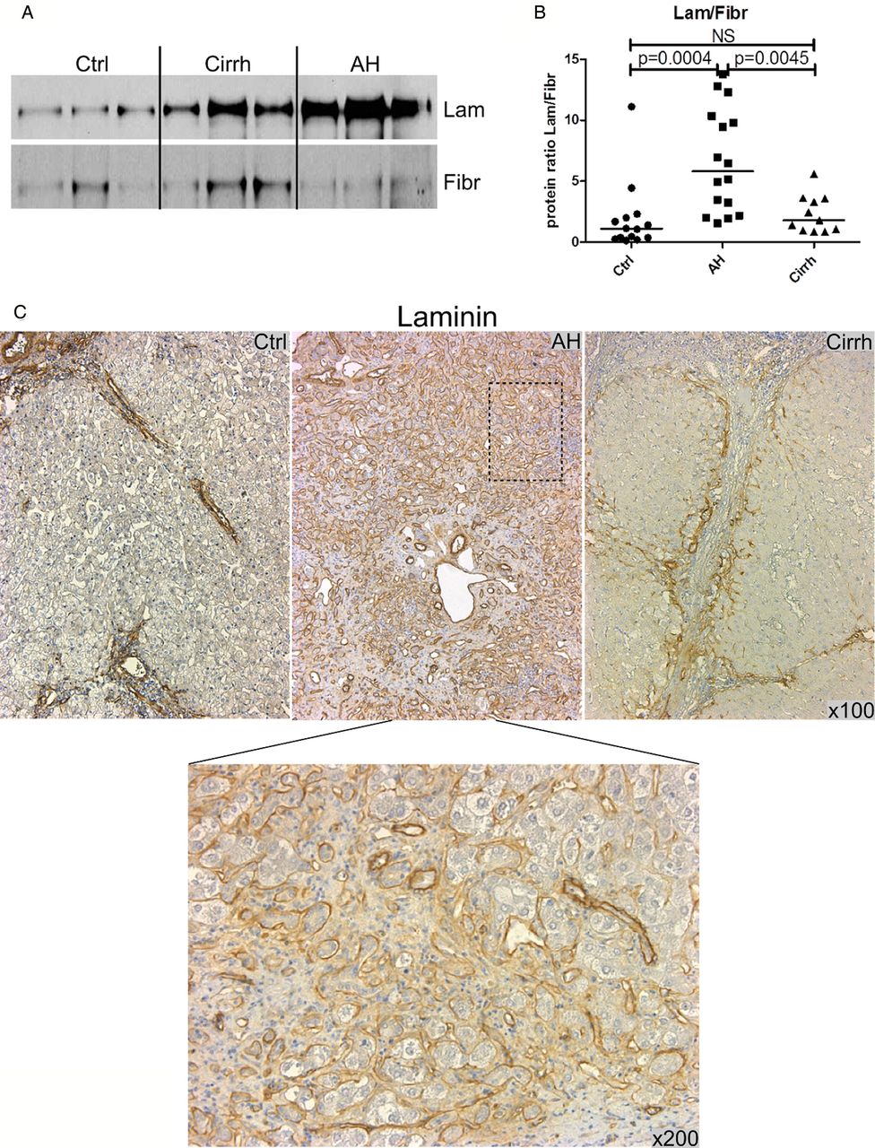
We next investigated differentiation of HPCs since they may differentiate into either hepatocytes or cholangiocytes. We had previously shown that expression of HPC markers can predict survival in patients with AH.8 We hypothesised that AH not responding to medical therapy is characterised by expansion of HPCs that poorly differentiate into mature hepatocytes. To test this hypothesis, we first assessed expression of cytokeratin 7 (KRT7) staining, a marker of the ductular reaction that is typically composed of HPCs engaged in cholangiocyte differentiation. As shown in figure 5A, livers from patients with AH were heavily populated by KRT7-positive HPCs that invade the whole parenchyma, which were less frequently seen in alcoholic cirrhosis and limited to fibrosis string. These results were confirmed with cytokeratin 19 (KRT19), which can also identify HPCs that are fated biliary cells (figure 5B). Overall, these results indicated that AH was characterised by a massive yet inefficient accumulation of HPCs having a predominant biliary phenotype. Finally, to gain insight into potential mechanisms favouring cholangiocyte differentiation of HPCs, we assessed the content of extracellular matrix proteins known to influence HPC fate. We assessed laminin, which promotes cholangiocyte differentiation, and fibronectin, a protein that favours hepatocyte differentiation.12 ,15 In alcoholic cirrhosis, protein expression of both laminin and fibronectin was significantly increased compared with normal controls (figure 6A). In contrast, AH displayed a marked increase in laminin content compared with both normal control and alcoholic cirrhosis, while fibronectin levels were not increased (figure 6A). Interestingly, expression of laminin surrounding HPCs was found, suggesting a close interaction between this extracellular protein and these cells (figure 6C); however, fibronectin staining appears less intense and more diffuse in AH liver (see online supplementary figure S5). The ratio between protein expression of laminin and that of fibronectin was extremely high in AH compared with that in alcoholic cirrhosis and normal controls (figure 6B), suggesting its specific role in altered differentiation of HPCs towards hepatocytes.

Cytokeratin 7 and 19 expression profile in liver parenchyma of patients. (A) Representative immunostainings for cytokeratin 7 (KRT7) in livers from control (Ctrl), alcoholic hepatitis (AH) and cirrhosis (Cirrh). Upper panel magnification ×100 and lower panel magnification ×400. Portal bile ducts are stained in Ctrl livers (arrowheads, left panel). In cirrhotic liver, we observe intense staining in the fibrotic string around regenerative nodules (RNs) (right panel). AH liver displays massive staining, which invades the entire parenchyma (middle panel). (B) Representative immunostainings for cytokeratin 19 (KRT19) in livers from Ctrl, AH and Cirrh. Upper panel: original magnification ×100; lower panel: original magnification ×400. The black dotted drawn boxes present in ×100 image represent the areas enlarged in ×400 images.

Laminin (Lam) and fibronectin (Fibr) exhibit different expression profiles in livers of patients. (A) Representative western blot of liver expression levels of Lam and Fibr in control (Ctrl), alcoholic hepatitis (AH) and cirrhosis (Cirrh). (B) Dot-plot shows the Lam/Fibr ratio determined after band intensity quantification of western blot analysis in livers of the three groups of patients. Results are expressed as the ratio of Lam/Fibr in relative units. Statistical significances are indicated. (C) Representative immunostainings for Lam in livers from Ctrl, AH and Cirrh. Original magnification: ×100 and ×200. The black dotted drawn box present in ×100 image represents the area enlarged in ×200 image.
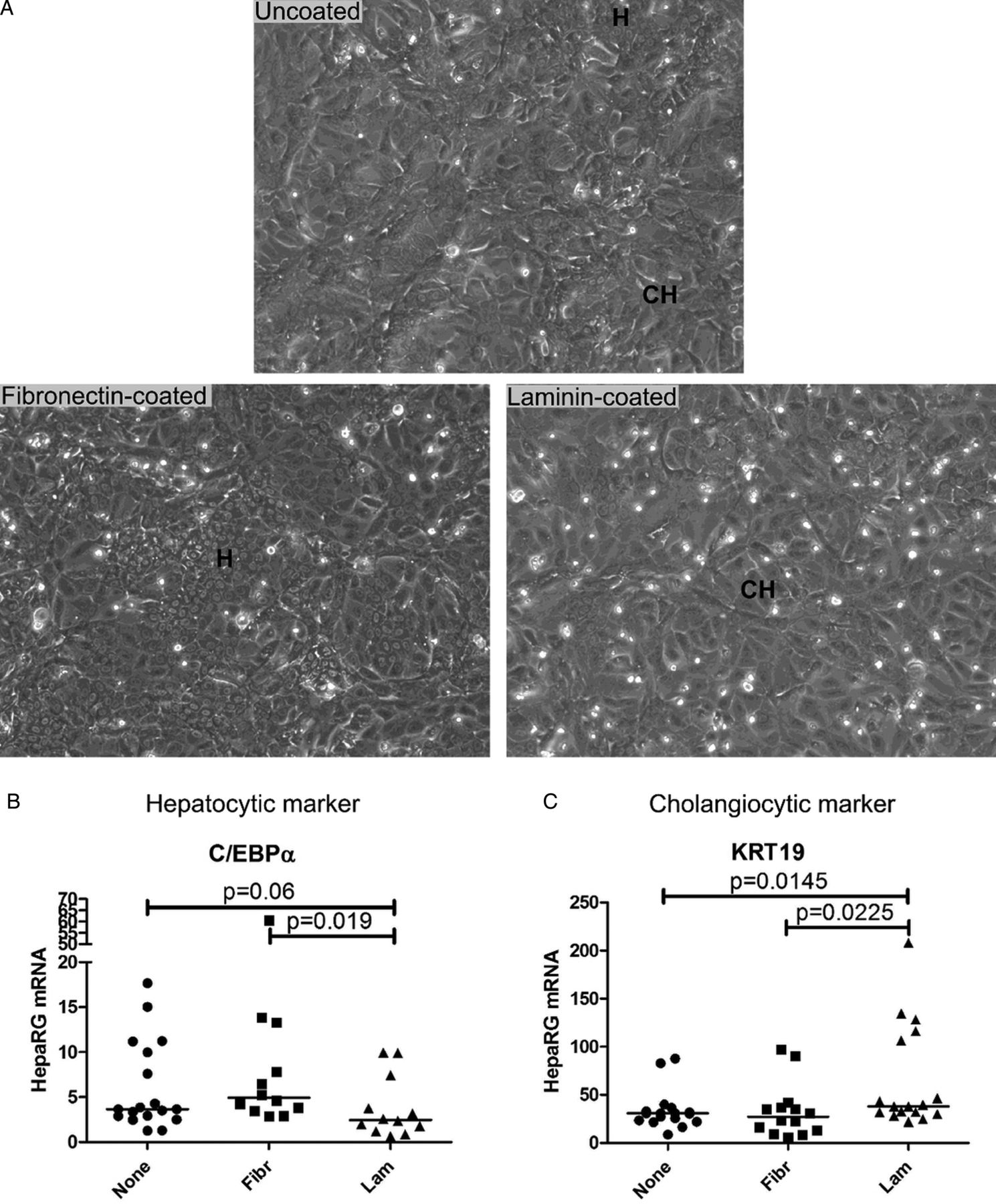
To further investigate the influence of laminin on the fate of bipotent cells of human liver origin, we have cultured HepaRG cells on different matrix. As shown in figure 6A, HepaRG culture on uncoated 6-well plate simultaneously displayed hepatocyte-like and biliary-like epithelial phenotypes roughly in equivalent proportion. Culture of HepaRG on fibronectin-coated plates led to enrichment of hepatocyte-like cells and conversely culture on laminin-coated wells led to enrichment in cholangiocyte-like cells (figure 7A). These data were confirmed by a significant decrease in a hepatocytic marker (figure 7B) and the increase in a cholangiocytic marker (figure 7C) in HepaRG cells cultured on laminin compared with those cultured on fibronectin.
Laminin promotes cholangiocyte phenotype of the bipotent human HepaRG cells. (A) Representative picture of HepaRG cells cultured for 15 days either in uncoated (upper panel), fibronectin-coated (lower left panel) or laminin-coated (lower right panel) 6-well plates. These different coatings influence the proportion of cells with hepatocytic (H) or cholangiocytic (CH) phenotypes. (B and C) Dot-plots show mRNA expression in HepaRG cells cultured for 15 days in uncoated (None), in fibronectin-coated (Fibr) or laminin-coated (Lam) 6-well plates for an early gene of hepatocyte lineage (CCAAT/enhancer binding protein α (C/EBPα)) (B) and a marker of cholangiocyte (cytokeratin 19 (KRT19)) (C). Results are expressed as relative unit. Each experimental well is represented, and the median is indicated as a solid line. Statistical significances are indicated.
Discussion
AH combines a necroinflammatory process, increased inflammatory mediators, cell death and its counterpart, liver regeneration. The present data completely shift the paradigm of relevant targets for improving outcome of patients with severe AH and lead us to rethink future development of molecules in this setting. In severe forms of the disease, inflammatory mediators seem to be less prominent than expected, whereas a substantial deficit in the capacity of HPCs to differentiate into hepatocytes appears crucial, as does a deficit in hepatocyte replication.
In addition to severely injured hepatocytes related, at least in part, to intense mitochondrial dysfunction, we observed a complex pattern of proinflammatory cytokines with unexpectedly low hepatic levels of two important drivers of liver regeneration, TNF-α and IL-6,16–19 and an expected increase in levels of IL-8, a key mediator of neutrophil recruitment.13 This decrease in TNF-α and IL-6 expression in liver parenchyma of patients with severe AH may explain, at least in part, the deleterious impact of anti-TNF-α therapies in randomised controlled trial patients with severe AH.3 ,4
In the same line, upregulation of TNF-α and IL-6 in patients with cirrhosis and severe liver dysfunction could be expected but was not observed in our samples. It should be kept in mind that our patients with cirrhosis had been abstinent for at least 6 months and did not have inflammatory lesions. Thus, alcohol withdrawal may have shut down the inflammatory stimuli leading to downregulation of cytokines as observed in animal models with chronic liver injury after removal of toxic insults such as CCl4.20 ,21
In the AH setting, upregulated NF-κB is associated with induction of IL-8, a cytokine implicated in the inflammatory process, whereas it seems unable to induce TNF-α and IL-6 cytokines mainly implicated in liver regeneration. However, the classical concept of NF-κB driving liver regeneration was established in models of liver regeneration after partial hepatectomy performed on healthy, not chronically injured liver.22 In AH, a disease that combines chronic injury, fibrosis, necrosis and inflammation, the transcription factor role of NF-κB may be selectively affected by epigenetic factors.23 ,24 Increased levels of bona fide NF-κB target genes such as NOS2, IL-8, CRP, lipocalin 2 (LCN2) and formyl peptide receptor 1 (FPR1) mRNAs and increased phosphorylated IκB support increased NF-κB activity in AH. However, NF-κB activity appears to be more complex because other bona fide genes such as TNF, IL-6 and NFKIA remain unchanged. In part this complexity may be related to the tolerance phenomenon but also to the fact that in vivo data reflect a combination of intracellular pathways from several cell types and intercellular networks, and crucial kinetic data may be missing.
Results showing both elevated levels of ‘non-tolerisable’ genes (LCN2 and FPR1) and unchanged levels of ‘tolerisable’ genes (IL-6, matrix metallopeptidase 13) in patients with AH suggest the presence of the lipopolysaccharide (LPS) tolerance phenomenon (see online supplementary figure S2B). This tolerance phenomenon has been illustrated in macrophages and is an adaptive response leading to selective inhibition of certain proinflammatory genes while other genes including antimicrobial effectors remain responders to LPS.25 It could partially explain the intriguing downregulation of IL-6 and TNF-α during NF-κB activation and chronic LPS exposure due to increased intestinal permeability.26 ,27 Further investigation of underlying mechanisms in the specific setting of AH is needed.
Alongside research linking alcoholic liver disease with mitochondrial dysfunction,28 we provided evidence that such a deficiency is a key feature of severe AH. Indeed, in this severe form of alcohol-induced liver injury, the mitochondrial defect is extremely severe in terms of low number and altered fitness, as shown by electron microscopy, increased amounts of 8-OHdG and decreased protein levels of Neil1 and OGG1, two key enzymes in mtDNA repair.29 ,30 Evidence from previous work suggests that the IL-6 defect in IL-6 knockout mice is related to a decreased capacity for mitochondrial DNA repair31 and that liver recovery after liver resection is stimulated by IL-6, particularly by inhibition of mitochondrial autophagy.32 Although the role of IL-6 in mitochondrial impairment was established in previous work, in the present study we did not specifically investigate the direct contribution of altered IL-6 pathways to such injury. Nevertheless, the present work clearly supports mitochondrial abnormalities as a key feature of severe forms of AH. LPS may also contribute to the marked mitochondrial abnormalities in AH based on experimental studies showing that LPS severely impaired mitochondrial function by damaging and depleting hepatic mtDNA.33
The present study demonstrates that severe AH is characterised by the absence of hepatocyte proliferation, which would seem at first glance to be counterbalanced by enhanced proliferation of HPCs, alternative cells known to restore functional liver mass.7 ,34 This induction of HPC proliferation is supported by the increases in TWEAK and its receptor Fn14, members of the TNF superfamily, that are potent growth factors for HPC proliferation prior to their differentiation into cholangiocytes and hepatocytes.35 It is noteworthy that previous studies had already pointed out the contributions of HPCs and the TWEAK pathway in AH.8 ,14 However, the present study shows that HPC proliferation is not only increased, but is deregulated, as demonstrated by major repopulation of liver parenchyma by HPCs. A striking feature of this parenchymal invasion is that HPCs, cells able to differentiate into cholangiocytes and hepatocytes, are mainly engaged in a biliary phenotype, as confirmed by KRT19 staining.36 ,37 This complex pattern in the fate of hepatocytes and HPC proliferation seems to play a central role in the inability to restore liver mass in patients with severe AH not responding to medical therapy.
The present study is the first to propose a role for laminin and fibronectin, components of the extracellular matrix implicated in differentiation of HPCs, in severe AH, as previously proposed in animal models and in humans suffering from non-alcohol-mediated liver injury.12 The substantial rise in the laminin/fibronectin ratio observed in AH interferes with the altered fate of HPCs since fibronectin favours HPC differentiation into hepatocytes and laminin into cholangiocytes as we have shown with the human HPC-like cells HepaRG. Further studies are warranted to gain deeper insight into the mechanisms leading to enrichment of laminin in patients with severe AH.
AH has to be viewed as an ongoing process with inflammation as the first phase leading to tissue injury followed by tissue repair and regeneration. Thus, an approach targeting liver regeneration alone is insufficient. The relative importance of inflammation, tissue repair and regeneration may vary from patient to patient because some may have an inflammatory profile that is sensitive to corticosteroid therapy while others have an inflammatory profile that is resistant to corticosteroids followed by defective regeneration. Stratification of patients according to their disease profile could improve overall success in treating patients with AH. The recent development of animal models of AH will provide more information on the chronology of the different events and their respective contributions.38 ,39
In summary, the present study strongly favours new therapeutic strategies aimed at restoring functional hepatocytes by improving hepatocyte integrity and targeting HPCs that mediate regeneration after reinstating differentiation of HPCs into mature hepatocytes.
Acknowledgments
The authors thank Linda Burkly (Department of Immunology, Biogen Idec, Cambridge, MA, USA) for providing mouse monoclonal antibodies against TWEAK and Fn14. The authors also thank Cécile Allet and Anne Loyens for their help in electron microscopy analysis and the “Service Commun de microscopie électronique” set-up by IFR 114 (IMPRT).
References
Supplementary materials
Supplementary Data
This web only file has been produced by the BMJ Publishing Group from an electronic file supplied by the author(s) and has not been edited for content.
Files in this Data Supplement:
- Data supplement 1 - Online supplement
Footnotes
Contributors LD, AL, GL, FA, FM, EG and AC generated data. LD, AL, GL and PM designed experiments. DB and EL performed histological analysis. ST, EB, CM and F-RP provided explanted livers. AL, SD, CM and PM selected patients. LD and AL analysed the data. LD prepared the manuscript. AL, SD, RB and PM edited the manuscript. LD, AL, RB and PM provided experimental funding.
Funding Financial support received from the 'Agence Nationale de la Recherche' ANR-06-PHYSIO-022, 'Association Française pour l'Etude du Foie' (AFEF) and NIAAA/NIH 1U01AA021908-01.
Competing interests None.
Patient consent Obtained.
Ethics approval Ethics Committee of the University Hospital of Lille.
Provenance and peer review Not commissioned; externally peer reviewed.