Article Text

Abstract
Objectives Proteases are key mediators of pain and altered enteric neuronal signalling, although the types and sources of these important intestinal mediators are unknown. We hypothesised that intestinal epithelium is a major source of trypsin-like activity in patients with IBS and this activity signals to primary afferent and enteric nerves and induces visceral hypersensitivity.
Design Trypsin-like activity was determined in tissues from patients with IBS and in supernatants of Caco-2 cells stimulated or not. These supernatants were also applied to cultures of primary afferents. mRNA isoforms of trypsin (PRSS1, 2 and 3) were detected by reverse transcription-PCR, and trypsin-3 protein expression was studied by western blot analysis and immunohistochemistry. Electrophysiological recordings and Ca2+ imaging in response to trypsin-3 were performed in mouse primary afferent and in human submucosal neurons, respectively. Visceromotor response to colorectal distension was recorded in mice administered intracolonically with trypsin-3.
Results We showed that stimulated intestinal epithelial cells released trypsin-like activity specifically from the basolateral side. This activity was able to activate sensory neurons. In colons of patients with IBS, increased trypsin-like activity was associated with the epithelium. We identified that trypsin-3 was the only form of trypsin upregulated in stimulated intestinal epithelial cells and in tissues from patients with IBS. Trypsin-3 was able to signal to human submucosal enteric neurons and mouse sensory neurons, and to induce visceral hypersensitivity in vivo, all by a protease-activated receptor-2-dependent mechanism.
Conclusions In IBS, the intestinal epithelium produces and releases the active protease trypsin-3, which is able to signal to enteric neurons and to induce visceral hypersensitivity.
- ABDOMINAL PAIN
- IRRITABLE BOWEL SYNDROME
- TRYPSIN
- NERVE - GUT INTERACTIONS
This is an Open Access article distributed in accordance with the Creative Commons Attribution Non Commercial (CC BY-NC 4.0) license, which permits others to distribute, remix, adapt, build upon this work non-commercially, and license their derivative works on different terms, provided the original work is properly cited and the use is non-commercial. See: http://creativecommons.org/licenses/by-nc/4.0/
Statistics from Altmetric.com
Significance of this study
What is already known on this subject?
Colonic tissues from patient with IBS release ‘trypsin-like’ activity.
Proteases in general and trypsins specifically can signal to enteric neurons.
Protease-activated receptor-2 activation can be activated on mouse sensory neurons and submucosal neurons.
In vivo, protease-activated receptor-2 activation causes visceral hypersensitivity.
What are the new findings?
The origin and the nature of one of the main proteases responsible for trypsin-like activity in tissues from patients with IBS have been unravelled: trypsin 3.
Intestinal epithelial cells are the major source of trypsin activity in colonic tissues from patients with IBS.
Among all the existing forms of trypsins, trypsin-3 is the protease responsible for IBS tissues-associated increased trypsin activity.
Trypsin-3 increases intestinal epithelium permeability. Trypsin-3 also signals to human submucosal neurons, mouse sensory neurons and causes visceral hypersensitivity, all through a protease-activated receptor-2-dependent mechanism.
Trypsin-3 could be considered as the endogenous agonist of protease-activated receptor-2 in the context of IBS.
How might it impact on clinical practice in the foreseeable future?
Trypsin-3 could be used as a marker of epithelial dysfunction in patients with IBS.
Specific inhibitors for trypsin-3 could be developed as new therapeutic options for the treatment of IBS hypersensitivity symptoms.
Introduction
IBS is the most common functional GI disorder.1 Patients with IBS suffer from abdominal pain, cramping and altered bowel habit. While the pathophysiology remains poorly understood, there is growing evidence that neurons innervating the intestine in patients with IBS are hyperexcitable, including both enteric neurons and extrinsic primary sensory afferents. Numerous studies suggest that a number of mediators that are present within tissues from patients with IBS underlie these neuronal changes, and among them, proteases seem to be particularly important.2–4
Studies reported that protease inhibitors completely inhibited neuronal activation induced by IBS tissue supernatants.3 ,4 However, the origin and the nature of proteases that could be responsible for neuronal hyperactivity in tissues from patients with IBS are still unclear. Identification of the origin and nature of such proteases would be an important step for potential therapeutic approach and drug development in the field of IBS. Here, we made the hypothesis that the intestinal epithelium could be an important source of proteases that are inducing neuronal signalling, and potentially hypersensitivity symptoms associated with IBS. Therefore, we have investigated proteolytic activity released by human cultured intestinal epithelial cells and tested their effect on sensory neuron activation. In humans, three serine protease (PRSS) genes encode trypsinogens: PRSS1 encodes trypsinogen-1 (cationic trypsin), PRSS2 encodes trypsinogen-2 (anionic trypsin) and PRSS3 encodes trypsinogen-3, where at least two isoforms with overlapping mature peptide sequences, formerly designated as mesotrypsinogen and trypsinogen IV, have been functionally characterised. The mature protein of PRSS3 gene uses the nomenclature of trypsin-3 protein, common to all transcripts of this gene. Here, we have determined that stimulated (by lipopolysaccharide (LPS) or epinephrine) intestinal epithelium released specifically on the basolateral side of trypsin-3, is able to signal to human enteric neurons and sensory neurons through a protease-activated receptor (PAR)-2 (PAR2)-dependent mechanism. In addition, trypsin-3 was able to induce increased epithelial permeability in vitro and visceral hypersensitivity in vivo, when delivered into the colon. Finally, in tissues from patients with IBS, we determined that the vast majority of trypsin-like proteolytic activity was associated with the epithelium, where trypsin-3 was upregulated compared with the levels of healthy controls. Our data demonstrated in IBS that the intestinal epithelium produces and releases active proteases, that is, trypsin-3 is able to signal to submucosal neurons, primary afferents and to induce visceral hypersensitivity.
Materials and methods
Patients
Colon tissues were obtained from patients with IBS and healthy controls at the Kingston General Hospital (Ontario, Canada) (ethic approval 6004988, collected between 2013 and 2016), Nantes (ethic approval DC-2008-402 collected between 2012 and 2016) and Toulouse (ethic approval DC-2015-2443 for the COLIC project collected in 2015 and 2016) Hospitals (France) (see online supplementary table S1). Patients with IBS were defined by the Rome III criteria. Descending colonic biopsies collected during colonoscopy procedures were used for immunohistochemistry and mRNA expression randomly. For immunohistochemistry, fresh biopsies were coated in optimal cutting temperature (OCT) compound (Dako) and stored at −80°C. For mRNA expression, complementary DNA (cDNA) samples generated from RNA extracts were used. For submucosal neuron imaging, samples of large bowel were harvested from non-pathologic zones of resections from six patients undergoing colectomy for colon cancer.
supplementary tables
Mice
Male C57BL/6 mice (6–10 weeks, Charles River Laboratories, Wilmington, Michigan, USA, or Janvier St Quentin-Fallavier, France) and male PAR2-deficient mice or wild-type littermates were used.5 Mice had free access to food and water and were subjected to 12 hours light/dark cycles. Animal protocols followed the Canadian Council of Animal Care Guidelines and were approved by Animal Care Committees at Queen's University (protocol 2016-1644), University of Calgary (protocol M08068-70) and Toulouse (protocol PI-U1220-NV19).
Rat models of visceral hypersensitivity
Adult Wistar rats (aged 8–9 weeks, Harlan Laboratory, Indianapolis, Indiana, USA), males (240–300 g) and females (175–220 g) exposed to limited bedding stress as neonates6 or naïve Sprague-Dawley rats (aged 7–8 weeks, 250–275 g) with free access to food and water and in 12 hours light/dark cycles were acclimated for 1 week before starting experiments. Experiments followed National Institutes of Health guidelines according to protocol #09026-11 and #9906-020 approved by the Institutional Animal Care and Use Committee of the Veteran Affairs Greater Los Angeles Healthcare System under the auspice of the Office of Laboratory Animal Welfare—Assurance of Compliance (A3002-01).
Two methods were used to induce visceral hypersensitivity: repeated water avoidance stress (WAS), (10 days, 1 hour/day)7 and the cortagine administration via intraperitoneal injection.8 Visceral sensitivity was assessed as previously described.7 ,8
Imaging
Section and live imaging were performed on Zeiss confocal LSM710 and ApoTome.2 microscopes, respectively.
Culture of sensory neurons from dorsal root ganglia
Dorsal root ganglia (DRG) neurons were isolated from mice.3 Cells were washed and incubated with Hank's Balanced Salt Solution (HBSS)+Ca2+, Fluo-4-AM (Invitrogen) and 20% pluronic F-127 (Invitrogen) during 30 min at 37°C followed by 30 min at room temperature before imaging.9 Supernatants from apical or basal compartments of Caco-2 cells culture, stimulated or not by LPS (Escherichia coli serotype K235, ATCC13027), were pre-incubated with the serine protease inhibitor FUT-175 (Futhan) (50 μg/mL) or the trypsin inhibitor leupeptin (100 μM) (all from Calbiochem) for 15 min. Additional recordings were made from DRG neurons after addition of trypsin-3 (10 nM, R&D) or its vehicle (HBSS+0.025% Brij35) in the presence or absence of specific PAR1 (SCH79797, Tocris Bio-Techne, UK) or PAR4 (ML-354, Tocris Bio-Techne) antagonists (all at 10 µM for 5 min). Neurons were identified by addition of high K+ (50 mM) solution at the end of the recording. Fluo-4 was excited at 475 nm, and fluorescence emission was collected at 490/515 nm. Changes in [Ca2+]i are reflected by Fluo-4 fluorescence intensity.
Human submucosal neuron imaging
To isolate submucosal ganglia from descending colon, we used previously described methods.10 ,11 The colon was placed in a cold oxygenated sterile HBSS (Sigma), cut along the mesenteric border and pinned flat with the mucosa facing upwards. Ice-cold HBSS was changed every 5 min. The submucosal plexus was dissected from the mucosal and underlying circular muscle, cut in small pieces (∼1.5×1.5 mm) and digested at 37°C in an enzymatic solution (1 mg/mL; Sigma) and collagenase (1.25 mg/mL; Sigma) for 45 min. The suspension was centrifuged, the ganglia were plated onto 96-well plates (Greiner), topped-up with neurobasal medium (Invitrogen) supplemented with 10% heat-inactivated fetal bovine serum (FBS), 1% antibiotic-antimycotic solution (Sigma) and nerve growth factor 25 ng/mL, and kept in an incubator at 37°C continuously gassed with 95% O2–5% CO2. 11 Ganglia were cultured for 3 days before performing Ca2+ imaging experiments.12 Tissues were loaded with 10 µM Fluo-4 AM (Molecular Probes, Invitrogen) for 30 min at 37°C, then rinsed with HBSS and transferred on the microscope stage. Recordings were made at room temperature to monitor Ca2+ flux after addition of trypsin-3 (0.5–1–10 nM) to the wells, in the presence or absence of specific PAR1 (SCH79797), PAR2 (GB83, Axon Medchem, The Netherlands) or PAR4 (ML-354) antagonists (all at 10 µM for 15 min). Neurons were identified by addition of high-K+ (75 mM) solution at the end of the recording. Fluo-4 was excited at 475 nm, and fluorescence emission was collected at 525/50 nm. Changes in [Ca2+]i are reflected by Fluo-4 fluorescence intensity.
PAR2 receptor internalisation in human submucosal neurons
Trypsin-3 (10 nM) was added to the submucosal tissue that was isolated from colonic biopsies and cultured in neurobasal medium supplemented with 10% heat-inactivated FBS, 1% antibiotic-antimycotic solution for 2 hours. The plexus was then washed in phosphate buffer saline (PBS), fixed in paraformaldehyde 4% and processed for immunostaining, using chicken antineurofilament 200 kDa (NF200, 1:500, ab134306-Abcam), to identify neurons and nerve fibres, and mouse anti-PAR2 antibody (1:200, SAM-11-LifeSpan BioSciences, Seattle, USA).13 Primary antibody incubation (overnight at 4°C) was followed by incubation with appropriate fluorescently labelled secondary antibodies (2 hours room temperature). The tissue was mounted on a microscope slide in Citifluor (Citifluor, Leicester, UK). PAR2 involvement specificity was investigated in the presence of the PAR2 antagonist GB83 (10 μM),14 added 30 min before trypsin-3.
Intestinal epithelial cell cultures
Caco-2 cells were grown to confluence as monolayers in Transwell plates (2×10⁵ cells per well) (Corning).15 After 21 days in culture (transepithelial electrical resistance of 350 Ω cm2),15 culture medium was replaced by OptiMEM (Life Technologies) and cells were exposed for 24 hours to trypsin-3 (0.5–10 nM) on the basolateral side, in the presence or absence of the (PAR2) antagonist (GB88). Paracellular permeability was measured by the passage of dextran-Fluorescein IsoThioCyanate (FITC) (3000 kDa, Sigma) from the apical to the basal medium, as previously described.16
Trypsin activity
Basal media were concentrated 3 times using Vivaspin 500 (Dutcher). Trypsin activity was measured in basal and apical medium with the substrate N-p-Tosyl-GPR-amino-4-methylcoumarin hydrochloride (0.1 mM) in 50 mM Tris, 10 mM CaCl2. Substrate degradation was calculated by the change in fluorescence (excitation: 355 nm, emission: 460 nm), measured over 30 min at 37°C on a microplate reader NOVOstar (BMG Labtech). OCT-included biopsies of control and patients with IBS were cryostat sectioned (8 μm thickness) and washed with PBS, 2% Tween-20. All samples were incubated overnight at 37°C with the substrate N-p-Tosyl-GPR-amino-4-methylcoumarin hydrochloride (50 μg/mL, Sigma) in 0.3% low melting agarose. Nuclei were stained with Topro3 (Invitrogen). Images were analysed with ImageJ software.
Reverse transcription, conventional and quantitative PCR
Total RNA was extracted with the Nucleospin RNA/Protein Kit (Macherey-Nagel, GmbH). DNAse-treated RNA was reverse transcribed using the Maxima First Strand cDNA Synthesis Kit for reverse transcription-quantitative PCR (RT-qPCR) (Thermo Scientific). Resulting cDNA samples were amplified by conventional PCR with Taq DNA polymerase (Invitrogen, USA) and sequence-specific primer pairs (see online supplementary table S2).17 For qPCR, cDNA was amplified with the SYBR Green Master I Kit (Roche) and sequence-specific primer pairs (see online supplementary table S2) in a LightCycler 480 Instrument (Roche). The analysis of stability of three standard housekeeping genes (HPRT1, GAPDH and TBP) by using RefFinder ranked HPRT1 as the overall most stable housekeeping gene in our experimental conditions. Thus, the relative level of mRNA expression for target genes was calculated with the method 2-DDCt17 by using hHPRT1 as reference gene. For conventional PCR and qPCR, negative controls consisted of samples from reverse transcription, where the enzyme was not added to reactions. The identity of amplicons from PRSS1, 2 and 3 was confirmed by automated DNA sequencing (Eurofins MWG Operon, GmbH).
Western blot analysis
Proteins from Caco-2 cells were extracted with the Nucleospin RNA/Protein Kit (Macherey-Nagel) as per manufacturer's instructions. Supernatant's proteins were precipitated by trichloroacetic acid/acetone, separated by sodium dodecyl sulfate-polyacrylamide gel electrophoresis (12%) and transferred onto a nitrocellulose membrane (Life Science). Membranes were incubated with antitrypsin-3 antibody (1/100, ab107430-Abcam) overnight at 4°C and secondary antibody conjugated with HRP (1/3000, w4018-Promega) and then visualised by chemiluminescence (Chemidoc XRS Bio-Rad).
Immunofluorescence in cell cultures and biopsies
Cell cultures were fixed using formaldehyde (4%) during 10 min followed by washes in 0.1 M glycine (2×10 min). Immunostaining was performed with antitrypsin-3 (1/500 ab107430-Abcam), anti-occludin (1/100 711500 Life Technologies), antizonula occludens (ZO)-1 (1/100, 617300 Invitrogen) and 670 phalloidin (PHDN1-cytoskeleton), to label actin network. Human colonic biopsies were included in OCT and tissue cryosections (5 µm thickness) were cut with cryostat (Leica Microsystems, GmbH). Tissue slices were costained with antibodies against trypsin-3 and the epithelial cell maker EpCAM/CD326 (1/500 VU1D9-Cell Signalling). Cells and tissue slices were then incubated with appropriate secondary antibodies conjugated to AlexaFluor 488 or 555 (Molecular Probes). Slides were mounted with Prolong Gold Antifading Reagent with 4',6'-diamidino-2-phenylindole (DAPI) (Molecular Probes), to counterstain cell nuclei. The mean fluorescence intensity corresponding to trypsin-3-immunoreactivity in epithelial cells, as delimited by EpCAM costaining, was quantified with ImageJ software.16
Electrophysiological recordings from nociceptive DRG neurons
Mouse DRG were dissected from T9 to L1 and were enzymatically dissociated, plated onto coverslips and cultured overnight, as described.4 Neurons were incubated with active trypsin-3 (10 nM) 20 min prior electrophysiological studies, the PAR2 antagonist GB83 (10 μM)14 was added 30 min before trypsin-3. Whole-cell perforated patch current-clamp recordings were made using amphotericin B (240 g/mL, Sigma) on small diameter neurons (<40 pF capacitance). Changes in excitability were quantified by measuring rheobase and numbers of action potentials discharged at twice rheobase. Recordings were performed using Multiclamp 700B, digitised by Digidata 1440A, were stored and processed using pClamp 10.2 software (all by Molecular Devices, Sunnyvale, California, USA). The recording chamber was continuously perfused with external solution (∼2 mL/min) at room temperature (23°C). Standard solutions used were (in mmol/L); pipette solution: K-gluconate 110, KCl 30, 4-(2-hydroethyl)-1-piperazineethane sulfonic acid (HEPES) 10, MgCl2 1 and CaCl2 2; pH adjusted to 7.25 with 1 M KOH; external solution: NaCl 140, KCl 5 HEPES 10, glucose 10, MgCl2 1 and CaCl2 2; pH adjusted to 7.3–7.4 with 3 M NaOH.
Colorectal distension
Recombinant human trypsinogen-IV was purchased and activated into trypsin-3 by enteropeptidase following commercial recommendations. Then, mice were submitted to intracolonic administration of trypsin-3 (10 U/mouse), followed by colorectal distensions at 0, 1, 3, 6 and 9 hours time points, as previously described.3 In another set of experiments, mice were intracolonically administered with increasing doses of trypsin-3 (0.1, 1 or 10 U/mouse), followed by colorectal distension 3 hours later. Mice from the control group were administered intracolonically with vehicle (10% v/v absolute ethanol and 10% v/v Tween-80).18 Similar experiments were repeated in PAR2-deficient mice and wild-type littermates.3
Statistical analysis
Data were expressed as mean±SEM, except for RT-qPCR, immunostaining and in situ zymography quantification of patient biopsies, where each dot represents one patient. Statistical analysis was performed using parametric t-tests, two-way or one-way analysis of variance and Bonferroni's post-test (see figure legends). GraphPad Prism V.5.0 software was used for analysis. Statistical significance was accepted at p<0.05.
Results
Epithelial proteolytic activity signals to sensory neurons
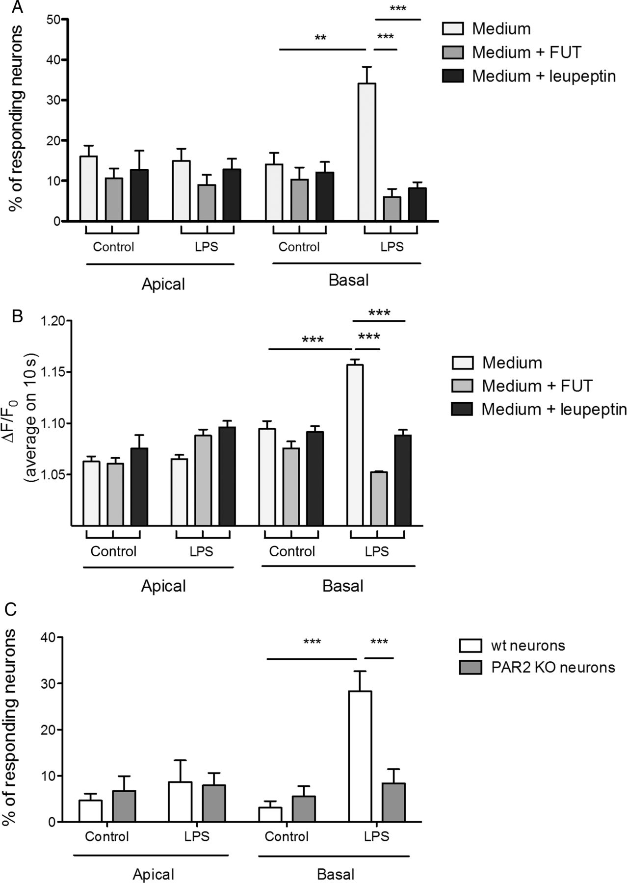
Culture media from the apical compartment of Caco-2 cells stimulated or not with LPS, induced very small amplitude Ca2+ response in a limited number of mouse sensory neurons (figure 1A, B). Similarly, culture media from the basal compartment of unstimulated Caco-2 cells had little effect on the Ca2+ signal in DRG neurons (figure 1A, B). However, culture media from basal compartment of LPS-stimulated Caco-2 cells induced a significant increase in the number of responding neurons (figure 1A), and in the amplitude of their response (figure 1B). This increase was completely suppressed by pre-incubation with the non-selective trypsin inhibitors (FUT or leupeptin) (figure 1A, B), and the effect of the LPS-stimulated supernatant was not observed using DRG neurons from PAR2 −/− mice (figure 1C). These experiments demonstrated that LPS stimulation of intestinal epithelial cells releases proteolytic activity specifically on the basal compartment, and that this activity can signal to sensory neurons, through the activation of PAR2.

Conditioned medium from intestinal epithelial cells activates dorsal root ganglia (DRG) sensory neurons by the release of trypsin-like serine protease. Effect of the serine protease inhibitor FUT or the trypsin inhibitor leupeptin on the percentage of responsive neurons (A) and on the Fluo-4 (ΔF/F0) measured Ca2+ levels (B) stimulated by apical or basal supernatants recovered from control or lipopolysaccharide (LPS)-treated Caco-2 cell monolayers. (C) Percentage of sensory neurons dissociated from DRGs of wild-type (wt) or PAR2 knockout (KO) mice responding to apical or basal supernatants recovered from control or LPS-treated Caco-2 cell monolayers. Data are expressed as mean±SEM and were compared using a one-way analysis of variance followed by Bonferroni's post-test (n=6, 2 wells per condition for this experimentation) **p<0.01, ***p<0.001.
Trypsin activity originates from intestinal epithelial cells in human colons
Residual trypsin activity was released in culture supernatants of intestinal epithelial Caco-2 cells, both in basal and apical compartments. This trypsin activity was significantly increased in the basal, but not the apical compartment after stimulation of Caco-2 cells by LPS (figure 2A). Thus, epithelial trypsin activity follows the same pattern as the epithelial proteolytic mediators activating primary afferents described above.

Intestinal epithelial cells released trypsin-like activity. (A) Trypsin-like activity measured in apical and basal supernatants recovered from control or lipopolysaccharide (LPS)-treated Caco-2 cells. Assembled data from seven independent experiments with 4–6 wells per test. Representative confocal photomicrographs of in situ zymography assays performed in colonic tissue slices from healthy controls and patients with IBS (scale bar: 50 μm), (B) from control (Ctrl) or water avoidance-induced hypersensitive rat (scale bar: 20 μm), (C) evidencing the level of trypsin-like activity. Graph representation of mean fluorescence intensity quantified from 6 to 12 patients per group. Data are expressed as mean±SEM and were compared using Student's t-test. *p<0.05, **p<0.01, ***p<0.005.
Using in situ zymography, we next investigated where trypsin activity was located in human colonic tissues. Very low trypsin activity was detected in colonic tissues from healthy controls (figure 2B, left panel), while in patients with IBS, increased activity was detected (figure 2B, middle panel), and this was observed predominantly in the epithelium, with similar activity at the base and the tip of the crypts (figure 2B, middle panel). The epithelial trypsin activity was significantly increased in tissues from all IBS subgroups (IBS-C, IBS-D and IBS-M) (figure 2B, right panel). Hypersensitivity was induced in rats by submitting them to WAS. Colonic tissues from rats with demonstrated hypersensitivity (not shown) were used for in situ zymography. Trypsin activity was also increased in the colon epithelium of hypersensitive rats compared with naïve controls (figure 2C).
Trypsin-3 is the predominant form of epithelial trypsin and is overexpressed in IBS
We then examined the forms of trypsin present in human intestinal epithelium and/or tissues from patients with IBS. We found that all three human trypsin genes: PRSS1, PRSS2 and PRSS3 forms were present in intestinal epithelial cells (figure 3A, B), and in human colonic tissues (figure 3B). However, only PRSS3 mRNA (codes for the trypsin-3 protein) expression was upregulated in epithelial cells after LPS exposure, or after addition of epinephrine to the basolateral side (figure 3A). Cortisol exposure of intestinal epithelial cells had no effect on PRSS1, 2 or 3 mRNA expression (see online supplementary figure S1). In human colon, PRSS3 was the predominant form of trypsin detected (figure 3B) and no difference was observed for PRSS1 and PRSS2 expression in IBS tissues versus healthy controls (see online supplementary figure S2A, B). However, PRSS3 expression was significantly upregulated in tissues from IBS-C (constipation-predominant IBS), compared with healthy controls, while changes in PRSS3 expression was not significant in IBS-D (diarrhoea-predominant IBS) or IBS-M (mixed-IBS), compared with healthy controls (figure 3C). We further investigated the splice variants of PRSS3 that would be expressed in colonic tissues, and we determined that PRSS3 variant 1 was the major transcript expressed in intestinal epithelial cells and in colonic tissue samples (see online supplementary figure S2C). Tryptase mRNA expression (amplicons from the TPSAB1, TPSB2 and TPSD1, the three tryptase genes) was not detected in Caco-2 cells stimulated or not with LPS (see online supplementary figure S2D).

Gene expression of trypsinogens A. Relative gene expression of PRSS1 (cationic trypsin: trypsin-1 precursor), PRSS2 (anionic trypsin: trypsin-2 precursor) and PRSS3 (trypsin-3 precursor) in control, lipopolysaccharide (LPS)-treated or epinephrine-treated Caco-2 cell monolayers. Data are expressed as mean±SEM and were compared using one-way analysis of variance followed by Bonferroni's post-test. *p<0.05, ***p<0.001. (B) Analytical agarose-gel electrophoresis of RT-PCR products performed with RNA extracted from human colonic biopsies (healthy control), isolated colonic crypts or Caco-2 cells. Amplicons were amplified with oligonucleotides specific for PRSS1, PRSS2, PRSS3 and HPRT1. (C) Relative mRNA expression of PRSS3 within colonic biopsies from healthy control and different IBS subtypes: IBS-C (constipated), IBS-D (diarrhoea), IBS-M (mixed). Data are expressed as mean±SEM and were compared using Student's t-test. **p<0.01 vs control (Ctrl).
supplementary figures
We confirmed the presence of the 33-kDa trypsin-3 protein in intestinal epithelial cells and its LPS-induced upregulation in those cells (figure 4A) as well as the epinephrine-induced upregulation (data not shown). In Caco-2 cells, trypsin-3 was associated with the plasma membrane colocalising with actin (figure 4B). We confirmed the secretion of trypsin-3 in culture supernatants of Caco-2 cells, at low level in apical supernatants (basal or LPS stimulated), but at significantly higher level in the basal compartment of Caco-2 cells after their stimulation with LPS (figure 4C).

Intestinal epithelial cells upregulate trypsin-3 secretion in inflammatory condition and in IBS. (A) Relative trypsin-3 quantification by western blot analysis from protein extract of control or lipopolysaccharide (LPS)-treated Caco-2 cells. (B) Confocal photomicrographs of control and LPS-treated Caco-2 cell monolayer evidencing trypsin-3-immunoreactivity (green) and actin cytoskeleton (red), scale bar: 25 μm. (C) Relative trypsin-3 protein quantification from apical and basal supernatants from control or LPS-stimulated Caco-2 cell monolayers. (D) Representative confocal photomicrographs of colonic tissue slices from control (ctrl) and patients with IBS (scale bar: 20 μm). (E) Colonic tissues from control or cortagine-induced hypersensitive rat (scale bar: 20 μm), showing trypsin-3-immunoreactivity (green) for D and E, epithelial cell labelling (EpCAM-positive cells, red) and nuclei counterstain (cyan) for D. (D) Mean fluorescence intensity for trypsin-3-immunoreactivity quantified specifically in epithelial (EpCAM-positive) cells. (F) Confocal photomicrographs showing the transversal view of a colonic crypt labelled for trypsin-3 (green), EpCAM (red) and nuclei (cyan), scale bar: 25 μm. The representative profile at the bottom of the panel shows the polarisation of trypsin-3-immunoreactivity towards the basolateral side of intestinal epithelial cells. Data are expressed as mean±SEM and were analysed by Student's t-test in A and D, and a one-way analysis of variance followed by a Bonferroni's post-test in C.
In human and rat tissues, epithelial trypsin-3 was just above detection level in healthy controls but was significantly upregulated in tissues from patients with IBS (figure 4D) and from hypersensitive rats (cortagine-treated) (figure 4E). All IBS subgroups (IBS-D, IBS-C and IBS-M) expressed significantly larger amounts of trypsin-3 compared with healthy controls (figure 4D, lower panel). We also found that trypsin-3 epithelial expression was polarised towards the basolateral membrane in these tissues (figure 4F). This suggests that trypsin-3 would be released from the basolateral membrane of intestinal epithelial cells in vivo, as found in the in vitro studies. Taken together, we confirmed at a protein level that trypsin-3 matched the trypsin activity detected in intestinal epithelial cells and tissues from patients with IBS.
Trypsin-3 increases epithelial permeability, signals to human and mouse neurons and causes visceral hypersensitivity
The addition of trypsin-3 (1 and 10 nM) to the basal compartment of Caco-2 monolayers caused an increased passage of dextran-FITC (figure 5A). It also caused a decreased staining and disorganisation of the epithelial tight junction protein ZO-1 and occludin, compared with control (trypsin-3 vehicle) conditions (figure 5B). Trypsin-3 increased excitability of mouse DRG neurons compared with controls (figure 6A–C). The mean rheobase of neurons was decreased by 30% and the mean action potential number at twice rheobase increased by 39% (n=23, p<0.05), compared with control neurons. Trypsin-1 (10 nM) and thrombin (50 nM) also increased neuronal excitability (figure 6C) to a similar level to that seen with trypsin-3 (10 nM).

Trypsin-3 increases cellular permeability in intestinal epithelial cells. (A) Dextran passage from apical to basal medium of Caco-2 cell monolayers after trypsin-3 exposure (0.5, 1 and 10 nM) on the basolateral compartment. Assembled data from three independent experiments with 4 wells per test. (B) Representative confocal photomicrographs of Caco-2 cell monolayers after trypsin-3 exposure on the basolateral compartment (0.5, 1 and 10 nM) showing immunodetection of cellular junction occludin (top) and zonula occludens (ZO)-1 (bottom) in green, scale bar: 50 μm. Data are expressed as mean±SEM and were analysed by one-way analysis of variance followed by Bonferroni's post-test, ***p<0.001.

Trypsin-3 evokes PAR2-dependent hyperexcitability of nociceptive dorsal root ganglia neurons and calcium signals in human submucosal neurons. (A) Representative traces of current clamp recordings showing the rheobase and the action potential discharge at twice the rheobase in control neurons (left panel), neurons incubated with trypsin-3 (10 nM) (middle panel) or neurons incubated with the PAR2 antagonist GB83 (10 μM) before trypsin-3 (right panel). (B) Mean data of the rheobase and the action potential number at twice rheobase for the control, and trypsin-3 with or without GB83 (10 μM). *p<0.05 and **p<0.01 compared versus control, ##p<0.01 compared versus trypsin-3. (C) Representative data showing the effect of trypsin and thrombin on the rheobase and action potential discharge is similar in magnitude to that observed with trypsin-3, shown in B. The PAR2 antagonist blocks the trypsin but not the thrombin effect on neuronal excitability. *p<0.05 compared with control, #p<0.05 compared with trypsin-1. One-way analysis of variance with Bonferroni's post-test. (D) Representative gray scale images of cultured human submucosal ganglia loaded with Fluo-4 and Ca2+ responses of three neurons (colour-coded numbers in the gray images) to trypsin-3 (10 nM). Matched colours are represented between cell numbering and traces. The identity of human neurons was confirmed by application of high-K+ solution (75 mM). Scale bar: 20 μm. (E) Average amplitude of trypsin-3 (0.5–10 nM)-induced [Ca2+]i rises in submucosal neurons in the presence or absence of the PAR2 antagonist GB83. Data are expressed as mean±SEM and were analysed by one-way analysis of variance followed by Bonferroni's post-test (n=6 subjects per group), *p<0.05, **p<0.01, °°p<0.01.
Trypsin-3 was able to induce Ca2+ transients in human submucosal neurons (figure 6D) in a dose-dependent manner (figure 6E).
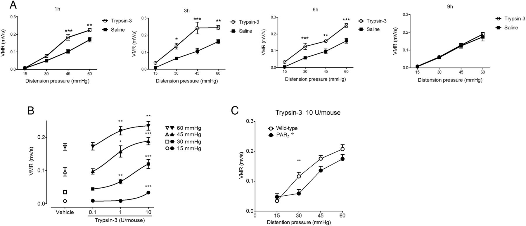
Trypsin-3 administered intracolonically, in a Tween-based saline solution (in order to allow the passage of trypsin-3 through the intestinal barrier) caused visceral hyperalgesia in response to colorectal distension, compared with baseline measures (figure 7A–C). This was evident by 1 hour and persisted for 6 hours (figure 7A). No effect was seen with vehicle alone and the trypsin-3 effect was dose-dependent (figure 7B).

Colorectal administration of trypsin-3 induces visceral hypersensitivity. (A) Kinetic visceromotor response (VMR) to intracolonic administration of trypsin-3 (10 U/mouse) 1, 3, 6 and 9 hours after its administration. (B) Visceromotor response to intracolonic administration of trypsin-3 (0.1, 1 or 10 U/mouse) or vehicle, 3 hours after intracolonic administration in wild-type or PAR2 −/− mice (C). Data are expressed as mean±SEM and were analysed by two-way analysis of variance followed by Bonferroni's post-test (n=10 per group). *p<0.05, **p<0.01 and ***p<0.001.
Mechanisms of trypsin-3-induced enteric effects
The effects of trypsin-3 in mouse sensory neurons were blocked by the PAR2 antagonist GB83, which also blocked the effect of trypsin-1 but had no effect on thrombin, a PAR1/PAR4 agonist (figure 6C). PAR1 or PAR4 antagonists had no significant effect on trypsin-3-induced rise in [Ca2+]i (see online supplementary figure S3A). In human colonic submucosal neurons, the trypsin-3-induced rise in [Ca2+]i was completely abolished by pre-incubation with the PAR2 antagonist GB83 (figure 6E), but was not modified by pre-incubation with PAR1 (SCH79797) or PAR4 (ML-354) antagonists (see online supplementary figure S3B). In PAR2-deficient mice, trypsin-3-induced visceral hypersensitivity was also significantly inhibited at the 30 mm Hg pressure (figure 7C). The whole area under the curve of trypsin-3-induced visceromotor response as a function of increasing pressures of distension was significantly lower in PAR2-deficient mice compared with wild-type (p<0.05, not shown), thereby confirming the notion that the enteric effects of trypsin-3, including in vivo visceral hypersensitivity, are mediated by PAR2 activation.
We further investigated in human colonic submucosal neurons, trypsin-3-induced potential changes in PAR2 expression, as a marker of PAR2 activation. In basal (unstimulated) conditions, PAR2 staining was clustered seemingly at the plasma membrane of submucosal neurons (figure 8, upper panel). After trypsin-3 stimulation, PAR2 expression radically changed, becoming either diffuse or in intracellular clusters (figure 8, middle panel). This suggested that PAR2 had been activated by trypsin-3 and had been internalised into submucosal neurons. Exposure to the PAR2 antagonist GB83 inhibited trypsin-3-induced changes in PAR2 staining (figure 8, bottom panel).

Trypsin-3 causes internalisation of PAR2 in human submucosal neurons. Representative images showing that the expression of PAR2 in human submucosal neurons changes in the presence of trypsin-3 (10 nM). Upper panel shows the clustered staining of PAR2 (arrowheads, red) on the plasma membrane of submucosal neurons (asterisks, NF200, green). Trypsin-3 (2 hours) caused internalisation of PAR2 (red) inside the neuron (asterisk, NF200, green). PAR2 staining appeared diffuse or clustered (arrowheads) inside the neuron. Pre-incubation with GB83 (10 μM, 30 min before trypsin-3) inhibited the internalisation of PAR2, which was expressed on the plasma membrane (arrowheads, red) of neurons (asterisks, NF200, green). Right panels show merged images. Scale bars: 20 μm.
Discussion
Increased proteolytic activity, and in particular trypsin activity is released by colonic tissues from patients with IBS. It is considered to be involved in hyperexcitability of both extrinsic and intrinsic enteric neurons, and in visceral hypersensitivity.2–4 However, the lack of knowledge on the origin and the nature of the protease(s) responsible for this trypsin activity has hampered further research on proteases as potential molecular targets for the treatment of IBS. The current study provides the first description of the expression and function of an epithelial form of trypsin: trypsin-3 and its potential importance in IBS. Trypsin-3 is presented as a valuable target for new therapeutic development, further highlighting the importance of epithelial biology in IBS.
Trypsin proteolytic activity is increased both in tissues from patients with IBS (figure 2B) and biopsy supernatants.3 Several potential sources could account for this increase. First, the microbiota could be a major source of trypsin activity, and indeed, such activity has been found in the faeces of patients with IBS.19 However, when the nature of the proteases present in human faeces has been investigated, only host proteases were identified.19 Second, pancreatic enzymes and in particular trypsins could explain the luminal presence of trypsin activity. But for both sources (microbiota or pancreas), it seems implausible that luminal proteases could penetrate the mucus layer, cross the epithelial barrier and reach the vicinity of enteric neurons, remaining active to potentially induce hyperactivity in those neurons. Therefore, we sought to investigate mucosal tissue sources for trypsin activity in this study, by using in situ zymography. We observed very low trypsin activity in colonic tissues from healthy controls, but this activity was markedly increased in tissues from patients with IBS, and was strongly associated with intestinal epithelial cells (figure 2B). We therefore considered the intestinal epithelium as a potential important source of trypsin activity. We demonstrated that LPS-stimulated intestinal epithelial cells release trypsin activity, specifically on the basolateral side, and that this activity was able to signal to sensory neurons by a PAR2-dependent mechanism. The polarised secretion of trypsin activity in LPS-treated intestinal epithelial cells constitutes a major breakthrough in our comprehension of the role of epithelial mediators in enteric neuron signalling. First, it demonstrates that stressed intestinal epithelial cells (LPS-induced or epinephrine-induced stress) can overexpress and release active trypsin-like enzymes. Second, it demonstrates that this release is oriented towards mucosa-submucosa, where enteric neurons and primary afferent nerve terminals are found. Our results further suggest that polarised expression of trypsin proteins also occurs in vivo in patient's tissues, as observed by the presence of trypsin-3 protein on the basolateral side of epithelial layers in tissues from patients with IBS (figure 4F).
When investigating the forms of trypsin that could be expressed by intestinal epithelial cells, we demonstrated the presence of transcripts from the three trypsin genes: PRSS1, PRSS2 and PRSS3. However, only PRSS3 mRNA was upregulated in LPS-stimulated or epinephrine-stimulated intestinal epithelial cells (figure 3A), and in tissues from patients with IBS (see online supplementary figure S2, and figure 3C). Interestingly, PRSS3 mRNA was significantly upregulated in patients with IBS (in agreement with the results of a previous study20), but in our study, this overexpression was significant only in the IBS-C subgroup (figure 3C), suggesting that only this condition is associated with transcriptional regulation of trypsin-3. The protein product of PRSS3, trypsin-3, matched the same pattern of overexpression in intestinal epithelial cells and in tissues from patients with IBS (figure 4), and is upregulated in all IBS subgroups: IBS-C, IBS-D and IBS-M. Taken together, our results clearly point to an increased presence of trypsin-3 in tissues from patients with IBS. However, differential regulatory mechanisms might be involved in patient subgroups with IBS, with some (IBS-C) being submitted to transcriptional regulation and others (IBS-D, IBS-M) being submitted to post-transcriptional regulation. Furthermore, we showed here that of the four different known alternative splice variants derived from PRSS3 transcription (1–4), PRSS3 variant 1 was the major transcript expressed in colonic tissues or in intestinal epithelial cells (see online supplementary figure S2C). Interestingly, we found that the protein product of PRSS3, trypsin-3 was expressed all along the colonic crypt (figure 4D), indicating that potentially most epithelial cell types express the protein. Low constitutive expression of trypsin-3 was detected in tissues from healthy controls (figure 4D). However, it might not be active there since very low trypsin-like activity was detected in tissues from healthy controls (figure 2B).
Very little is known about the physiological or pathophysiological functions of trypsin-3. Work by the group of E Radisky reported that trypsin-3 targets multiple endogenous human canonical inhibitors, therefore presenting trypsin-3 as a gatekeeper in the protease web.21 ,22 We demonstrated that active trypsin-3 was able to signal both to primary afferent nerves and to submucosal enteric neurons. In both cases, our data show that trypsin-3 signals to PAR2 in neurons. This finding is consistent with previous studies reporting that PAR2 can be activated by trypsin-3.23 Although our findings are in keeping with previous evidence that proteases in IBS tissues signal to neurons by activating PAR2,3 ,4 ,24 specific evidence for the role of trypsin-3 in IBS supernatants cannot be directly tested as trypsin-3 inhibitors are not yet available. However, we did show that PAR2 was present on human submucosal neurons and was internalised following exposure to trypsin-3 (figure 8). This clearly suggests that PAR2 can be activated in human submucosal neurons, and that trypsin-3 is a potential endogenous agonist of PAR2 on this cell type in humans. Previous reports have demonstrated that PAR2-activating peptides induced low Ca2+ signals in human submucosal neurons, leading the authors to conclude that PAR2 was minimally activated in human submucosal neurons.25 However, recent studies have highlighted the complex pharmacology of PARs and in particular the fact that these receptors can be cleaved at multiple sites, triggering different intracellular signalling pathways.13 ,14 ,26 Canonical PAR2 activation leading to β-arrestin-dependent receptor internalisation has been well documented.14 In the present study, we provide evidence that such signalling occurs in human submucosal neurons in response to trypsin-3 exposure. We also report that other PARs (PAR1 and PAR4) are not implicated in trypsin-3-induced signalling to mouse sensory neurons or human submucosal neurons, at least on the amplitude of the response of neurons to trypsin-3 (see online supplementary figure S3). Interestingly, the data generated in PAR2-deficient mice demonstrated the implication of PAR2, but trypsin-3-induced visceral hypersensitivity was not fully inhibited in PAR2-deficient mice (figure 7C), suggesting that other mechanisms than PAR2 are involved. One possible explanation could be a PAR2-independent effect of trypsin-3 on barrier function. This effect could either be direct: trypsin-3 inducing the cleavage of tight junction proteins, or indirect, trypsin-3 degrading the endogenous protease inhibitors, which maintain proteolytic homeostasis and barrier function. In-depth analysis will be necessary to completely decipher trypsin-3 mechanisms of action on visceral hypersensitivity.
Our data also demonstrate increased expression of trypsin-3 in rats with cortagine-induced visceral hypersensitivity (figure 4E) and show that intracolonic administration of trypsin-3 recapitulates visceral hypersensitivity (figure 7). In these studies, we used a barrier breaker to allow trypsin-3 access to the lamina propria, recreating the postulated in vivo conditions, where trypsin-3 signals from the basolateral aspect of epithelial cells. This hyperalgesic effect of trypsin-3 was observed at concentrations as low as 1 U/mouse. This activity is comparable to that detected in supernatants of patients with IBS,3 and in supernatants of LPS-treated intestinal epithelial cells. Together, these data suggest that epithelial trypsin-3 basolateral release could well be sufficient to activate neurons, and participates to visceral hypersensitivity symptoms. Another possible pathway for trypsin-3 to participate to visceral hypersensitivity is by affecting barrier function. Indeed, our results demonstrated that trypsin-3 basolateral exposure to intestinal epithelial cells leads to an increased permeability and a decreased expression and organisation or tight junction proteins (figure 5). At this point, it is impossible to determine the relative contribution of trypsin-3-induced increased permeability and its action on neuron signalling in the genesis of hypersensitivity symptoms. It might well be that both participate to generate visceral hypersensitivity, which could highlight trypsin-3 as a relevant target for the treatment of IBS.
In summary, these results point to colonocytes as a novel source of proteases that activate nociceptive and enteric nerves in patients with IBS and that participate to barrier dysfunction. We reveal that epithelial protease regulation might have profound implications in IBS, particularly when considering signalling from the lumen. Conceptually, most models in IBS have focused on capacity of the epithelium to release serotonin and on its role in barrier function. Here, we demonstrate that proteolytic homeostasis in intestinal epithelium is also a major signalling pathway in IBS and that luminal stimuli may be one important trigger leading to mucosal proteolytic changes. Thus, epithelial trypsin-3 may be a potential new target for IBS therapeutic intervention.
Acknowledgments
The authors thank the Image Core Facility of the CPTP, Toulouse-Purpan, headed by Sophie Allart, and the ANINFIMIP EquipEx facility headed by Professor E Oswald, and supported by the French government through the Investments for the future programme (ANR-11-EQPX-0003). We also thank Dr Eric Espinosa (CPTP, INSERM U1043, Toulouse, France) for the kind donation of human primary mast cells and Ms Muriel Quaranta (IRSD, INSERM U1220, Toulouse, France) for the isolation of human colonic crypts. The authors are also thankful to Nabila Moussaoui and Mandy Biraud for providing tissues from hypersensitive rats, and to Tony Durant for help with the patient's samples.
References
Footnotes
CR-F, AD-S, CC, CD and NV contributed equally.
Contributors CR-F, AD-S, CC, CL, JOJ, NC and J-PM have acquired data, have performed statistical analysis and have participated to manuscript drafting. PVB, MN, EC, SK, GP, DB, YT, ML, SV and LA have provided technical and/or material support. SV, CD and NV have participated to study concept and design, analysis and interpretation of data, drafting of the manuscript. CD and NV have supervised the study and have obtained funding.
Funding This work was supported by the Agence Nationale de la Recherche (R12177BB to NV), the Region Midi-Pyrénées (to CRF), the European Research Council (ERC-2012-StG-20111109) (to NV). The Toulouse Hospital for the COLIC project CC is a postdoctoral fellow of the Fonds voor Wetenschappelijk Onderzoek (FWO, Belgium). National Institutes of Health (NIH) grants DK088937 (to ML), NIH P30 DK41301 (models of GI function and disease core), NIH R01 DK57238 and NIH P50 DK064539 (both to YT) were also supporting parts of this study.
Competing interests None declared.
Patient consent Obtained.
Ethics approval French human ethics committee, Canada human ethics committee.
Provenance and peer review Not commissioned; externally peer reviewed.