Article Text

Abstract
BACKGROUND Gut mucosal blood flow measurement is used to study a variety of disorders and possibly extrinsic neural function.
AIMS To determine optimal measurement criteria and validate this technique as a measure of level of activity of extrinsic autonomic gut innervation.
METHODS In 26 healthy volunteers a laser Doppler mucosal probe was applied 10 cm from the anus. Response to inhaled salbutamol 200 μg and ipratropium 40 μg, intravenous metoprolol 2.5 mg, and direct sacral nerve electrostimulation (in nine incontinent patients) was also studied.
RESULTS The coefficient of variation for subjects studied under identical conditions on two, three, and four days was 0.06, 0.05, and 0.06, respectively. Mean mucosal blood flow increased after a standard meal. Blood flow decreased for 15 minutes after smoking and returned to baseline at 30 minutes. Fasted measurements at 0900, 1200, 1600, and 2200 were similar. There was a negative correlation between blood flow and body size but not age. Follicular phase mucosal flow was less and more reproducible than luteal. Mucosal blood flow was highest in men and lowest in postmenopausal women. Inhaled salbutamol did not change blood flow; ipratropium significantly reduced, and metoprolol and sacral nerve stimulation increased flow.
CONCLUSIONS Measurement of gut mucosal blood flow by laser Doppler flowmetry is highly reproducible. Eating, smoking, body size, sex, ovulatory status, and menstrual phase influence blood flow. Changes in mucosal blood flow induced by autonomically active drugs and nerve stimulation confirm the role of the mucosal microcirculation as a measure of extrinsic nerve activity.
- laser Doppler flowmetry
- rectum
- autonomic nervous system
- blood flow
Abbreviations used in this paper
- CV
- coefficient of variation
- LDF
- laser Doppler flowmetry
Statistics from Altmetric.com
In many functional gastrointestinal disorders it is unknown whether the symptoms and physiological abnormalities relate to a process which is intrinsic to the gut or an alteration in the extrinsic autonomic innervation. Existing measures of autonomic activation relate to the cardiovascular system, skin, and circulating hormones.1 However, their level of activity may bear no relation to level of gut activation, because of the specialised pathways from the brain which innervate the different organ systems. In view of the fact that gastrointestinal mucosal blood flow is thought to be regulated at least in part by the extrinsic nerve supply,2 ,3 we have postulated that assessment of mucosal blood flow may provide a sensitive quantitative measure of the level of activity of the nerves innervating the gut. Mucosal blood flow is also influenced by local neural and endocrine factors, but given a steady state situation the variation in blood flow may be accounted for by differences in extrinsic nerve activity.
Laser Doppler flowmetry (LDF) has found clinical application in a variety of vascular disorders such as skin grafting,4Raynaud’s phenomenon,5 and cerebral hypoperfusion.6 In the gut, use of this technique has been limited to the study of perfusion in inflammatory or vascular diseases and to study anastomotic blood flow.7 ,8 Erosive duodenitis,9 ulcerative colitis,10 and the ileal pouch11 have all been shown to be associated with reduced mucosal perfusion, although other studies in ulcerative colitis have shown increased flow.12 ,13 The possible application of this technique to study the neural regulation of the microcirculation, as opposed to the effects of inflammation or trauma, opens up a new means of studying functional disorders.
This study aimed to establish the reproducibility of this technique, to determine which physiological variables affect mucosal flow, and lastly to confirm that mucosal regulation is influenced by extrinsic autonomic nerve activity.
Materials and methods
PRINCIPLES AND TECHNIQUE OF LASER DOPPLER FLOWMETRY
A DRT4 laser Doppler flowmeter (Moor Instruments, Devon, UK) was used, which produces a low intensity beam almost exclusively of monochromatic coherent 780 nm light generated by a near infrared laser diode source and delivered by a fibreoptic probe. LDF measures the frequency shift in light reflected from a moving object. In tissue, red blood cells account for most of the moving structures, and the speed of their movement determines the frequency of light which is reflected. Reflected light is detected by a photocell and the signal processed to determine the frequency shift. As the volume under observation is relatively constant apart from slight changes with vasodilatation, the volume flow (or “flux”) usually approximates to ml of blood per minute per 100 g tissue. The approximate area of measurement is 1 mm2 at up to 1 mm depth from the tip of the probe. The DRT4 uses a bandwidth of 14.9 kHz and has an inbuilt microprocessor which calculates the flux over any desired time period. The probe (DP6A) also monitors the temperature of the surface under study. With routine application of the probe to the mucosa there is minimal change in flux. The outside diameter of the probe was 2.1 mm and was a two fibre probe (fibre diameter: 200 μm core, 500 μm with cladding; fibre separation 500 μm). Movement artefact is also eliminated by the built in software which averages recorded values over 0.1 second time intervals.
BLOOD FLOW MEASUREMENT PROTOCOL
Patients were examined in the left lateral position with no prior bowel preparation in a room with a constant ambient temperature of 22°C. Examination with a rigid sigmoidoscope using minimal insufflation confirmed that the rectum was empty. The probe was then placed against the mucosa 10 cm above the lower limit of the anal margin under direct visualisation after air had been released. Stable readings were invariably obtained after 30 seconds. Studies in our laboratory over 30 minutes had revealed no change in flux measurement after stabilising at 30 seconds. A recording was taken for three minutes after a stable recording was obtained. Four recordings of blood flow were made at 90° circumferentially, and the mean of these taken as the mucosal flux.
Rigid sigmoidoscopy could conceivably influence autonomic function, as a result of acute stress. However, this procedure was the same in every subject, and during all experiments, and should therefore not have served as a variable.
For those studies in which inhaled autonomically active drugs were used, skin blood flow was measured simultaneously with rectal mucosal blood flow. Skin blood flow was recorded using a probe placed on the skin of the S2 dermatome.
SUBJECTS
Twenty six healthy volunteers (seven men; mean age 37 years, range 18–61) were studied on two to four consecutive days. None had gastrointestinal, cardiovascular, or respiratory disease on history or examination. The only regular medications consumed were anticonvulsants taken by two subjects. In the six volunteers who smoked regularly all studies were performed at least six hours after the last cigarette as it is known that smoking reduces mesenteric arterial and mucosal blood flow.14
NUMBER OF MEASUREMENTS REQUIRED TO ACHIEVE REPRODUCIBILITY
The initial study was designed to determine the number of measurements required at a fixed site to obtain optimum reproducibility of the technique, that is, the minimum coefficient of variation (CV = standard deviation/mean). The CV is an index of the amount of variation between readings taken under identical circumstances. Twelve healthy volunteers (six men, median age 32.4 years, range 25–51; six women, median age 31.7 years, range 27–41) were studied. Seven to 10 (mean 8.8) flow measurements were made every three to four minutes over a 30 minute period. Four readings were sufficient to produce a coefficient of variation less 0.10 and hence for the rest of the study four measurements were taken at each site.
DAY TO DAY REPRODUCIBILITY
All 26 healthy volunteers were studied on two to four consecutive days: six were studied on two days, 12 on three days, and eight on four days.
EFFECT OF FOOD INGESTION ON BLOOD FLOW AND REPRODUCIBILITY
Five men were studied five times on the same day before, and two, four, six, and eight hours after a meal. Blood flow returned to baseline after four hours, which then determined the shorter duration of subsequent studies on the effect of food.
To investigate the effect of food, 18 subjects (six men) were studied after a six hour fast and 30 and 60 minutes after a standard meal of 400 ml warmed cream of chicken soup (Heinz) on two consecutive days.
EFFECT OF SMOKING ON BLOOD FLOW AND REPRODUCIBILITY
Four men and two women, all of whom were established smokers, were studied at baseline and 5, 15, 30, and 60 minutes after smoking a medium tar cigarette. All studies were performed after a minimum six hour fast.
EFFECT OF TIME OF DAY ON BLOOD FLOW
Seven women and seven men had measurements performed at 0900, 1200, 1600, and 2200 in order to identify any diurnal variation. The first two measurements were taken on a separate day to the latter two measurements. Subjects had been fasted for a minimum of four hours prior to these measurements. Other variables were also kept constant, such as phase of the menstrual cycle and abstinence from smoking.
EFFECT OF MENSTRUAL CYCLE ON BLOOD FLOW
Fifteen women were studied between two and four times in both the follicular (mean 3.6) and luteal phases (mean 3.5) of the menstrual cycle. The follicular phase was defined as within 14 days of commencement of menstruation, and the luteal phase was defined as the following two weeks. Four other postmenopausal women were studied on three occasions. All studies were performed after a minimum six hour fast.
EFFECT OF SEX AND BODY SIZE ON MUCOSAL FLOW AND REPRODUCIBILITY
Comparison of baseline fasted mucosal blood flow values was made between men and women in either phase of the menstrual cycle. The association between body mass index and rectal mucosal blood flow was also investigated.
EFFECT OF INHALED SALBUTAMOL AND IPRATROPIUM ON RECTAL MUCOSAL BLOOD FLOW
We also wished to determine whether extrinsic nerve activity could be altered using inhaled pharmacologically active autonomic drugs, and whether this could be reflected in mucosal blood flow changes. We hypothesised that if a rapid response was observed, this would be much more likely to relate to effects on extrinsic nerves, rather than on intrinsic gut nerves. Double blinded, placebo controlled, randomised studies of the effect of inhaled salbutamol and ipratropium were conducted. Twenty two healthy volunteers (seven men, mean age 33 years) were studied on four separate occasions. Two sets of two studies were performed, each set comparing active drug and placebo. Inhalers were not labelled and used a spacing device to ensure adequate lung exposure to the inhaled drug. One set of studies compared a metered 200 μg dose of salbutamol with placebo, and another set of studies compared a metered 40 μg dose of ipratropium with placebo. A 60 minute washout period was allowed between each administration of inhaled drug. Mucosal flux was measured 30 seconds after inhalation. Simultaneous measurement of blood flow in both the rectum and skin was undertaken to ascertain that a sufficient dose of inhaler had been consumed in order to alter at least dermal blood flow.
Salbutamol is a β2 agonist. It was chosen because of the limited availability of inhaled β agonists, although it was recognised that the gut has a predominantly β1innervation. However, the skin has a β2 innervation, so it was possible to use an effect on skin blood flow as a marker of absorption. Ipratropium is a muscarinic antagonist, which might be expected to diminish the positive cholinergic drive to mucosal blood flow.
EFFECT OF INTRAVENOUS METOPROLOL ON MUCOSAL BLOOD FLOW
Eight healthy volunteers (seven men, mean age 33 years) underwent a double blind, placebo controlled, randomised study of the effect of 2.5 mg intravenous metoprolol on mucosal blood flow. Measurements of mucosal blood flow, heart rate, and blood pressure were made at baseline, and 5, 10, 15, 30, 45, 60, and 120 minutes after administration of the β1 adrenoceptor antagonist. The placebo and active drug administrations were performed two weeks apart, and all patients were fasted for a minimum of six hours.
EFFECT OF SACRAL NERVE STIMULATION ON MUCOSAL BLOOD FLOW
Nine female patients (mean age 46 years, range 36–55) with faecal incontinence and anatomically intact sphincters who were undergoing an experimental therapy of sacral nerve stimulation15 underwent measurement of rectal mucosal blood flow. Readings were obtained as the stimulation current was increased in a stepwise fashion (1 mA increments) until discomfort threshold was reached. The stimulation was at 20 Hz and a pulse width of 200 μsec, using a monopolar electrode.
STATISTICAL METHODS
Data were normally distributed and are presented as mean (SEM). Variation of repeated measurements made in the same session at the same site were assessed using the coefficient of variation (CV). The two tailed paired t test was used to analyse the effect of meals, the time of day, phase of the menstrual cycle, and autonomically active drugs. Correlations between variables were analysed by bivariate analysis. Regression was analysed by variance analysis. The Wilcoxon rank test was used to compare differences between the premenopausal and postmenopausal subjects, as the standard deviation in the former was greater than in the latter. All flux values are given as the mean (SEM). Probability values of less than 0.05 were regarded as significant.
Results
DETERMINATION OF OPTIMUM NUMBER OF MEASUREMENTS
Table 1 shows the relation between number of blood flow measurements and the CV. A CV of 10% or less is generally regarded as acceptable.7 ,8 Therefore no additional benefit was gained by performing more than four measurements at a site, and further studies consisted of determining the mean of four recordings per site.
Relation between number of flux readings made at one site and the coefficient of variation (CV) of the measurements
DAY TO DAY REPRODUCIBILITY
The weighted mean CV between flux values measured on different days was 0.05. The CV was 0.07 for subjects studied on two occasions, 0.05 for those studied on three occasions, and 0.06 for those studied on four occasions, confirming that this technique had good day to day reproducibility.
EFFECT OF MEALS ON MUCOSAL BLOOD FLOW
The first study in five men was designed to determine the effect of a meal on mucosal flux. This showed that for at least two hours after a meal mucosal flux remained elevated with values similar to those immediately postprandially. Flux values were 208.2 (17.6) flux units (FU) at baseline, 259.0 (21.6) at 30 minutes, 267.5 (27.9) at 60 minutes, and 258.4 (23.1) at two hours. All these postprandial values differed significantly from baseline (p=0.03, p=0.03, and p=0.05 respectively). The CV was significantly smaller when fasted compared with after meals: 0.10 fasted, 0.19 at 30 minutes, 0.18 at 60 minutes, and 0.18 at two hours (p<0.05 for postprandial values compared with fasted).
A larger study was then conducted with 18 subjects to determine the time required for mucosal flux to return to baseline after a meal. Mucosal flux values four, six, and eight hours after a meal were 196.2 (12.9), 187.4 (15.1), and 195.8 (16.5) respectively, none of which significantly differed from each other, and none of which significantly differed from the baseline level of 200.4 (p=0.29, p=0.18, and p=0.28 respectively).
EFFECT OF SMOKING ON MUCOSAL BLOOD FLOW
It was shown that following a cigarette there was a temporary reduction of mucosal flux compared with baseline. Flux values compared with baseline 199.4 (16.2) FU were 171.9 (16.8) at five minutes (p<0.01), 175.4 (17.8) at 15 minutes (p<0.03), 205.7 (22.0) at 30 minutes (p=0.68), and 203.8 (22.2) at 60 minutes (p=0.75) after the cigarette.
EFFECT OF TIME OF DAY ON MUCOSAL BLOOD FLOW
There were no differences in mucosal circulation identified with measurements taken on the same subjects at different times of the day. Values at 0900, 1200, 1600, and 2200 were 197.5 (11.2), 198.8 (9.0), 196.4 (10.9), and 201.1 (11.7) FU respectively, none of which differed significantly from each other.
VARIABILITY OF MUCOSAL FLUX DURING THE MENSTRUAL CYCLE
Table 2 shows mucosal flux in different phases of the menstrual cycle and in postmenopausal women. Mucosal flux in the follicular phase was significantly lower than in the luteal phase, and follicular phase measurements were significantly more reproducible. Thirteen of the 15 subjects showed a difference between the two phases of the menstrual cycle.
Variation of mean mucosal flux and coefficient of variation (CV) with sex, phase of the menstrual cycle, and menopause
In the four postmenopausal women the flux values were lower and the range of flux values was much tighter, resulting in a smaller CV in this group. In view of the small number studied this was not tested for statistical significance.
DIFFERENCES IN MUCOSAL FLUX BETWEEN MALES AND FEMALES
Rectal mucosal flux was significantly higher in men than women in the follicular phase of the menstrual cycle, but not in the luteal phase due to the wide range of values in the latter (table 2). The coefficient of variation of flux was similar in men to that in women in the follicular phase of the menstrual cycle.
RELATION BETWEEN BODY SIZE AND MUCOSAL FLUX
No correlation was identified between body mass index and rectal mucosal blood flow in the group of 26 healthy volunteers (r=0.29, p=0.10). Within sex comparisons also revealed no relation between body mass index and mucosal flux (men: r=0.31, p=0.09; women:r=0.21, p=0.18).
RELATION BETWEEN AGE AND MUCOSAL FLUX
There was no correlation between age and rectal mucosal blood flow in the group of 26 healthy subjects (r=−0.25, p=0.19). Within sex comparisons also revealed no relation between age and flux (men:r=−0.17, p=0.24; women:r=−0.43, p=0.09).
EFFECT OF INHALED BRONCHODILATORS ON MUCOSAL BLOOD FLOW
In the 22 healthy individuals there was a significant reduction in mucosal blood flow in response to inhaled ipratropium compared with placebo (mean reduction 15.7% with ipratropium versus 2.5% with placebo, p<0.01).
Inhaled salbutamol did not significantly affect rectal mucosal blood flow in healthy individuals (mean increase 2.3% with salbutamol versus 0.2% with placebo, p=0.20). However, skin blood flow was significantly reduced by inhalation of salbutamol compared with placebo (mean reduction 13.1% with salbutamol versus 0.8% with placebo, p<0.01).
EFFECT OF INTRAVENOUS METOPROLOL ON MUCOSAL BLOOD FLOW
Intravenous metoprolol induced a reduction of heart rate (by a mean of 21% with metoprolol versus 2% with placebo, p<0.001) and mean arterial pressure (by a mean of 12% with metoprolol versus 2% increase with placebo, p<0.003). It simultaneously produced a significant increase in colonic mucosal blood flow, which peaked (increased flow by 30% compared with placebo) at 15 minutes after administration and persisted for 30 minutes (fig 1).
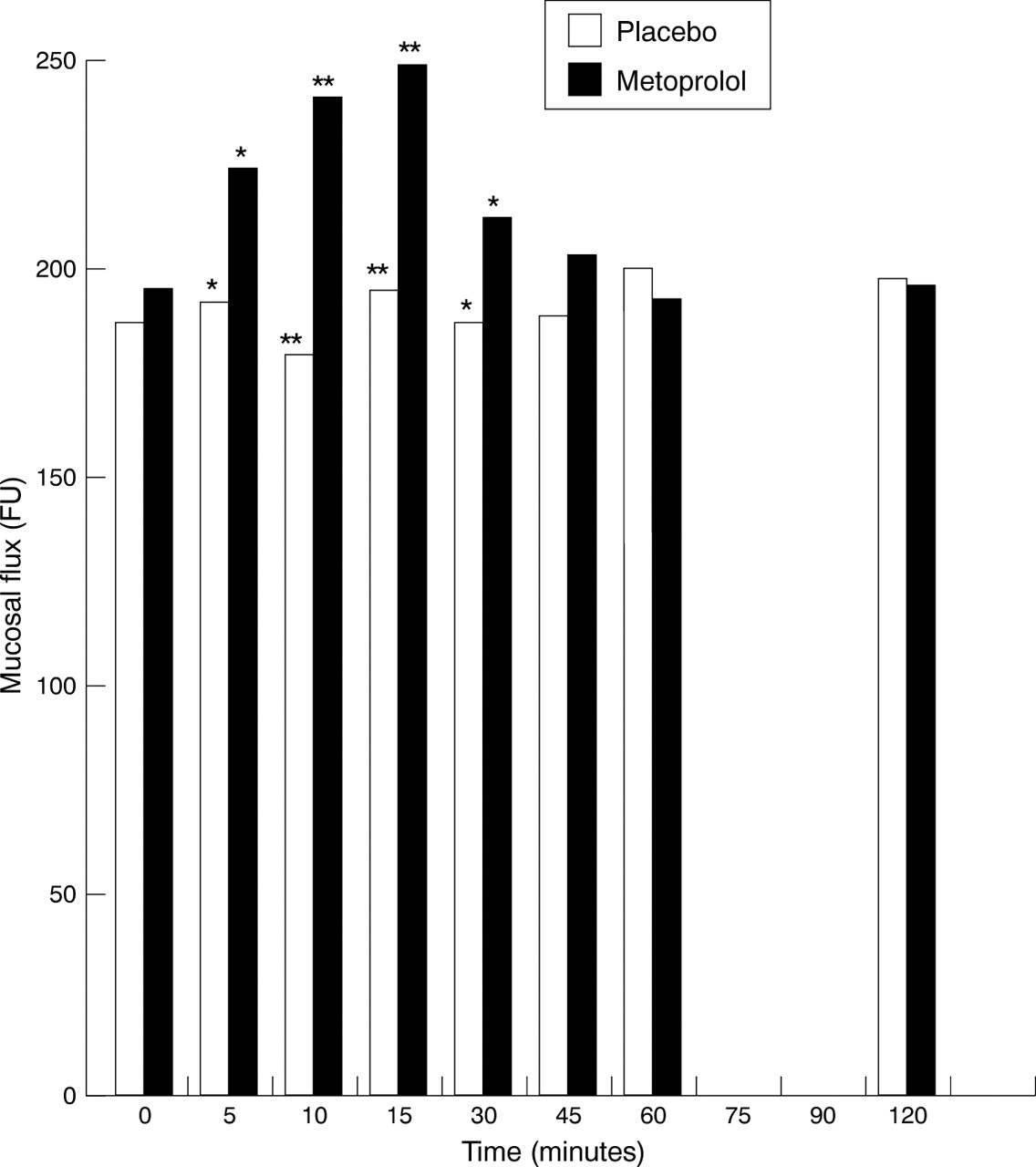
Effect of 2.5 mg intravenous metoprolol and placebo on rectal mucosal blood flow in eight healthy volunteers. Bars represent mean data.*p<0.05, **p<0.01.
EFFECT OF SACRAL NERVE STIMULATION ON MUCOSAL BLOOD FLOW
The mean discomfort threshold for the patients was 5.4 mA (4.0–7.0 mA) and in all patients, as stimulation current was increased, there was a corresponding increase in mucosal blood flow until a “plateau” level of mucosal vasodilatation was reached (fig2). The mean stimulation current to achieve the plateau was 4.0 mA (3.0–5.0 mA).

Typical graph of effect seen of direct sacral nerve stimulation on rectal mucosal blood flow in a patient with faecal incontinence.
Discussion
This study has shown that rectal mucosal blood flow can be reproducibly measured. Factors which affect this measurement have also been determined, allowing for optimal study conditions to be defined.
LDF offers a non-invasive means of studying circulation in a variety of microvascular beds. It has particular application to the gut mucosa, which receives the highest vascular perfusion of any organ, as measured by the rate of oxygen consumption.16 The technique has been used to study colonic blood flow as an index of organ viability after anastomosis7 ,17 ,18 and prolonged hypoperfusion.19 In patients with ulcerative colitis it has been used to study colonic mucosal blood flow but with widely discrepant results.10 ,12 It is therefore important to assess the optimal conditions for mucosal flux measurement. The probe we have used allows highly reproducible measurements to be made, and we have defined the circumstances required to obtain consistent results.
Four readings taken circumferentially at 90° to each other at 10 cm from the anal verge provided results which differed only slightly from each other. The CV was comparable or better than that recorded with LDF in other microvascular beds.8 ,20 ,21
Previous animal and human studies have shown LDF to be at least as accurate a measure, or even a better measure of microcirculatory flow than other established techniques such as tonometry,22 reflectance spectrophotometry,22 ,23 pulse oximetry, and ultrasound flowmetry.24 Key advantages of LDF over these other methodologies include the fact that it is less invasive and relatively free of movement artefact. When applied to a mucosal site a steady state trace was invariably obtained within 30 seconds. Values obtained were highly reproducible with only minimal variation from one day to another, suggesting that a single measurement is an accurate reflection of tissue perfusion. The test was safe and well tolerated by all subjects.
This study has established for the first time which variables may affect rectal mucosal blood flow in healthy subjects. One report describing the methodological aspects of the technique7was limited to the study of older patients with a variety of gastrointestinal and cardiovascular diseases who had undergone bowel preparation and opiate premedication.
Minimum variability of recorded flux was observed in fasted patients. Eating induced a significant rise and increased variability in mucosal blood flow, and so all subsequent studies were performed in the fasted state. A four hour fast was found to be adequate for flux values to return to baseline values. Our findings in relation to smoking complement those of Zhang and Leung14 who observed a dose related reduction of flux with cigarette smoke in anaesthetised rats.
Rectal microcirculatory flux was not shown to be subject to diurnal variation. This agrees with the results of animal studies using radioisotope and particle dilution techniques.2 ,16 Diurnal variations in blood flow are therefore more likely to relate to meals than to intrinsic circadian rhythm.
In keeping with previous observations of cyclic changes in pelvic blood flow,25 we found variations in rectal mucosal blood flow during the menstrual cycle. Flow was increased and more variable during the luteal phase. Postmenopausal women had a significant reduction of blood flow compared with women in either phase of the menstrual cycle. In the absence of ovarian activity, as occurs in postmenopausal women, peripheral blood flow is relatively lower at rest than that found in premenopausal women. These observations highlight the importance of hormonal influences in modulating pelvic blood flow26 and the need to control for this variable when investigating rectal blood flow. The increased rectal microvascular flux seen in men compared with women may be related to the endocrine environment. The peripheral blood flow changes that we have observed in relation to the ovarian cycle are likely, at least in part, to be due to the secretion of oestrogen and progesterone. Both these hormones are strongly vasoactive but their effects are often difficult to interpret, varying with the site of action, duration of exposure, and species in animal studies.27 Age had no specific demonstrable effect on mucosal blood flow in this group of healthy volunteers who had no cardiovascular disease.
Rectal mucosal blood flow was shown to be increased by an intravenous β adrenoceptor antagonist. Inhalation of a β adrenoceptor agonist produced no alteration of mucosal blood flow, but did affect skin perfusion. This suggests that an adequate blood level was achieved to affect peripheral β2 adrenoceptors. The most likely explanation for the absence of gut response is a low β2receptor density in gut blood vessels. A previous study in the human sigmoid colon28 showed that metoprolol increased motor activity. Coupled with our findings this would suggest the presence of a tonically active β1 adrenoceptor inhibitory pathway which controls colonic mucosal blood flow and motility. Although β2 adrenoceptors have been identified in colonic tissue of rats,29 ,30 it is unknown whether human colonocytes have a similar receptor population. Our data have failed to show a role for them in controlling mucosal blood flow.
We have produced evidence from both pharmacological and nerve stimulation studies that extrinsic nerves play a major role in controlling mucosal blood flow. However, we do not believe that extrinsic nerves are the only factor influencing mucosal blood flow. Inflammation, and possibly other factors, may cause a notable change in the local neural regulation of blood flow. However, given a steady state situation in which intrinsic factors are likely to be similar in different subjects, mucosal blood flow seems to be a good reflection of the level of activity of extrinsic nerves. We cannot exclude the possibility that some of the altered mucosal blood flow that one might observe in patients with a motility disorder is directly related to changes in the activity of intrinsic nerves. We have not addressed this aspect of control in these studies.
We have shown that there is a reduction of colonic mucosal blood flow in response to inhalation of ipratropium, a muscarinic receptor antagonist. It is known that in mammalian species31muscarinic receptors are involved in colonic secretion and our findings argue in favour of a tonically active cholinergic muscarinic mechanism which increases mucosal blood flow. We cannot exclude the fact that some of these changes were produced by a direct effect of the drug on intrinsic gut neurones, although the rapid response makes this unlikely.
The vasodilator response of the colonic mucosa to direct sacral nerve stimulation suggests that there is a cholinergic mediated vasodilatory input to the mucosa which is effective via sacral spinal paraympathetic efferents. The response also confirms that mucosal blood flow alterations are brought about by nerve activation, hence providing strong support for the validation of rectal mucosal flux as an index of gut specific autonomic tone.
In conclusion, measurement of laser Doppler rectal mucosal blood flow is a reproducible technique. Sex, ovulatory status, menstrual phase, absence of smoking, and eating should be controlled for. The bowel need not be prepared providing that the mucosa being studied has been visualised. There is also evidence that mucosal blood flow measurements can be used as a tool to investigate the activity of intra-abdominal autonomic tone.
Acknowledgments
We are grateful to Dr C Dore for statistical advice, and to Dr A Roy for practical and technical help in performing this work.
Abbreviations used in this paper
- CV
- coefficient of variation
- LDF
- laser Doppler flowmetry