Article Text

Abstract
Background and aims: Crohn’s disease, characterised by chronic T helper 1 (Th1) inflammation and dysmotility of the gut, is most prevalent in developed countries. Parasitic infections are most prevalent in developing countries and induce a T helper 2 (Th2) immune response. We hypothesised that this Th2 immune response protects against Th1 gut inflammation.
Methods: The parasite Schistosoma mansoni induces a transient Th2 immune response in the semipermissive rat host. 2,4,6-Trinitrobenzene sulphonic acid (TNBS) induced colitis is an experimental model of Th1-like gut inflammation. The effect of concurrent infection with S mansoni on the course of TNBS induced colitis was assessed using macroscopic and microscopic damage scores, histology, myeloperoxidase (MPO) activity assay, cytokine production assay, and by studying in vitro contractility of longitudinal and circular colonic muscle strips.
Results: TNBS induced colitis that spontaneously healed after four weeks. Concurrent infection with S mansoni significantly reduced the duration of TNBS induced colitis to two weeks, as shown by macroscopic and microscopic damage scores and by a faster decrease in colonic MPO activity. TNBS increased colonic interleukin 2 (IL-2) production whereas S mansoni increased splenic IL-4 and IL-2 levels. Contractility of longitudinal and circular muscle strips was maximally inhibited one week after TNBS and normalised after three weeks. After four weeks, longitudinal muscle strip contractility was significantly increased. Concurrent infection with S mansoni normalised longitudinal muscle contractility after one week whereas circular muscle contractility remained inhibited.
Conclusions: Concurrent infection with S mansoni significantly attenuates TNBS induced colitis in the rat. Inflammation induced disturbances in contractility of longitudinal and circular colonic muscle strips may outlast the inflammatory reaction.
- Schistosoma mansoni
- inflammatory bowel disease
- intestinal motility
- irritable bowel syndrome
- rat
- ACh, acetylcholine
- Emax, maximal contractile response
- CSA, cross sectional area
- IFN-γ, interferon γ
- IL, interleukin
- MPO, myeloperoxidase
- Th, T helper
- TNBS, 2,4,6-trinitrobenzene sulphonic acid
Statistics from Altmetric.com
- ACh, acetylcholine
- Emax, maximal contractile response
- CSA, cross sectional area
- IFN-γ, interferon γ
- IL, interleukin
- MPO, myeloperoxidase
- Th, T helper
- TNBS, 2,4,6-trinitrobenzene sulphonic acid
The incidence of Crohn’s disease is highest in the developed world,1 and reported only sporadically in the developing countries of Middle Africa and South America,2–4 which are endemic areas for intestinal parasitism.5,6 There is a positive correlation between the incidence of Crohn’s disease, a higher socioeconomic status, and high domestic hygiene in childhood.7–9 The incidence of Crohn’s disease is lower in countries with higher infant mortality.10 Genetic and environmental factors, childhood infections, and alimentation may explain this phenomenon.11
One can hypothesise that concurrent infection with helminths prevents the development of Crohn’s disease. In animals, infection with the parasite Schistosoma mansoni alters the host’s immune response to other (non)-parasite antigens.12 Concurrent infection with S mansoni accelerated the elimination of a second parasite through a T helper (Th) cell mediated mechanism.13 Coinfection of mice with Heligmosomoides polygyrus and Helicobacter felis significantly attenuated Helicobacter induced gastritis.14 Experimentally induced colitis in mice is significantly attenuated if mice are preinfected with Trichinellaspiralis or pretreated with dead eggs from S mansoni worms.15,16
The hypothesis that helminths protect the infected host against other immunologically mediated diseases is based on the mutual interaction between Th1 and Th2 lymphocytes and their respective cytokines: Th1 cytokines suppress the expansion and/or effector functions of Th2 lymphocytes and vice versa.17 This immunological balance may be of benefit for diseases that are associated with a distinct Th1 or Th2 immune response. Helminths such as S mansoni induce Th2 polarisation of the immune status, as shown by the increased production of interleukin (IL)-4, IL-5, and IL-10.18–21 On the other hand, based on the increased production and secretion of cytokines such as interferon γ (IFN-γ), IL-2, and IL-12, the chronic mucosal inflammation in Helicobacter induced gastritis and Crohn’s disease is Th1 polarised.14,22–25
In the present study, we investigated whether infection with S mansoni ameliorates the course of a Crohn’s disease-like Th1 mediated gut inflammation in rats. S mansoni infection elicits strong T cell mediated immune responses in humans, mice, and rats.18,19–21,26 The mouse is a fully susceptible host to S mansoni and develops a chronic disease that is similar, although not identical, to that seen in infected humans. The rat however is a semipermissive host, which eliminates the parasite within four weeks after the primary infection through a T cell dependent mechanism.27 Whereas the mouse immune system mainly reacts against antigens from parasite eggs, the rat immune system reacts against antigens from developing worms.18 Being a semipermissive host, the rat does not develop disease after infection but its immune status is transiently polarised towards Th2.28,29
In the this study, we used the well characterised model of 2,4,6-trinitrobenzene sulphonic acid (TNBS) induced colitis30–32 that induces a Th1-like inflammation in rats and mice.33–35 To investigate the hypothesis that infection with helminths renders the gut less prone to the development of colitis, we studied the time course of TNBS induced colitis in control rats and in S mansoni infected rats. As gut inflammation alters gastrointestinal motility in humans and animals,36–38 we also studied the effect of concurrent infection with S mansoni on the contractility of longitudinal and circular muscle strips of the TNBS inflamed colon.
MATERIALS AND METHODS
Induction of Th2 polarised immune status
Transcutaneous infection of rats with S mansoni has been described previously.29,39 Briefly, male Wistar rats (180–220 g) were fasted for 24 hours with free access to drinking water. Under sodium pentobarbital (20 mg/kg intraperitoneally) anaesthesia, a metal ring was placed on the abdomen. The ring was filled with 4 ml of water containing 2×10³ cercariae of a Puerto Rican strain of S mansoni. After 20 minutes, the water was collected to count the remaining cercariae: 96.6 (0.8)% of all cercariae penetrated the abdominal skin of the rat within 20 minutes.
Induction of Th1 mediated colitis
Induction of TNBS colitis has previously been described in detail.40,41 Briefly, fasted male Wistar rats were anaesthetised with sodium pentobarbital (20 mg/kg intraperitoneally). Then, 0.5 ml of a 15 mg TNBS in 10% ethanol solution was injected intracolonically (8 cm proximal to the anus) using a flexible cannula. After removal of the cannula, rats were held upside down for 60 seconds and transferred back to their cages.
Experimental protocols
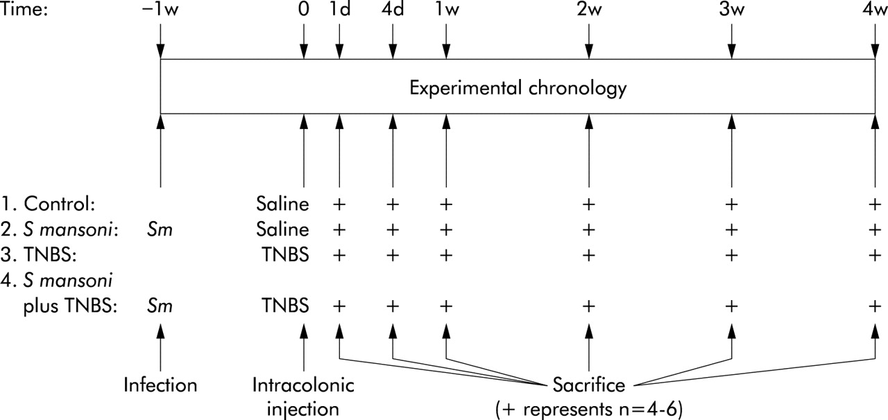
Rats were randomly divided into one of four groups (fig 1). Rats from the control group received an intracolonic injection of 0.5 ml saline. Rats from the TNBS group received an intracolonic injection of 0.5 ml TNBS. Rats from the S mansoni group were infected with 2000 cercariae of S mansoni one week before receiving an intracolonic injection of 0.5 ml saline. Rats from the S mansoni+TNBS group were infected with 2000 cercariae of S mansoni one week before receiving an intracolonic injection of 0.5 ml TNBS. Experiments were performed one day, four days, one week, two weeks, three weeks, and four weeks after intracolonic TNBS injection. Each experiment at any time interval for any experimental group was repeated at least four to six times. The day before the experiment, all animals were fasted overnight with free access to drinking water.

Experimental chronology of the study. Experiments were performed one day, four days, one week, two weeks, three weeks, and four weeks after intracolonic injection. Each experiment at any time interval for any experimental group was repeated at least four to six times.
The local ethics committee of the University of Antwerp approved all experiments.
Tissue preparation
Rats from respective groups were killed by decapitation and bleeding. The distal colon (∼4.0 cm) and spleen were removed and placed in ice cold Krebs-Ringer solution. The proximal ∼1.0 cm of the colonic segment was used for histology. The remaining segment was opened and rinsed. After scoring the gross morphological damage, the most distal ∼1.0 cm was used for the myeloperoxidase (MPO) assay. The middle ∼2.0 cm was used to determine the mucosal content of different cytokines and to prepare muscle strips for contractility experiments. The spleen was cut in two for determination of MPO and cytokine content.
Macroscopic damage score
Macroscopically visible damage of the opened colonic segment was blindly scored on a 0–10 scale using the scoring system for TNBS induced colitis in rats described by Wallace and Keenan.41 A score of 0 represents no visible damage whereas overall colitis has a maximal score of 10.
Histology and microscopic damage score
The colonic segment was fixed in 4% formaldehyde and embedded in paraffin. Morphometric analysis was performed on both haematoxylin-eosin and Sirius red stained 4 µm transverse sections in a blinded fashion, using a colour image analysis system (PC-image Colour; Foster Findlay Associates, Newcastle upon Tyne, UK). The thickness of the circular and longitudinal muscle layer was determined at three points around the circumference separated by ∼120°. The average of the three measurements was used for statistical analysis. The thickness of muscle layers is expressed in µm. In addition, the extent of colonic inflammatory damage was assessed using the scoring system described by Ameho and colleagues.42 A score of 0 represents no histological damage whereas extensive colitis with necrosis into the muscularis propria involving 50% of the specimen has a maximal score of 6.
MPO activity assay
Tissue MPO activity, which is directly related to the number and activity of myeloid cell infiltrate in inflamed tissue, was assayed to monitor the degree of inflammation.43 MPO activity was measured in the colon and spleen, as described in detail previously.43,44 Colonic and splenic MPO activity is expressed in units per g tissue.
Cytokine content of colonic mucosa and spleen
Tissue samples were blotted dry, weighed, and placed in ice cold Tris-EDTA buffer containing 0.05% sodium azide, 1% Tween 80, 2 mmol/l phenylmethylsulphonyl fluoride, and 1 µg/ml antipain, aprotinin, leupeptin, and pepstatin A at a ratio of 100 mg tissue per ml of buffer.45 Tissue samples were minced, homogenised, and centrifuged at 11 000 g for 10 minutes at 4°C. The supernatants were collected and filtered through a 0.45 µm pore filter. Splenic and colonic levels of IFN-γ, IL-2, and IL-4 were assayed by four member solid phase sandwich ELISA kits (Biosource International, California, USA) according to the manufacturer’s instructions. Colonic and splenic cytokine levels are expressed as ng protein per g colonic mucosa or spleen.
Contractility studies
Mucosa free circular and longitudinal muscle strips (length ∼1.0 cm) of the distal colon were mounted in organ baths (5 ml) filled with Krebs-Ringer solution (37°C, aerated with 5%CO2/95%O2). The strips were connected to a strain gauge transducer (Statham UC2) for recording of isometric tension. The length of the strips was measured after establishing their optimal point of length-tension relationship.46 At the end of the experiment, muscle strips were blotted dry and weighed.
Concentration-response curves to acetylcholine (ACh 0.1 nmol/l–10 mmol/l) were constructed. Contractions were expressed in g contraction per mm2 cross sectional area (CSA) of the respective muscle layer. CSA was determined as follows47,48:

Muscle thickness ratio was determined on histological colonic sections as the ratio of the circular or longitudinal muscle layer thickness to the thickness of the total muscularis externa, as explained in detail previously.48 The density of the muscle was assumed to be 1.05 mg/mm3.47 Contractions to ACh are presented as pD2 (−log mol/l) and Emax (g contraction/mm2 CSA) values.
Drugs used
All drugs were from Sigma Chemical Co. (St Louis, Missouri, USA) except hydrogen peroxide 30% and sodium azide which were from Merck (Darmstadt, Germany).
Presentation of results and statistical analysis
Macroscopic and microscopic damage
The macroscopic damage score was expressed as a whole number from 0 to 10.41 The microscopic histological damage score was expressed as a whole number from 0 to 6.42 These non-parametric values are shown as median (25% and 75% percentiles) for the number of rats indicated. For statistical analysis, the Kruskal-Wallis test followed by the Mann-Whitney test was used to compare the results of the four different groups. A p value of <0.05 was considered significant.
MPO activity, cytokine assay, and contractility studies
Values are shown as mean (SEM) for the number of rats indicated. For statistical analysis, one way ANOVA followed by the Student-Newman-Keuls test was used to compare the results of the four different groups. A p value of <0.05 was considered significant.
RESULTS
Gross morphological damage
Intracolonic injection of TNBS induced linear ulcerations surrounded by hyperaemic mucosa, leading to thickening of the colonic wall. Figure 2 shows the time course of the gross morphological macroscopic damage score41 of the distal colon in control rats, in S mansoni infected rats, in TNBS treated rats, and in rats infected with S mansoni followed by TNBS treatment. Control rats and S mansoni infected rats did not show any macroscopic signs of colonic inflammation during the course of the study. Intracolonic injection of TNBS resulted in an immediate inflammatory reaction, which was maximal at day 1 (median score 5.0 (3.5–6.0); n = 6) and which was still evident but reduced to a mild residual inflammatory infiltrate four weeks later. Concurrent infection with S mansoni significantly reduced the inflammatory response to TNBS at day 1 (median score 3.0 (2.0–4.0); p<0.05, n = 6) and shortened the duration of colitis to two weeks.

Macroscopic damage score of the colon in the four groups of rats: control rats; rats infected with Schistosoma mansoni alone; rats given an intracolonic injection of 2,4,6-trinitrobenzene sulphonic acid (TNBS) alone; and rats with concurrent infection with S mansoni plus intracolonic injection of TNBS. Results are expressed as boxplots of the median. *p<0.05 compared with the TNBS group (Kruskal-Wallis test followed by a post hoc Mann-Whitney test, n = 4–6).
Histology
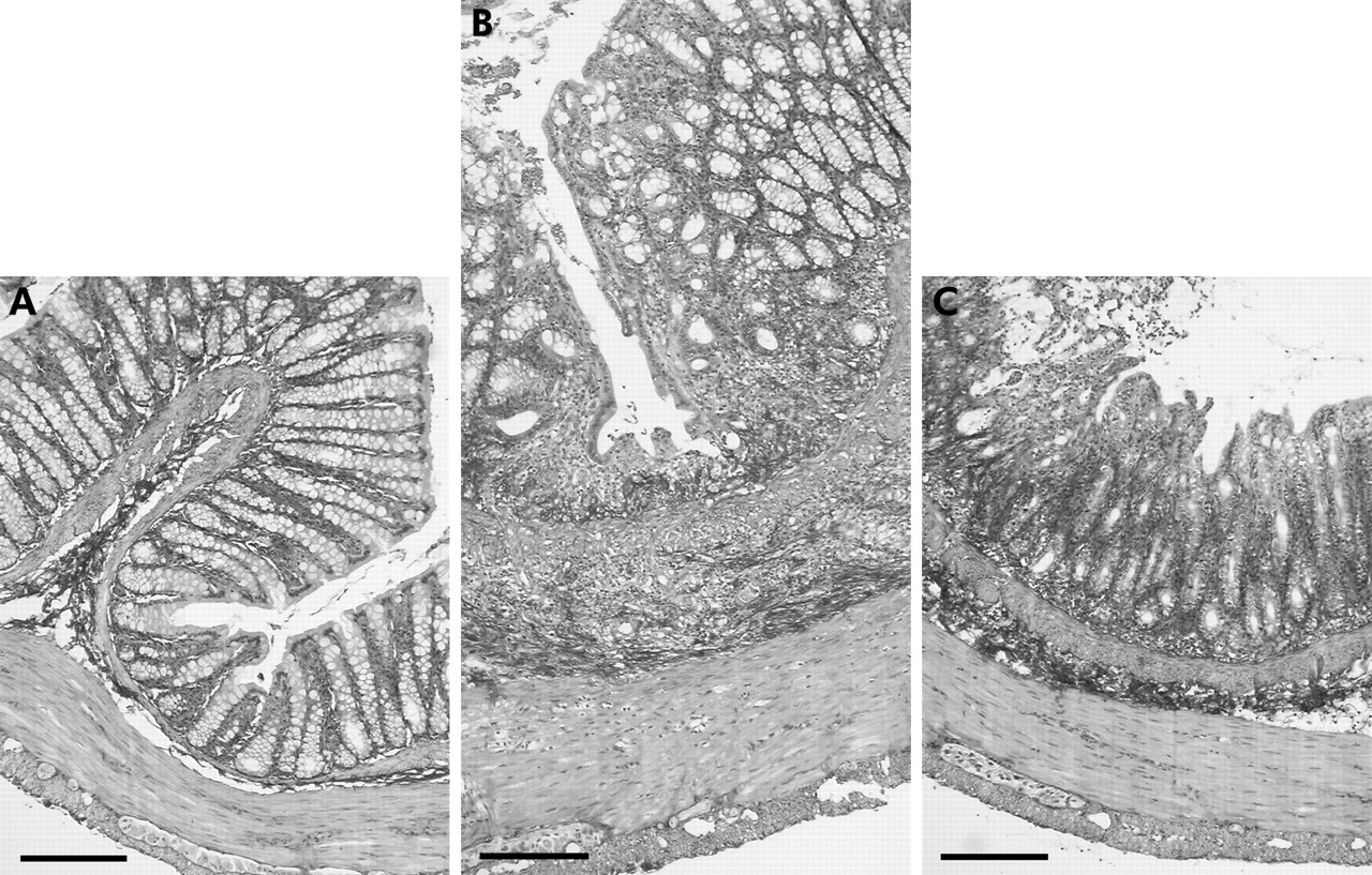
No histological damage was observed in the colon of control and S mansoni infected rats. Compared with the colon of control rats, TNBS induced a distortion of the colonic mucosal architecture and an inflammatory infiltration within the oedematous lamina propria after one week (fig 3). The infiltrate extended into the circular muscle layer. Concurrent infection with S mansoni accelerated the mucosal regeneration and reduced the inflammatory infiltrate (figs 3, 4). Validation of microscopic damage according to Ameho and colleagues42 showed that the inflammatory damage in TNBS treated rats was maximal at day 4 (median score 5.0 (4.0–5.5); n = 6) and was spontaneously reduced but still evident four weeks after TNBS treatment (median score 1 (0.5–1.5); n = 6). The inflammatory damage in S mansoni infected rats that were treated with TNBS was maximal at day 1 (median score 4.5 (4–5.5); n = 6). Concurrent infection with S mansoni significantly shortened the duration of the TNBS induced inflammatory infiltrate. A mild residual mucosal infiltrate was still observed three weeks after TNBS treatment but no signs of histological damage were observed four weeks after TNBS treatment in S mansoni infected rats.

Cross section of the distal colon of a control rat (A), a rat four days after intracolonic injection of 2,4,6-trinitrobenzene sulphonic acid (TNBS) (B), and a Schistosoma mansoni infected rat four days after intracolonic injection of TNBS (C). The images are printed at the same final magnification. Note disruption of the epithelial barrier, distortion of the mucosal villi, and transmural inflammatory infiltrate after intracolonic injection of TNBS. Concurrent infection with S mansoni markedly attenuated these histological signs of inflammation. Bar represents 200 µm (Sirius red staining, ×100).
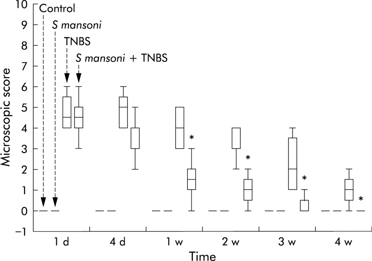
Microscopic damage score validated on histological sections of the colon in the four groups of rats: control rats; rats infected with Schistosoma mansoni alone; rats given an intracolonic injection of 2,4,6-trinitrobenzene sulphonic acid (TNBS) alone; and rats with concurrent infection with S mansoni plus intracolonic injection of TNBS. Results are expressed as boxplots of the median. *p<0.05 compared with the TNBS group (Kruskal-Wallis test followed by a post hoc Mann-Whitney test, n = 4–6).
MPO activity assay
Colonic MPO activity in control rats was 1.5 (0.2) U/g mucosa (fig 5A) and did not significantly change throughout the course of the study. Colonic MPO activity in S mansoni infected rats was 1.7 (0.2) U/g mucosa without significant alterations throughout the course of the study. One day after TNBS treatment, colonic MPO activity was increased 10-fold to 17.2 (5.2) U/g mucosa. This gradually decreased to normal values three weeks after intracolonic injection of TNBS. Concurrent infection with S mansoni did not significantly modulate MPO activity one day after TNBS treatment but accelerated normalisation of the TNBS induced increase in MPO activity. After four days, mucosal MPO activity of S mansoni infected rats with TNBS induced colitis was significantly lower (12.3 (1.7) U/g mucosa) compared with rats with TNBS induced colitis alone (18.4 (4.3) U/g mucosa). One week after TNBS treatment in S mansoni infected rats, colonic MPO activity (3.1 (1.3) U/g mucosa) was not different from time control values (2.0 (0.5) U/g mucosa) whereas it was still 8.9 (3.5) U/g mucosa in rats with TNBS induced colitis alone.

Myeloperoxidase (MPO) activity of the colonic mucosa (A) and spleen (B) in the four groups of rats: control rats; rats infected with Schistosoma mansoni alone; rats given an intracolonic injection of 2,4,6-trinitrobenzene sulphonic acid (TNBS) alone; and rats with concurrent infection with S mansoni plus intracolonic injection of TNBS. *p<0.05 compared with controls and S mansoni infected rats; †p<0.05 compared with controls and TNBS alone (one way ANOVA followed by Student-Newman-Keuls test, n = 4–6). Results are expressed as units per gram tissue colonic mucosa or spleen. Splenic MPO activity was significantly increased in all S mansoni infected rats, independent of intracolonic injection of TNBS or saline. Differences were independent of the time course of infection.
Splenic MPO activity was significantly increased in all S mansoni infected rats, independent of intracolonic injection of TNBS or saline (fig 5B). The differences were independent of the time course of infection. Mean splenic MPO activity of control rats was 8.9 (0.3) U/g spleen (n = 30). This was not significantly different from mean splenic MPO activity in rats after TNBS treatment (10.3 (0.4) U/g spleen, n = 28). However, in S mansoni infected rats with intracolonic injection of either saline or TNBS, mean splenic MPO activity was significantly increased to 12.1 (0.8) U/g spleen (p<0.05, n = 24) and 12.5 (0.8) U/g spleen (p<0.05, n = 24), respectively.
Cytokine content of colonic mucosa and spleen
Infection with S mansoni induced a transient increase in splenic IL-4 content, irrespective of whether rats received intracolonic injection of saline or TNBS (fig 6C). Also, splenic IL-2 content significantly increased after infection with S mansoni (fig 6B). However, subsequent intracolonic injection of TNBS attenuated splenic IL-2 production. No significant alterations in splenic IFN-γ content were noted (fig 6A).

Interferon γ (IFN-γ) (A), interleukin (IL)-2 (B), and IL-4 (C) production in the spleen in the four groups of rats: control rats; rats infected with Schistosoma mansoni alone; rats given an intracolonic injection of 2,4,6-trinitrobenzene sulphonic acid (TNBS) alone; and rats with concurrent infection with S mansoni plus intracolonic injection of TNBS. *p<0.05 compared with controls and TNBS alone; †p<0.05 compared with rats infected with S mansoni alone (one way ANOVA followed by Student-Newman-Keuls test, n = 4–6).
Colonic IL-2 levels were significantly increased four days and one week after intracolonic injection of TNBS (fig 7B). Concurrent infection with S mansoni prevented this transient rise in IL-2 secretion and tended to induce a rise in colonic IL-4 content, although this increase was not statistically significant (fig 7B, 7C). Although a slight rise in colonic IFN-γ production was noted, the increase was not statistically significant (fig 7A).

Interferon γ (IFN-γ) (A), interleukin (IL)-2 (B), and IL-4 (C) production in the colonic mucosa in the four groups of rats: control rats; rats infected with Schistosoma mansoni alone; rats given an intracolonic injection of 2,4,6-trinitrobenzene sulphonic acid (TNBS) alone; and rats with concurrent infection with S mansoni plus intracolonic injection of TNBS. No significant differences were found in colonic IL-4 production (one way ANOVA followed by Student-Newman-Keuls test, n = 4–6). *p<0.05 compared with controls, S mansoni infected rats, and S mansoni with intracolonic injection of TNBS rats (one way ANOVA followed by Student-Newman-Keuls test, n = 4–6).
Contractility study
In circular and longitudinal muscle strips of control rats, ACh induced concentration dependent contractions (fig 8). Contractility per contractile unit of circular muscle was half of the contractility per contractile unit of longitudinal muscle (fig 8). The mean Emax value for ACh in longitudinal muscle strips of control rats was 40.6 (2.5) g/mm2 (n = 6) and the mean pD2 value was 8.2 (0.6) (n = 6). In circular muscle strips these values were 20.6 (0.9) g/mm2 (n = 6) and 4.6 (0.2) (n = 6), respectively.

Cumulative concentration-response curves to acetylcholine (ACh) in longitudinal and circular muscle strips of the colon of control rats. Contractions are expressed as g contraction per cross sectional area (CSA) of the longitudinal and circular muscle layers. Emax represents the maximal contractile response. pD2 represents the negative logarithm of the molar concentration inducing 50% of the maximal contractile response.
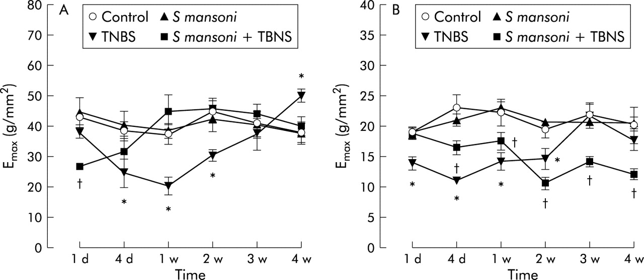
For the longitudinal muscle layer, the change over time in Emax values for ACh in the four different groups are shown in fig 9A. Intracolonic injection of TNBS significantly reduced the maximal contractile response to ACh. Inhibition was maximal one week after TNBS treatment. After this initial decrease, the Emax value gradually increased and normalised after three weeks. It increased further after four weeks to become significantly different from the Emax value in strips of age matched controls. Concurrent infection with S mansoni caused immediate inhibition of the Emax value for ACh one day after intracolonic injection of TNBS. However, at later time points, the Emax value was not different from control values. S mansoni infection alone had no effect on the Emax value for ACh in longitudinal muscle strips. pD2 values for ACh in control rats were not significantly different in the other treatment groups and they did not change significantly over time (data not shown).

Time course of the maximal contractile response (Emax) values for acetylcholine in longitudinal (A) and circular (B) muscle strips in the four groups of rats: control rats; rats infected with Schistosoma mansoni alone; rats given an intracolonic injection of 2,4,6-trinitrobenzene sulphonic acid (TNBS) alone; and rats with concurrent infection with S mansoni plus intracolonic injection of TNBS. *p<0.05 compared with the other groups; †p<0.05 compared with the other groups (one way ANOVA followed by the Student-Newman-Keuls test, n = 4–6).
For the circular muscle layer, the change over time in Emax values for ACh in the four different groups is shown in fig 9B. Intracolonic injection of TNBS significantly reduced the maximal contractile response to ACh. Inhibition was maximal four days after TNBS treatment and normalised after three weeks. Concurrent infection with S mansoni led to a gradual inhibition of the Emax value for ACh starting four days after TNBS treatment. The Emax value remained significantly lower than the Emax value of age matched controls throughout the course of the experiment. S mansoni infection alone had no effect on the Emax value for ACh in circular muscle strips. The mean pD2 value for ACh (4.6 (0.2); n = 6) in control rats remained unchanged throughout the course of the experiment (data not shown). However, the decrease in Emax values due to TNBS treatment was associated with an increase in the pD2 value to 5.7 (0.3) (p<0.05 v controls, n = 4) one day after induction of colitis. Concurrent infection with S mansoni also led to a significant increase in the pD2 value for ACh one day after intracolonic injection of TNBS (6.1 (0.2); p<0.05 v controls, n = 4).
DISCUSSION
The incidence of Crohn’s disease and helminthic infections seems to be inversely correlated.1–4,6 Genetic and environmental factors, childhood infections, and alimentation may explain this phenomenon.11 Dysregulation of the mucosal immune system was shown to play a pivotal aetiopathological role in the development of Crohn’s disease.23–25 People living in endemic areas such as Middle Africa and South America are infected with helminths early in life. As helminths induce strong Th2-like immune responses, it is possible that these people become less prone to develop Crohn’s disease due to the anti-inflammatory actions of Th2 cytokines. Crohn’s disease and TNBS induced colitis in mice and rats are both Th1-like forms of gut inflammation33–35 whereas infection with S mansoni in rats is associated with a Th2-like immune response.28,29 Th1 cytokines suppress the expansion and/or effector functions of Th2 lymphocytes and vice versa.17 Using the rat model of TNBS induced colitis, we showed that helminthic infection with S mansoni attenuates gut inflammation.
In the present study, TNBS induced colitis in rats spontaneously healed after four weeks, as shown by the standardised gross morphological and histological damage scores41,42 and by the colonic MPO activity assay. As we used low concentrations of TNBS and ethanol, the severity and duration of colitis was less severe compared with previous reports.40 Concurrent infection with S mansoni significantly attenuated the course of TNBS induced colitis. The macroscopic damage score was normalised after two weeks, the microscopic damage score was normalised after three weeks, and colonic MPO activity was normalised as early as one week after intracolonic injection of TNBS. Similar downregulation of MPO activity was reported in mice with colitis that were preinfected with Trichinella spiralis.15 Histology showed a reduced inflammatory infiltrate. This may be based on reduction of the initial inflammatory insult or on acceleration of the healing process. This is in line with the observation that intraperitoneal injection of dead schistosome eggs protects mice against experimental colitis.16
As the colonic histological damage score and MPO activity were similarly increased one day after intracolonic injection of TNBS with or without concurrent infection with S mansoni, we would assume that the initial inflammatory response itself was not reduced. However, the gross morphological damage score of S mansoni infected rats with TNBS induced colitis was significantly lower from the first day onwards.
The beneficial effect of schistosomiasis on TNBS induced colitis may be based on immunological alterations. It was previously shown that S mansoni induces a Th2 immune response in the semipermissive rat host.28 In general, the systemic immune response to S mansoni in rodents can be followed by measuring cytokine production in the lungs, liver, spleen, or mesenteric lymph nodes.28 In the present study, we evaluated the systemic immune response by analysing cytokine production in the spleen. Infection of rats with S mansoni induced a transient rise in splenic levels of the Th2 cytokine IL-4. However, splenic IL-2 levels—a Th1 cytokine—also gradually rose during the course of the infection whereas IFN-γ levels remained unchanged. This is in accordance with previous observations showing that splenic production of Th2 cytokines is increased more abundantly compared with Th1 cytokines in S mansoni infected rats.28,29 A rise in Th1 cytokines after infection with S mansoni is unexpected as this parasite is known to elicit a Th2 immune response in the infected host.18–21 According to Cêtre et al, the unexpected increase in splenic IL-2 without an increase in IFN-γ might be due to lymphoproliferation after an antigenic stimulation.28 Although in the present study no quantitative method was used to count splenic lymphocytes, proliferation of leucocytes within the spleen is suggested by the overall increase in splenic MPO activity after infection with S mansoni, irrespective of the presence of TNBS induced colitis. The reason for the isolated increase in splenic IL-2 in the rat after S mansoni infection remains largely unexplained. The rat, in contrast with the mouse, is a semipermissive host for S mansoni. The rise in splenic IL-2 levels in the S mansoni infected rat may therefore be species dependent as murine splenic cells produce IFN-γ after infection with S mansoni.17–19 Interestingly, TNBS treatment abrogated this increase in splenic IL-2, indicating that topical colonic treatment with TNBS also resulted in systemic immune effects.
In the rat colon, TNBS treatment induced a transient rise in colonic IL-2 levels and unexpectedly only a slight increase in colonic IFN-γ production but this was not statistically significant. Colonic IFN-γ levels were a multiple of IL-2 and IL-4 levels in control animals. The high basal level of colonic IFN-γ may be the reason why the expected increase after TNBS treatment was not substantial. Although IL-2 and IFN-γ are considered prominent Th1 cytokines, other studies also failed to show an increase in IFN-γ production during intestinal inflammation.49,50 In addition, levels of IFN-γ may also vary depending on the phase of the inflammatory response.51 In the present study, colonic IL-2 levels were clearly increased after TNBS treatment, and colonic IFN-γ levels tended to increase, indicating that TNBS induced Th1 colitis. The rise in colonic IL-2 production was completely abrogated when rats were infected with S mansoni. Although our results suggest that differential production of cytokines may play a role in the attenuation of TNBS induced colitis after infection with S mansoni, further studies with specific cytokine blocking agents are needed to unravel the exact underlying mechanisms.
Gut inflammation leads to disturbed gastrointestinal motility.36–38 The model of TNBS induced colitis is widely used to investigate motility disturbances occurring in the inflamed rat colon. In vivo, an initial increase in colonic transit and decrease in colonic myoelectrical activity is noted within the first hours after TNBS installation, followed by a progressive decrease in transit and increase in colonic myoelectrical activity.52,53 In vitro studies showed increased contractility of the circular smooth muscle layer four hours after intracolonic injection of TNBS whereas after one week contractility was decreased, irrespective of the type of contractile stimulus.47,54
In the present study, we investigated the contractile responses of circular and longitudinal muscle layers to ACh during the course of TNBS induced colitis. Although the circular muscle layer encompasses the majority of the total muscle mass, its contractile capacity per contractile unit is only half the capacity of the longitudinal muscle layer. Contractility of longitudinal muscle strips was time dependently decreased during colitis, with maximal inhibition one week after intracolonic injection of TNBS. After three weeks, the contractile response was normalised and at the time of complete mucosal healing contractility of the longitudinal muscle layer was significantly increased. This hypercontractility supports the hypothesis that gastrointestinal inflammation may cause prolonged dysmotility55,56 which may lead to symptoms such as post-infectious irritable bowel syndrome.57–59 Concurrent infection with S mansoni not only attenuated the course of the inflammatory reaction, it also abrogated the TNBS induced contractility disturbances of the longitudinal muscle layer. Contractility of circular muscle strips was also time dependently decreased during TNBS induced colitis. Similar to the longitudinal muscle layer, circular hypocontractility was normalised after three weeks. However, no post-inflammation hypercontractility was found. On the contrary, concurrent infection with S mansoni prolonged the inhibition of circular muscle contractility. These results illustrate that normalisation of inflammation induced dysmotility of the gut does not necessarily follow the time course of the mucosal healing process. It also illustrates that circular and longitudinal muscle layers can behave differently in response to gut inflammation. It is known that the longitudinal and circular muscle layers of the gastrointestinal tract show several differences in innervation, contractile pattern, and signal transduction pathways under normal conditions.60 However, only few studies have compared the behaviour of both smooth muscle layers during inflammation. Martinolle et al showed that one week after induction of ileal inflammation, receptor mediated contractile responses of the longitudinal smooth muscle layer were differently modulated compared with the circular smooth muscle layer, suggesting alterations of specific receptor pathways.61 Shi and Sarna showed that ileal inflammation differently modulated the role of M2 and M3 subtypes of muscarinic receptors of longitudinal and circular smooth muscle cells.60 In the present study, we also found differential behaviour of both smooth muscle layers. Firstly, the contractile capacity per contractile unit of the circular muscle layer was only half the contractile capacity of the longitudinal muscle layer. Secondly, the disturbed contractile response of the longitudinal smooth muscle layer in TNBS treated rats was normalised more rapidly when rats were infected with S mansoni. However, this was not observed in the circular muscle layer. Accelerated normalisation of the longitudinal muscle layer may be related to the accelerated healing of the inflamed mucosa, resulting in a reduced action of inflammatory mediators on the contractile apparatus. Another explanation is that a progressive increase (although not statistically significant in our study) in IL-4 production in the inflamed colonic mucosa of S mansoni infected rats has a beneficial effect on the disturbed muscle contractility. Little is known of the effect of IL-4 on smooth muscle cell contractility. Possibly, the differential response of both smooth muscle layers is related to a differential action of IL-4 or other Th2 cytokines.
In conclusion, we showed that concurrent infection of the semipermissive rat host with S mansoni significantly attenuates the course of TNBS induced colitis. The mechanism of action may be based on the immunomodulatory interaction of Th1 and Th2 cytokines but this needs further confirmation. Inflammation induced contractility disturbances of both the circular and longitudinal muscle layers outlasts the mucosal inflammatory reaction.
Acknowledgments
The authors thank Gunther Vrolix for his excellent technical assistance with Schistosoma mansoni infection of rats. This work was supported by the Belgian Interuniversity Poles of Attraction Programme (grant No P5/20, Services of the Prime Minister-Federal Agency for Scientific, Technical and Cultural Affairs) and by the Crohn en Colitis Ulcerosa Vereniging vzw, Belgium.
REFERENCES
Linked Articles
- Commentary