Article Text

Abstract
BACKGROUND Trefoil factors (TFFs) are peptides produced by mucus-secreting cells in the gastrointestinal tract. A functional association between these peptides and mucus, leading to stabilisation of the viscoelastic gel overlying the epithelia, has been suggested. Both oral and parenteral administration of the peptides increase the resistance of the gastric mucosa.
AIM To study the effect in rats of oral and parenteral porcine trefoil factor 2 (pTFF2) on the healing of gastric and duodenal ulcerations and to clarify the distribution and metabolism of orally administered pTFF2 in the gastrointestinal tract.
METHODS Gastric ulcers were induced in female Sprague-Dawley rats by indomethacin and duodenal ulcers by mercaptamine. The rats were treated for up to seven days with oral or subcutaneous pTFF2. Ulcer size after treatment was assessed by stereomicroscopy after whole mount staining with periodic acid-Schiff stain. 125I-labelled pTFF2 was given orally to rats, and tissues were investigated by gamma counting of samples and by autoradiography of paraffin embedded sections.
RESULTS pTFF2 accelerated gastric ulcer healing after both oral and subcutaneous administration. Duodenal ulcers were aggravated by both treatments. After oral administration of 125I-pTFF2, intact peptide was recovered from the superficial part of the mucus layer in the stomach; it passed through the small intestine but was degraded in the caecum. Only a minor part of the labelled pTFF2 entered the colon and was excreted in the faeces. Most of the label was excreted in the urine.
CONCLUSIONS Oral as well as parenteral pTFF2 accelerates the healing of gastric ulceration and aggravates duodenal ulcers. Oral pTFF2 binds to the mucus layer of the stomach and the small intestine but does not reach the colonic mucosa.
- trefoil factors
- spasmolytic polypeptide
- ulcer healing
- gastric ulcer
- duodenal ulcer
- rat
Abbreviations used in this paper
- TFF
- trefoil factor
- pTFF
- porcine TFF
- hTFF
- human TFF
- rTFF
- rat TFF
Statistics from Altmetric.com
The trefoil factors (TFFs) constitute a family of mucin associated gastrointestinal peptides that create a characteristic three leafed structure because of the existence of domains in which six cysteine residues form three disulphide bonds.1-3 They are produced in exocrine usually mucus-secreting cells in the gastrointestinal tract,4-8 and there is increasing evidence that they are important in the maintenance of the integrity of the gastric and intestinal mucosa. The peptides are extremely resistant to degradation by proteases9; they are co-secreted with mucus and are supposed to be involved in oligomerisation of mucin polysaccharide molecules, processes that lead to the formation of a gel-like high viscosity mucus.10-12
Today the mammalian TFF family includes three peptides: TFF1, also named breast cancer associated peptide or pS2,6 and TFF2, or spasmolytic polypeptide (SP), are both present mainly in the stomach4; TFF3, or intestinal trefoil factor (ITF), is present mainly in the intestinal goblet cells.7 ,8 ,13
To elucidate the role of the TFFs in the gastrointestinal tract, their ability to protect the gastric mucosa against experimental ulceration has been investigated. Babyatsky et al 14 showed that oral human TFF2 (hTFF2) and rat TFF3 (rTFF3) given before ulcer induction protected against both ethanol induced and indomethacin induced gastric injury. Playfordet al 15 surprisingly found that even small doses of hTFF2 given subcutaneously to rats prevented the formation of indomethacin induced gastric ulceration, indicating that TFF2 also can exert its effect in an endocrine manner, which may imply the presence of a receptor for the peptides. In fact, a putative receptor for TFF3 has recently been characterised,16 ,17and it has been shown that parenterally administered porcine TFF2 (pTFF2) is taken up by mucus-secreting cells in the stomach and secreted into the mucus layer in the same way as endogenous TFF2.18
In the study of Babyatsky et al,14 there was no or very little effect of intraperitoneally administered peptide, and Playfordet al 15 found that oral hTFF2 had no effect. Thus there are conflicting results with respect to the protective effect on the gastric mucosa of both oral and systemic TFF.
In this study we compared oral and systemic pTFF2 not with respect to the protective effect, but with respect to the healing of gastric and duodenal ulcerations, and we describe the metabolism and distribution in the gastrointestinal tract of orally administered pTFF2 in order to elucidate the possible role of oral TFFs in gastrointestinal disorders.
We report that pTFF2 accelerates gastric ulcer healing after both oral and subcutaneous administration. Only very small doses are necessary to obtain an effect of oral treatment. Surprisingly, duodenal ulcers are aggravated by both treatment regimens. Orally administrated pTFF2 binds in the mucus layer of the stomach and also in the intestine. It seems to be degraded in the caecum, and only very small amounts enter the colon.
Materials and methods
ULCER INDUCTION
Female Sprague-Dawley rats weighing 180–200 g were used throughout the study. They were fed a standard laboratory diet (Altromin 1314, Lage, Germany). The study was approved by the Danish National Committee for Animal Studies.
Duodenal ulceration was induced with mercaptamine.19 For ulcer induction, the rats were fasted for five hours in raised mesh bottom cages and thereafter given 30 mg/kg mercaptamine hydrochloride (Sigma Chemical Co, St Louis, Missouri, USA) subcutaneously. Gastric ulceration was induced by means of indomethacin and restraint.20 Rats were fasted for 12 hours and given indomethacin 20 mg/kg subcutaneously. They were restrained by being placed in Bolmann cages for three hours.
The rats were killed after being anaesthetised with barbiturate (Brietal, methohexital; 50 mg/kg intraperitoneally; Eli Lilly, Indiana, USA). The stomach and duodenum were removed, the distal end of the oesophagus and the duodenum were ligated, and the organs were fixed by intraluminal injection of 10% formalin for 10 minutes. Thereafter the stomach was opened along the greater curvature and suspended on a polyethylene plate and fixed for a further 24 h. The specimens were then flushed with water and stained as whole mounts with periodic acid-Schiff (PAS).21 After staining they were rinsed with tap water and stored in 70% ethanol. The surface of the gastric and duodenal mucosa, immersed in 70% ethanol, was investigated and photographed with a Wild photomacroscope (fig 1). Determination of the number and size of the ulcers was performed using an image analysis program (NIH Image 1.59) run on a Power Macintosh (8500/120) computer with a high resolution camera (Hamamatsu C2400, Hamamatsu Photonics KK, Hamamatsu, Japan).
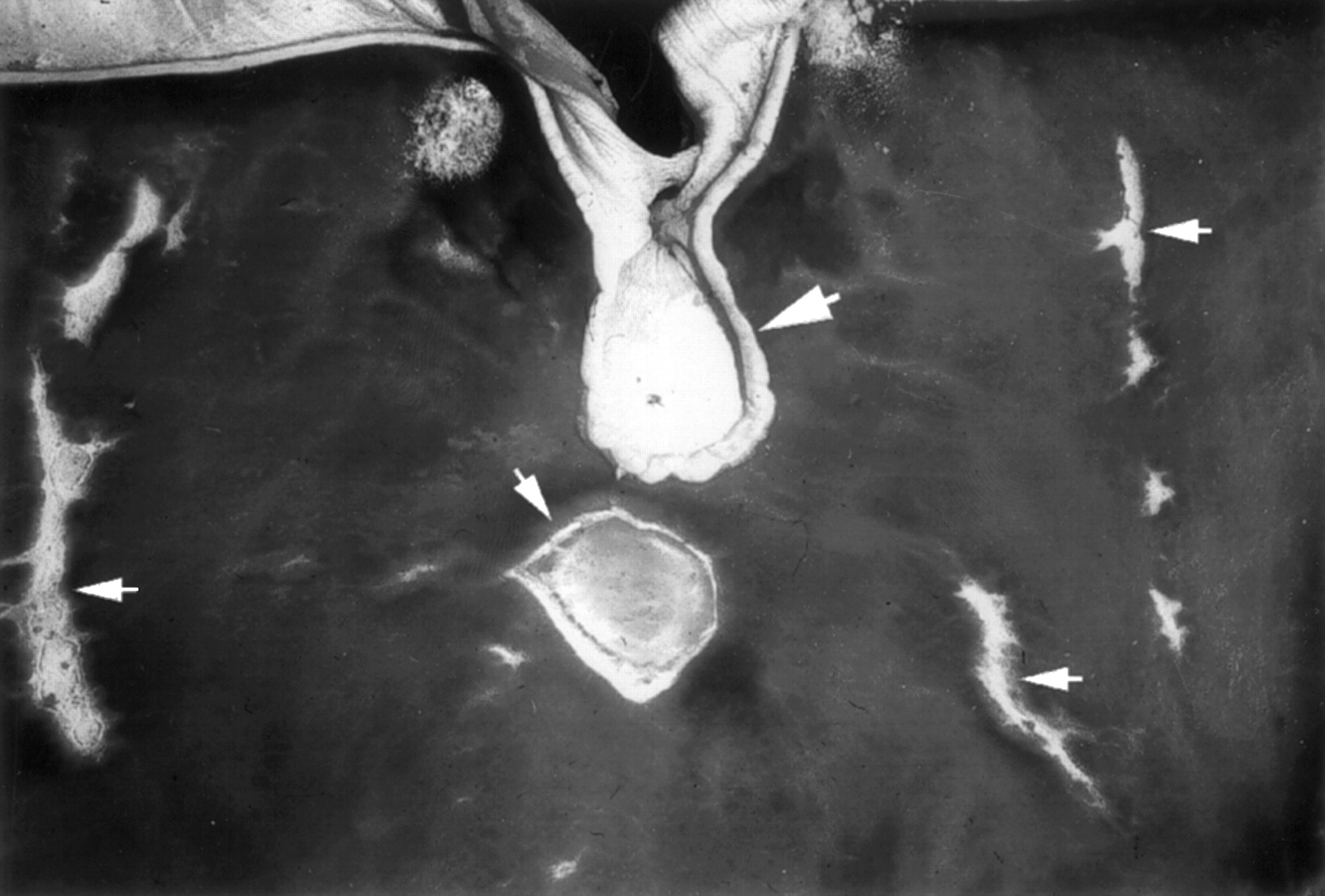
Rat stomach with indomethacin induced ulcers after whole mount staining with periodic acid-Schiff. The specimen has been photographed with a Wild photomacroscope. The borderline of the rumen is in the upper part of the photograph. The gastro-oesophageal junction is indicated by a large arrow and the ulcers are seen as well defined white areas (small arrows). Original magnification × 4.
ULCER HEALING
Gastric and duodenal ulceration was induced as described above. Treatment was not initiated until 48 hours after ulcer induction to make sure that ulceration had developed fully at the onset of treatment.
Oral treatment with pTFF2
Gastric ulceration was induced with indomethacin in 14 rats. Seven were given pTFF2 in the drinking water (2 mg pTFF2 in 20 ml water per rat per day). They drank all the water, and to ensure that they were not dehydrated, they were allowed to drink water ad libitum for one hour every morning. Controls received tap water following the same schedule. The rats were killed after seven days of treatment (nine days after ulcer induction).
Duodenal ulceration was induced in 32 rats. Half of them received pTFF2, 3 mg in 20 ml water per rat per day as described above, and the rest received tap water. The rats were divided into groups of eight and killed after one or three days of treatment.
Subcutaneous treatment with pTFF2
Gastric ulceration was induced with indomethacin in 84 rats. The rats were divided into four groups of 21. One group received 50 μg/kg pTFF2 in 1 ml saline subcutaneously three times a day, one received 1 mg/kg three times a day, one served as controls which were given 1 ml saline × 3, and one served as positive controls for the effect of treatment and received omeprazole, 3 mg × 2 as intramuscular injections. The rats were killed in groups of seven after one, three, or seven days of treatment.
Duodenal ulceration was induced with mercaptamine in 42 fasted rats. They were divided into three groups of 14. One group received 50 μg/kg pTFF2 in 1 ml saline subcutaneously three times a day, one received 1 mg/kg three times a day, and one served as controls which were given 1 ml saline × 3. The rats were killed in groups of seven after one or three days of treatment.
METABOLISM OF 125I-LABELLED pTFF2
Preparation of 125I-pTFF2
pTFF2 (100 μg), highly purified as previously described,6 was iodinated by Amersham International, Amersham, Bucks, UK, with Na125I, using hydrogen peroxide and lactoperoxidase.7 The specific radioactivity obtained was 222 kBq/μg (6 μCi/μg). The iodinated peptide was diluted to a radioactive concentration of 100 μCi/ml with 5% lactose, 0.25% radioimmunoassay grade bovine serum albumin, and 0.3 TIU/ml aprotinin in 50 mM sodium phosphate buffer, pH 7.4. Before use125I-pTFF2 was diluted in 0.154 mM saline with 1% albumin to a final concentration of 0.5 μg/ml (about 1 × 10⁶cpm/ml).
Tissue distribution and metabolism of oral 125I-TFF2
Twenty female Sprague-Dawley rats were fasted for 12 hours and given 950 μl 125I-pTFF2 by stomach tube. A sample of 50 μl was counted with a gamma spectrometer (Wallac LKB, Crownhill, UK) in order to calculate the amount of radioactivity given to each rat. The rats were killed in groups of four after 30 minutes, 2, 4, 12, or 24 hours. Rats in the last two groups were kept in individual metabolic cages (Techniplast, model 1700; Paolo, Malta) in order to collect urine and faeces. When killed the rats were anaesthetised by an intraperitoneal injection of 50 mg/kg methohexital (Brietal), connected to an animal respirator (Harvard Apparatus, Holliston, Massachusetts, USA), and the thorax was opened. A 1 ml sample of blood was drawn from the right cardiac ventricle and urine was collected from the bladder. The vascular system was then perfused through the left cardiac ventricle (outlet through the right atrium) with 150 ml saline to ensure removal of blood from the organs, and thereafter fixed by perfusion for two minutes with ice cold freshly prepared 4% glutaraldehyde in 0.04 M phosphate buffer, pH 7.4. The stomach, duodenum, the small intestine divided into 10 cm sections, the caecum, and the colon, all with their contents, were counted for radioactivity together with samples of known weight of liver, kidney, skin, muscle fat, thyroid, urine, faeces, and blood in a gamma spectrometer. The radioactivity was calculated as the percentage of the total radioactivity administered to each rat present in each organ or as the percentage of total radioactivity per gram of tissue in each organ.
The tissue localisation of radioactivity was investigated by autoradiography. The tissues were postfixed for 24 hours in 70% ethanol, dehydrated, and embedded in paraffin. Histological sections 5 μm thick were coated with Ilford K2 liquid autoradiographic emulsion diluted 1:1 in 300 mM ammonium acetate, placed in light-tight boxes, and stored at −80°C for seven weeks. The autoradiographs were developed in an Ilford developer for five minutes at 17°C and fixed in Kodak fixer for five minutes. The sections were stained slightly with haematoxylin and examined by light microscopy.
Biochemistry
Degradation of the 125I-pTFF2 was examined by trichloroacetic acid precipitation. Samples of unfixed tissue were taken from the last segment of small intestine including the luminal contents from the group of rats killed after 120 min. Also urine collected from rats killed after 24 hours was investigated.
Urine and tissue samples were kept frozen at −20°C for analysis of peptide bound label by trichloroacetic acid precipitation. Samples (500 μl tissue extract, 500 μl urine diluted 1 + 249 with phosphate buffer, or 500 μl 125I-pTFF2 diluted with phosphate buffer) were counted in a gamma spectrometer. A 1 ml volume of ice cold 20% trichloroacetic acid was added to each sample. The samples were mixed and incubated on ice for 30 minutes. The pellet obtained after centrifugation at 2100 g for 20 minutes at 4°C was counted. The fraction precipitated with trichloroacetic acid was calculated by dividing the counts present in the pellet by the counts present in the sample.
Statistical analysis
Results are expressed as means (SD). The Mann-Whitney non-parametric test was used to test differences between groups. Two-tailed tests were employed. The level of significance chosen was 5%.
Results
ORAL TREATMENT
In rats with gastric ulcers, seven days of treatment with pTFF2 in the drinking water reduced both the area of ulceration and the number of ulcers compared with the controls (fig 2). In the pTFF2 treated group, the ulcerated area was 191 (149) mm2 v 508 (239) mm2 (mean (SD); p<0.05) in the untreated group, and the number of ulcers were 14.7 (6)v 42.8 (33) respectively.

(A) The effect of oral treatment with porcine trefoil factor 2 (pTFF2) on the healing of gastric ulceration expressed as the total area of ulceration in mm2 in the stomach after seven days of treatment (mean (SD), n = 7). *p<0.05 v controls. (B) The effect of oral treatment for one and three days on the healing of duodenal ulceration expressed as ulcerated area in mm2(mean (SD), n = 8). *p<0.05 v controls.
In rats with duodenal ulceration, there was no beneficial effect of oral treatment with pTFF2. After three days of treatment, the ulcerated area was 18.6 (9) mm2 v 17.3 (11) mm2 in the treated vuntreated group and after five days 20.6 (14) mm2 v 8.1 (6) mm2 (p<0.05). Thus there was a reduced healing rate in the pTFF2 treated group.
SUBCUTANEOUS TREATMENT
In rats with gastric ulceration, all treatment groups had an increased healing rate compared with the controls after three days of treatment, but the differences were not statistically significant. After seven days, the ulcerated area in the controls was 212 (32) mm2 v 133 (57) mm2after treatment with 50 μg pTFF2, 128 (22) mm2 after treatment with 1 mg pTFF2, and 102 (41) mm2 after treatment with omeprazole (fig 3). The differences between the controls and treated groups were statistically significant (p<0.05). The healing rates after the low and high dose of pTFF2 and after omeprazole did not differ significantly from each other.

(A) Effect of subcutaneous treatment with porcine trefoil factor 2 (pTFF2) and omeprazole for three and seven days on the healing of gastric ulceration expressed as the total area of ulceration in mm2 in the stomach (mean (SD), n = 7). *p<0.05 v controls. (B) Effect of subcutaneous treatment for three days on the healing of duodenal ulceration expressed as ulcerated area in mm2 (mean (SD), n = 7). *p<0.05 v controls.
In rats with duodenal ulceration, the healing rate was significantly reduced by treatment with subcutaneously administered pTFF2. After three days of treatment, the ulcerated area in controls was 9.4 (6) mm2 v 16.6 (4) after treatment with 50 μg pTFF2, and 16.8 (6) mm2 after treatment with 1 mg pTFF2 (p<0.05). One ulcer had perforated in the controls, whereas nine ulcers had perforated in the groups of pTFF2 treated rats. Thus pTFF2 treatment seemed to aggravate the duodenal ulcers.
METABOLISM OF125I-pTFF2
When 125I-pTFF2 was given orally to rats, 90% and 80% respectively of the radioactivity was present in the gastrointestinal tract after 30 minutes and two hours (table 1). After four hours, the content in the gastrointestinal tract had decreased to 40% and after 12 hours to 10%. After 24 hours only 6% was present in the gastrointestinal tract and 6% in the faeces, indicating that about 90% of the radioactivity had been absorbed. This is in agreement with the presence of radioactivity in blood (11% after two hours and 9% after four hours). Some 24% of the radioactivity was excreted in the urine after 12 hours and 52% after 24 hours. Only 0.05% of this could be precipitated with trichloroacetic acid, indicating that the radioactivity did not represent intact pTFF2. More than 90% of the sample of 125I-pTFF2 that was administered to the rats was precipitated by trichloroacetic acid (mean 94%, n = 3).
Distribution of percentage radioactivity after oral125I-labelled porcine trefoil factor 2 over time
In the gastrointestinal tract, the stomach was almost clear of radioactivity after four hours. Only 3.5% was retained after four hours, 2% after 12 hours, and 1% after 24 hours (fig 4, table 2). Very little of the orally administered pTFF2 was bound in the duodenum (never more than 0.2%). After two hours, the 125I-pTFF2 present in the terminal part of the small intestine was still intact, as judged from precipitation with trichloroacetic acid. The amount of radioactivity precipitated was 85 (2)% (mean (SD), n = 3). The small intestine contained 45% of the radioactivity after 30 min and 30% after two hours.

Percentage of total 125I-pTFF2 radioactivity given orally to each rat present in the various parts of the gastrointestinal tract after 0.5, 2, 4, 12, and 24 hours. The small intestine is divided into 10 cm segments. Results are mean (SD) (n = 4).
Distribution of percentage radioactivity after oral125I-labelled porcine trefoil factor 2 in the gastrointestinal tract over time
In the caecum, about 25% was present after two hours, while 53% was still proximal to this, in the stomach and small intestine. After four hours, there was 23% in the caecum and 11% proximal to this, but still only 7% in the colon, indicating that the radioactivity had been absorbed in the caecum or in the colon. After 12 hours, 0.5% was left in the caecum, 4% proximal to this, and 5% in the colon. The content of radioactivity in the colon never exceeded 7%.
Tissue samples from the rats given oral 125I-pTFF2 were studied by autoradiography. In the stomach most of the radioactive grains were localised in the luminal contents, especially after 30 minutes and 120 minutes. A high density of grains was localised also in the superficial part of the surface mucus gel layer (fig 5), but there was no uptake in the surface epithelial cells or other parts of the mucosa. After four and 12 hours, some grains could still be detected in the outer layer of the mucus, whereas after 24 hours no or very few grains remained.

Autoradiograph showing grains in the superficial part of the mucus layer (arrows) in the stomach, four hours after oral administration of 125I-labelled porcine trefoil factor 2. Original magnification × 550.
In the duodenum, there were grains in the luminal contents, especially after 30 minutes. Some grains were visible in the mucus layer, mainly in the thin layer covering the apex of the villi and in the space between the luminal parts of the villi. There were no grains in the surface epithelium. Grains could still be seen in the mucus layer after two and four hours. There were similar findings in the remaining small intestine, those in the distal part being most pronounced after 120 minutes.
In the caecum, grains were seen in the luminal contents after two hours and were more pronounced after four hours. There were grains in the mucus layer and also some in the surface epithelium and in the lamina propria.
In the colon, there were very few grains in the luminal contents and no grains in the mucus layer.
Discussion
Previous studies have shown that trefoil peptides can protect the gastric mucosa after both oral and subcutaneous administration. Babyatsky et al 14 found that hTFF2 and rTFF3 (0.5–15 mg/rat) given before induction of gastric damage by ethanol or indomethacin protected the mucosa in a dose dependent manner at doses above 1 mg/rat. After intraperitoneal administration of the same doses, there was an effect only at the maximum dose. Playford et al 15found that extremely small doses of hTFF2 (3 or 6 μg/rat) given subcutaneously to rats prevented indomethacin induced gastric ulceration. Oral administration of 12–50 μg had no effect. Thus Babyatsky et al found a significant effect of oral TFF and, apart from very large doses, no effect of injected TFF, whereas Playford et al found an effect of injected TFF in very small doses and no effect of oral TFF (a small dose). Thus previous findings are somewhat contradictory, but suggest a protective effect of both oral and parenteral TFF.
When the ability of a compound to protect the gastric mucosa against development of ulcers is investigated, the effect may be influenced by the way the damage is induced—for example, antacids would directly protect against acid induced mucosal damage, etc. Once the ulcer is established, the process of healing is likely to be independent of the way the damage has been induced. In the clinical situation, the mucosal damage has usually already developed, and treatment is given in order to accelerate the process of healing.
In this study, we have shown that pTFF2 given orally as well as subcutaneously from day two after ulcer induction—that is, when the ulcers are fully developed and the process of spontaneous healing has started—can accelerate the healing of gastric ulcers in the rat. The dosage used in the oral study was comparable with the doses used by Babyatsky et al to prevent gastric mucosal damage. The ulcerated area was decreased by 62% in comparison with controls after seven days of treatment. After subcutaneous injection of pTFF2, the ulcerated area was decreased to the same extent (38 and 40%) by the two doses used (50 μg and 1 mg × 3). The lower dose is comparable with that used by Playford et al. Thus the findings confirm the results of both Playfordet al and Babyatsky et al.
The functional role of locally secreted trefoil peptides in the luminal secretions of the gastrointestinal tract is increasingly well consolidated. The trefoil peptides are localised to exocrine usually mucus-producing cells.4 ,5 ,7 ,8 ,22 High concentrations of TFF2 have been measured in rat gastric mucus,5 and immunoreactive TFF2 has been detected in the mucus layer.5 In the damaged mucosa, the trefoil peptides have been co-localised with epidermal growth factor, which is also secreted in an exocrine manner in the “ulcer associated cell lineage”.23-26 The role of TFFs has been further substantiated by studies in transgenic and knockout models. Transgenic mice overexpressing TFF1 have an increased resistance to indomethacin induced damage,27 and mice lacking TFF3 have impaired mucosal healing and develop extensive colitis after oral administration of dextran sulphate.28 The colitis was improved by local application of rTFF3.
The mechanism of the protective and healing effect of the trefoil peptides in the gastrointestinal mucosa is still not fully elucidated. Early studies showed that oral pTFF2 inhibited pentagastrin stimulated acid secretion, but an effect on acid secretion has not been confirmed by recent studies.15 ,29 One theory is that the trefoil peptides are involved in oligomerisation processes of the mucin glycoprotein molecules, which increases the viscosity of the mucus,10-12 but this has not been finally proved. In vitro, all three TFFs stimulate cell migration,15 ,29 ,30and it has recently been shown that hTFF2 interacts with gastric mucin in a manner that decreases proton permeation through gastric mucus both in vitro and in vivo.31 The protective effect of intragastrically administered TFF against gastric injury in rats14 suggests that orally administered TFF is actually incorporated into the mucus layer.
In this study we have shown an effect of oral pTFF2 on gastric ulcer healing. When pTFF2 is administered parenterally, it is excreted almost unmetabolised in the urine,18 indicating that circulating pTFF2 is not degraded by enzymatic activity in the kidneys or other organs. When pTFF2 is given orally, it seems not to be absorbed without being degraded, as virtually none of the radioactivity found in urine could be precipitated with trichloroacetic acid. This supports the notion that orally administered TFF2 promotes its effect locally. The binding of oral pTFF2 to the mucus layer of the gastric mucosal surface was confirmed by autoradiography. The fraction of one oral dose of 125I-pTFF2, which remained in the stomach beyond its emptying time, was rather low. After four hours only 3.5% was left in the stomach and after 12 and 24 hours 2% and 1% respectively. An explanation for the rather large dose of oral TFF2 necessary to obtain a protective effect14 may be that oral TFF is applied to the luminal superficial part of the mucus layer and may induce complex formation and thereby a superficial membrane without penetrating into the inner mucus layer. The TFF taken up by the superficial layers will be desquamated when the mucus layer is renewed.
The reason why duodenal ulcers are aggravated by oral pTFF2 is not obvious. It seems not to be related to the damaging agent used, as treatment was not initiated until 48 hours after ulcer induction. At this time, mercaptamine induced ulcers are fully developed and the process of spontaneous healing has started.20 One explanation may be that the increase in viscosity in the mucus of the stomach reduces the amount of mucus from the stomach that is forwarded to the duodenum and perhaps also the ability of stomach mucus to adhere to the duodenal surface. It must be taken into consideration that the duodenum has a reduced mucus-producing capacity if the submucosal, mucus- and TFF2-producing Brunner’s glands are destroyed by the process of ulceration.
Orally administered pTFF2 seems to be degraded and absorbed in the caecum. Two hours after oral administration of 125I-pTFF2, 80% is still present in the gastrointestinal tract: 23% of this is in the stomach, 30% in the small intestine, and 26% in the caecum. Thus after two hours only a minor proportion has been degraded and absorbed from the gastrointestinal tract. Some 85% of the contents in the distal part of the small intestine could be precipitated with trichloroacetic acid, indicating that only a minor part of the pTFF2 had been degraded at this point. This is an interesting observation which indicates the possible usefulness of TFFs also for oral treatment of small intestinal disorders in which the mucosal integrity is of pathophysiological importance. As in the stomach, only a minor proportion of oral pTFF2 remains in the small intestine after four hours or more. After four hours, only 40% of the radioactivity was left in the gastrointestinal tract, indicating that the remainder had been excreted or absorbed. As 125I-pTFF2 in the distal small intestine seems to be intact and enters the caecum, and very little leaves the caecum for the colon, pTFF2 most probably is fermented in the caecum by bacteria and the degradation products absorbed. This is confirmed by the finding that, after 24 hours, only 6% is left in the gastrointestinal tract, and 6% has been excreted in faeces, whereas 52% of the radioactivity is now present in the urine and 27% in the thyroid. This suggests that a beneficial effect of orally administered TFF2 in the colon is unlikely. Topical rTFF3 had an effect on experimental colitis in TFF3 knock-out mice28, but in that study the rTFF3 was introduced by rectal instillation.
The mechanism of action of parenterally administered TFF2 in the defence of the gastric mucosa is yet not fully explained. The first reports on the effects of pTFF2 described an inhibitory effect on intestinal motility and on gastric acid secretion,32 but this has not been confirmed by recent investigations.15 ,29 In vitro studies have shown an effect on cell migration,15 ,30 ,33 but this has not been extended to in vivo studies. We have previously shown a specific binding of parenterally administered pTFF2 to mucous neck cells and pyloric glands of the stomach, and the main part of this seemed to end up in the mucus layer without being degraded.18 Endogenous trefoil peptides are secreted from the gastric glands and the surface epithelium together with mucus and thereby end up in the most recently secreted inner part of the mucus layer. If a significant part of the injected pTFF2 ends up in the mucus layer in the same way as endogenous TFF2, this may offer an explanation for the mechanism of the protective and ulcer healing effect of parenteral pTFF2. The rather low dosage that is necessary to obtain the effect may be possible because parenteral pTFF2 has a prolonged half time in blood, because it is not degraded and because it is not taken up by the liver.18
In conclusion, we have shown that the healing of gastric ulceration is accelerated after both oral and parenteral administration of pTFF2. Oral pTFF2 is taken up by the mucus layer in the stomach and the small intestine but seems to be degraded and absorbed in the caecum, and therefore it is not available to the colonic mucosa.
Acknowledgments
This study was supported by the Biotechnology Center for Cellular Communication and the Danish Medical Research Council. The technical assistance of Jette Schousboe and Jette Hansen, and the photographic assistance of Grazyna Poulsen are warmly acknowledged.
Abbreviations used in this paper
- TFF
- trefoil factor
- pTFF
- porcine TFF
- hTFF
- human TFF
- rTFF
- rat TFF