Article Text

Abstract
BACKGROUND Noxious intestinal distention elicits a reflex depressor response in the sodium pentobarbitone anaesthetised rat, which can be used as an index of visceral nociception. 5-HT3 receptor antagonists inhibit this reflex. Repeated colorectal distention (CRD) induces Fos like immunoreactivity (Fos-LI) in the rat spinal cord.
AIMS To examine the effect of the 5-HT3 receptor antagonist alosetron on the depressor response to CRD, and on Fos expression in the lumbosacral spinal cord.
METHODS Male rats were anaesthetised with sodium pentobarbitone, and mean arterial blood pressure monitored during repeated colorectal balloon inflation before and after treatment with alosetron or saline. Rats anaesthetised with urethane and treated with alosetron or saline underwent a repeated CRD paradigm, after which the lumbosacral spinal cord was removed and processed for visualisation of Fos-LI.
RESULTS CRD elicited reproducible, volume dependent falls in arterial blood pressure, and repeated distention-effect curves were constructed. Alosetron (1–100 μg/kg intravenously) inhibited the depressor response to CRD in a dose related manner, with an ID50 value of 3.0 μg/kg. Following repeated CRD, numbers of Fos-LI neurones were significantly increased to 1246 (total in 12 sections at 120 μm intervals from L6 to S1) compared with 49 in sham distended animals. Pretreatment with alosetron (100 μg/kg) significantly reduced numbers of Fos-LI neurones to 479.8.
CONCLUSION The 5-HT3receptor antagonist alosetron inhibits the depressor response to CRD in a potent and dose dependent manner. It also inhibits CRD induced Fos-LI in the spinal cord. These results suggest that 5-HT3receptors are involved in visceral nociceptive transmission, perhaps located on primary afferent or spinal neurones.
- colorectal distention
- alosetron
- Fos
- 5-HT3
- spinal cord
- pseudoaffective reflex
Statistics from Altmetric.com
In man, distention of the gastrointestinal tract elicits visceral pain, as well as sensations including fullness and discomfort.1 2 In healthy subjects, visceral pain occurs in response to potentially tissue damaging distention pressure; however, the irritable bowel syndrome (IBS) has been described as a hypersensitivity of the gut, as many patients show reduced thresholds to pain and associated sensations in response to rectal balloon distention, which is not due to altered compliance.3 4Recent studies have shown that 5-HT3 receptor antagonists may modify gut sensation in patients with IBS. Alosetron has been reported to reduce pain scores significantly, and to increase rectal compliance in patients with IBS.5-8 Granisetron has also been reported to reduce rectal sensitivity in patients with IBS9 although ondansetron seems to be ineffective at reducing pain scores in similar studies.10 11
In animals, noxious intestinal distention elicits a range of pseudoaffective responses including vasomotor, visceromotor, and respiratory responses,12-14 which can be used as an index of visceral nociception. The vasomotor response consists of a transient increase or decrease in arterial blood pressure, which is dependent on state and type of anaesthesia.14 This reflex is abolished by neonatal or perineural capsaicin,15 16 and is blocked by morphine in a naloxone reversible manner.13 Noxious intestinal distention also elicits an aversive response in conscious rats, as measured in a passive avoidance paradigm.14 Thus, intestinal distention can be considered to be an appropriate stimulus for studies of visceral nociception.
The immediate early gene c-fos is expressed in discrete areas of the central nervous system following chemical (cyclophosphamide, acetic acid, formalin, capsaicin) or mechanical (distention, ligation) visceral stimulation.17-23Repetitive colorectal distention (CRD) inducesc-fos expression in rat spinal cord, the number of Fos-like immunoreactive neurones increasing with stimulus intensity.24 Numbers of Fos-like immunoreactive nuclei are greatest in the lumbosacral spinal segments following CRD, which corresponds to afferent projection from the pelvic nerve.25 Systemic morphine has been shown to attenuatec-fos expression in the rat spinal cord following noxious colorectal distention.26
In animal studies, a number of 5-HT3 receptor antagonists have been reported to inhibit the vasomotor response to noxious intestinal distention.27 28 In the pentobarbitone anaesthetised rat, ondansetron, granisetron, and tropisetron have been shown to inhibit dose dependently volume related hypotensive responses that are also sensitive to inhibition by morphine, in a naloxone sensitive manner.28 In the present study, we have extended these studies to investigate the effect of the potent and selective 5-HT3 receptor antagonist alosetron,29 and we also report the effect of alosetron on repetitive noxious CRD evoked spinal c-fos in the urethane anaesthetised rat. Thus we aim to establish a mechanism of action of 5-HT3 receptors specific to visceral nociceptive neurotransmission. A preliminary account of this work has been presented in abstract form.30
Methods
All experiments were performed using male Wistar rats (220–340 g) with food and water ad libitum. All procedures were carried out in accordance with “Principles of laboratory animal care” (NIH publication No. 85-23) and Home Office guidelines (Animals (Scientific Procedures) Act 1986).
COLORECTAL DISTENTION—DEPRESSOR RESPONSE
Twenty nine rats were anaesthetised with sodium pentobarbitone (60 mg/kg intraperitoneally; 3–5 mg/kg intravenously). The trachea, right external jugular vein, and carotid artery were cannulated. The carotid cannula was connected to a chart recorder via a blood pressure transducer for continuous measurement of blood pressure. Body temperature was kept constant at 36–37°C using a homeothermic blanket. A 1 cm long latex balloon was inserted intrarectally so that the tip of the balloon was 2 cm from the anal verge. The balloon was connected via a double barrelled cannula to a pressure transducer, and to a saline filled syringe for inflation/deflation of the balloon (the cannula was secured to the base of the tail with tape). The animals were left for 20 minutes, in order to obtain a stable recording baseline. The balloon was rapidly inflated with increasing volumes of saline (0.5–2.5 ml) for 30 seconds at five minute intervals and resultant blood pressure changes recorded. The value taken was the maximum decrease in mean arterial pressure obtained during the 30 second distention period. Three distention-response curves were constructed in this manner, allowing 10 minutes between successive curves. Alosetron (1–100 μg/kg) or saline was administered 10 minutes prior to commencement of the third curve. A single dose only was tested in each animal.
COLORECTAL DISTENTION—SPINALc-fos EXPRESSION
A total of 19 rats in four groups were included in this study: control CRD with intravenous saline (n=5), alosetron (30 μg/kg; n=5), alosetron (30 μg/kg; n=5), and sham distended (n=4). Rats were anaesthetised with urethane (1.5 g/kg intraperitoneally). The trachea was intubated and the right external jugular vein was cannulated for drug administration. A 4 cm long latex balloon was inserted intrarectally until the tip of the balloon was 5 cm from the anal verge. The balloon was connected via a cannula to a barostat (Bioengineering, GlaxoWellcome Research and Development), and was inflated to 80 mm Hg for 30 seconds, every two minutes for a total of 120 minutes. In control (sham distended) animals, the balloon was not inflated. Within 30 minutes following the end of the distention period, the rats were perfused intracardially with 150 ml phosphate buffered saline followed by 300 ml neutral buffered formalin (Pioneer Research Chemicals Ltd).
The lumbosacral spinal cord (L4–S2) was removed and postfixed in fresh fixative for four hours at 4°C, then transferred to 30% sucrose in 0.1 M phosphate buffer overnight, again at 4°C for cryoprotection. Sections (40 μm) were cut on a freezing microtome; every fourth section was collected. Sections were stained for Fos-like immunoreactivity using the avidin-biotin technique, following a method similar to that described previously by Boissonadeet al.31 Briefly, following preincubation with 10% normal rabbit serum (NRS) diluted in phosphate buffered saline (PBS) containing 0.2% Triton (PBST) for one hour, sections were incubated in an antibody raised in sheep against Fos (anti-Fos polyclonal antibody, 1/5000, Genosys Biotechnologies Inc., Cambridge, UK; diluted in PBST containing 5% NRS) for 44–48 hours at 4°C. The sections were rinsed, and incubated for 30 minutes in a solution containing biotinylated rabbit antisheep IgG (1/300, 30 minutes; Vector Laboratories Inc., Burlingame, California, USA), then incubated with the avidin-biotin complex (Vectastain Elite kit, Vector Laboratories Inc.). The reaction product was visualised using diaminobenzidine solution containing H2O2and nickel chloride (DAB substrate kit, Vector Laboratories Inc.); the sections were then mounted on gelatin coated glass slides, dehydrated, and coverslips applied. To assess antibody specificity, diluted Fos antibody was preincubated with Fos peptide (10 nmol/ml; Genosys; for 24 hours at 4°C) prior to the procedure above. In addition, incubation with the primary antibody was omitted for some sections. In either case, no significant staining was observed.
Numbers of Fos-like immunoreactive (Fos-LI) nuclei were counted in 12 consecutive sections across the L6–S1 border as identified by morphology; the total number of Fos-LI nuclei in 12 sections was used for subsequent data analysis. The grey matter was divided into four regions, similar to those described previously26 for assessing regional distribution of Fos-LI nuclei (see fig 1). Images of the spinal sections were taken using an image analysis package (Leica Q600 running QWIN software; Leica UK Ltd, Milton Keynes, UK).


(A) Bilateral Fos-like immunoreactivity (Fos-LI) in a section of L6–S1 spinal cord following repeated (2 h, 80 mm Hg for 30 s every 120 s) colorectal distention. (B) Areas used to count regional Fos-LI: area 1, superficial laminae I and II; area 2, laminae III and IV; area 3, lateral laminae V–VII (intermediolateral horn) and lateral ventral horn; area 4, medial laminae V–VII and X, and medial ventral horn.
COMPOUNDS
Alosetron (2,3,4,5 tetrahydro-5-methyl-2-[5-methyl-1-H-imidazol-4-yl-methyl]-1h-pyrido[4,3-b]indol-1-one maleate; GlaxoWellcome) was dissolved in saline (0.9%, wt/vol sodium chloride) and was administered intravenously 10 minutes prior to the onset of the test procedure in both experimental groups (600 μl dose volume). Urethane was purchased from Sigma and dissolved in saline (33% wt/vol).
DATA AND STATISTICAL ANALYSIS
Results are given as mean (SEM) from n observations. For the depressor response experiments, the ID50 for alosetron was calculated as the dose required to inhibit the response to a 1.5 ml distention by 50% (geometric mean with 95% confidence limits), and statistical significance was determined using Student's pairedt test. In the immunocytochemistry study, significance was determined using one way analysis of variance (ANOVA) followed by Dunnett's post hoc test for multiple comparisons. In both cases, p<0.05 was considered significant.
Results
DEPRESSOR RESPONSE
The pentobarbitone anaesthetised rats displayed a mean arterial blood pressure of 108.3 (6.1) mm Hg (n=12). CRD (0.5–2.5 ml) elicited volume dependent falls in mean arterial blood pressure which were rapid in onset, and persisted for the duration of the distention period, despite some reduction (10–15%) in intraballoon pressure, returning to control values within two minutes after the distention was stopped. Some animals (26% in this study) did not display a depressor response to CRD; these animals were excluded. Preliminary studies showed that sequential distention-effect curves could be constructed, the second and third of which were highly reproducible (fig 2). In all subsequent experiments, the first distention-effect curve acted as a primer, the second as control, and the third curve was constructed following drug administration.
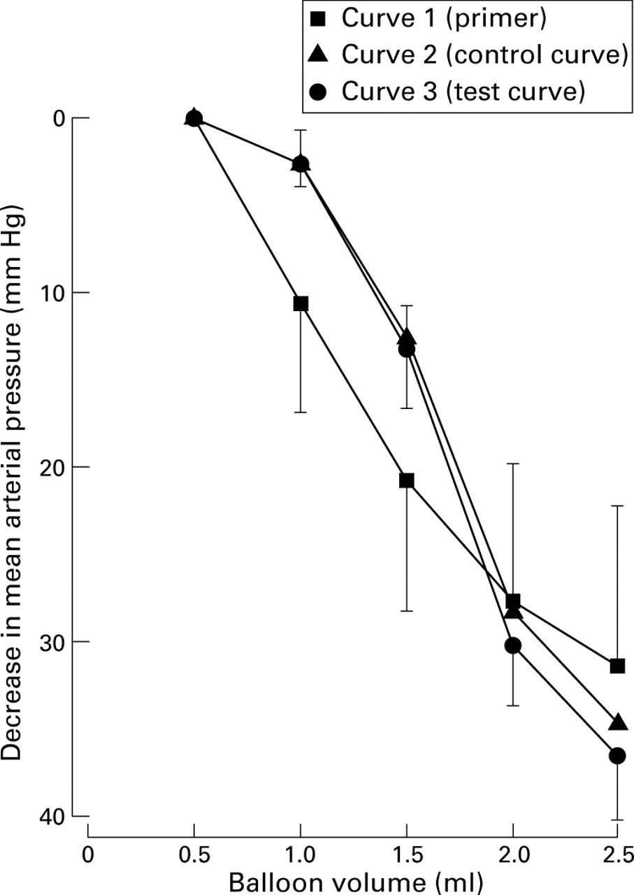
Colorectal distention (30 s every 5 min) evokes volume related decreases in mean arterial pressure which are highly reproducible (n=4).
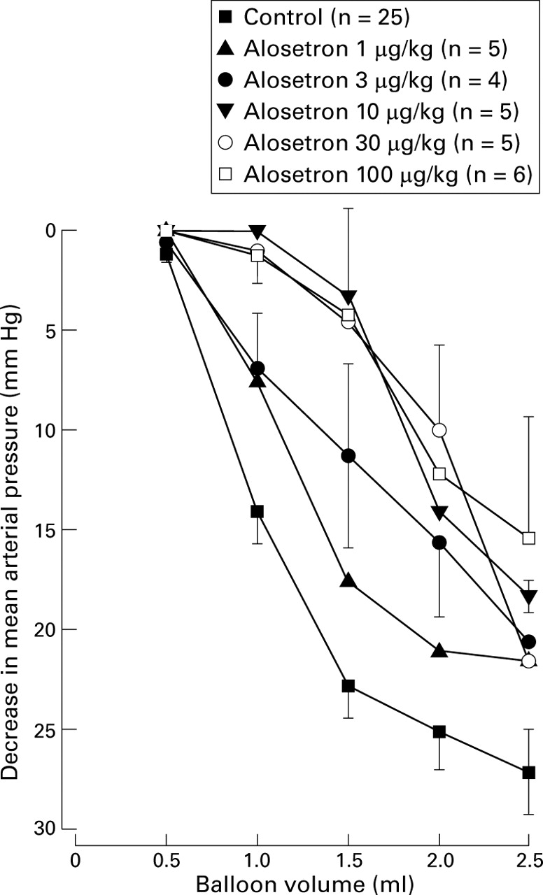
The 5-HT3 receptor antagonist alosetron (1–100 μg/kg) inhibited the depressor response to colorectal distention in a dose related manner (fig 3). The response to 1.5 ml CRD was significantly inhibited at doses of 3–100 μg/kg (p<0.05), giving an ID50 of 3.0 μg/kg (0.4–9.3). At 10 μg/kg, alosetron reduced the response to 1.5 ml CRD to 3.3 (4.4) mm Hg compared with 21.8 (1.7) mm Hg control response (p<0.05, n=5). There was no further inhibition at doses up to 100 μg/kg (4.2 (2.4) mm Hg compared with 22.9 (2.3) mm Hg control response; p<0.05, n=6). Reduction of the response to 2.5 ml distention was greatest at 100 μg/kg (15.4 (6.1) mm Hg versus 27.1 (2.1) mm Hg control response; p<0.05, n=6). Alosetron had no effect on basal blood pressure (112.4 (6.8) mm Hg at 100 μg/kg versus 108.3 (6.1) mm Hg in control animals; n=6).

Influence of alosetron (1–100 μg/kg) on the hypotensive response to colorectal distention.
Fos EXPRESSION
Repetitive CRD significantly increased bilateral expression of Fos-LI compared with sham distended animals (from 49 (24) to 1246 (146); p<0.05, n=4–5). This expression was located in discrete areas of the spinal grey matter (fig 1): Fos-LI nuclei were located in superficial laminae I and II of the spinal cord (area 1, 27.6% of total), lateral laminae V and VI, including the intermediolateral horn (area 3, 31.3%), and the area surrounding the central canal (medial lamina VII and X; area 4, 30.2%). There were fewer Fos-LI nuclei in laminae III and IV (area 2, 10.9%), and staining in the ventral horn region of areas 3 and 4 was relatively sparse. In sham distended animals, total numbers of Fos-LI were too low to assess differences in regional distribution.
In animals pretreated with 100 μg/kg alosetron, total numbers of Fos-LI nuclei were significantly reduced (479.8 (122); p<0.05, n=5). At a lower dose (30 μg/kg), alosetron did not significantly reduce the total number of Fos-LI nuclei (993 (89), n=5). Regionally, alosetron inhibited Fos-LI in specific areas of the spinal cord (see fig 4), with reductions of 56% in area 1, 63% in area 3, and 68% in area 4 (100 μg/kg, p<0.05). There was no significant inhibition of Fos-LI in area 2. At 30 μg/kg, alosetron did not significantly reduce numbers of Fos-LI nuclei in any region, although there was a reduction of 35% in area 1. Figure 5 shows the distribution of Fos-LI nuclei in control and drug treated animals.
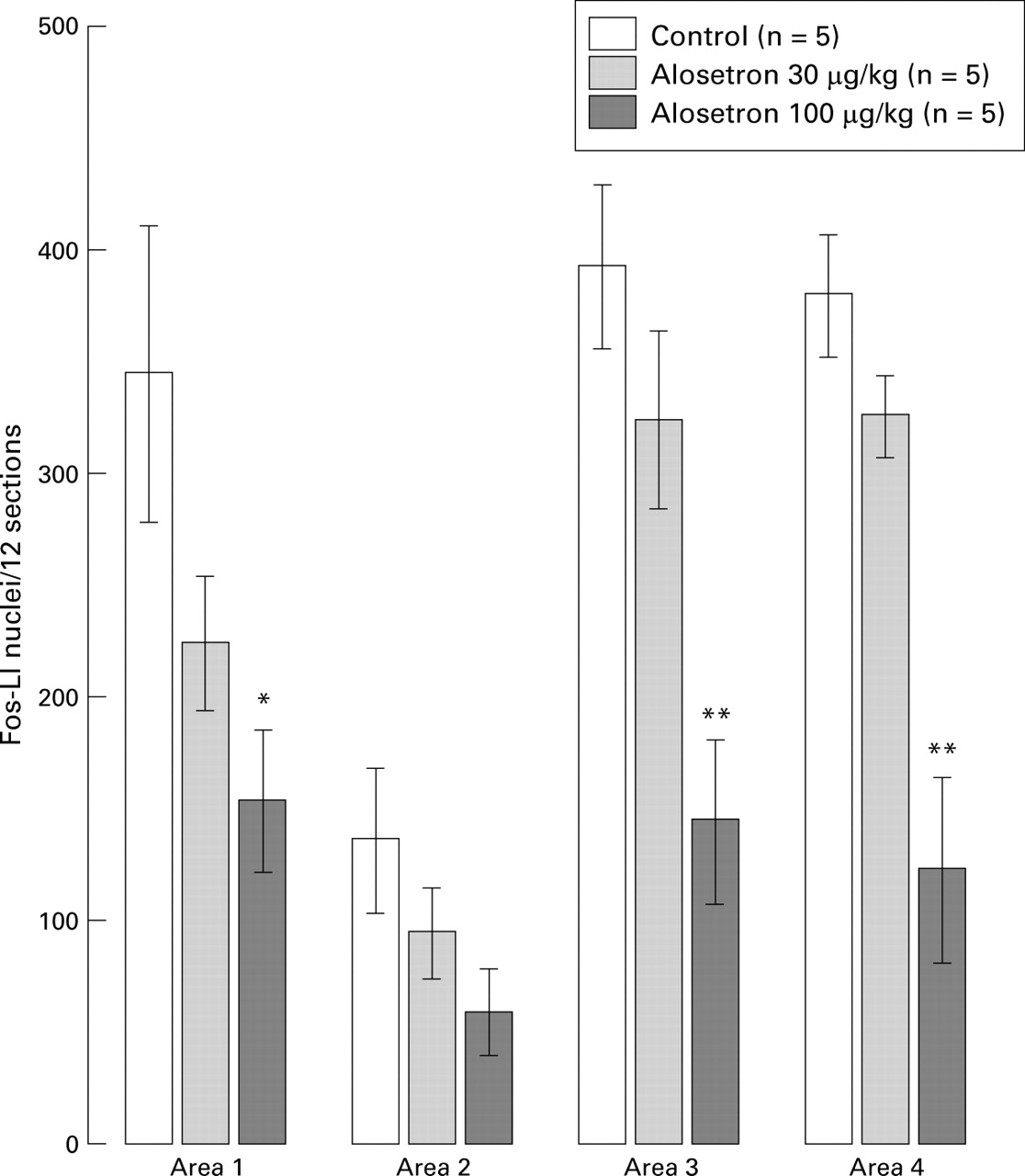
Alosetron reduces the number of Fos-like immunoreactive (Fos-LI) nuclei (in 12 sections across L6–S1) evoked following 2 h (80 mm Hg, for 30 s every 120 s) repeated colorectal distention. *p<0.05, **p<0.01, significant reduction of Fos-LI.

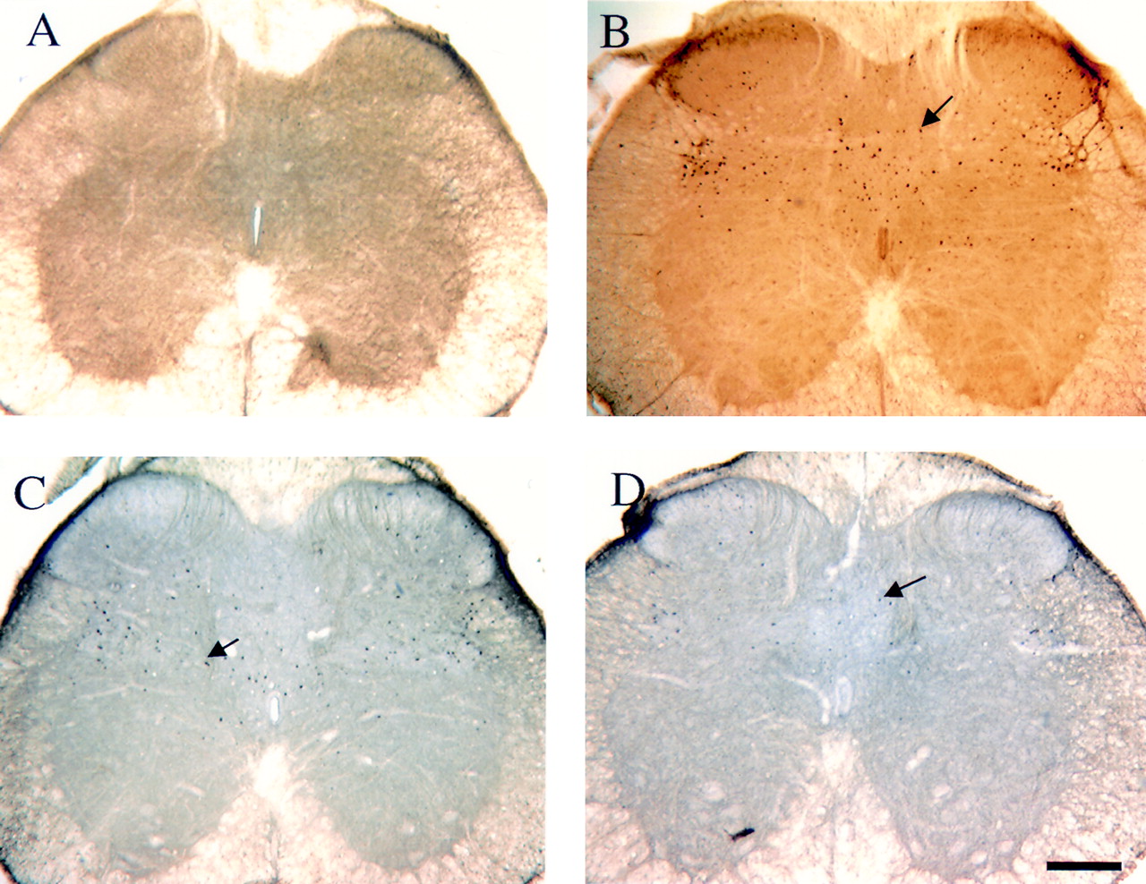
Representative bitmap images illustrating distribution of Fos-like (Fos-LI) immunoreactivity in all treatment groups: (A) sham distended; (B) control colorectal distention (CRD); (C) CRD/alosetron, 30 μg/kg; (D) CRD/alosetron 100 μg/kg. Horizontal scale bar, 150 μm; arrows indicate examples of Fos-LI nuclei.
Discussion
We have shown that responses to CRD using two experimental paradigms are blocked by the 5-HT3 receptor antagonist alosetron. In the pentobarbitone anaesthetised rat, noxious CRD elicits reproducible depressor responses, consistent with previous studies in both the small and large intestine.13 15 We have also shown that reproducible distention-effect curves obtained in this study can be used to assess drug effects. Of the animals tested, 74% displayed a vigorous depressor response to CRD, which is similar to previous findings.15 27 In the present study, the response parameters were somewhat dependent on the depth of anaesthesia. In urethane anaesthetised rats, however, where such reflexes are reported to be unaffected by anaesthetic levels, a similar proportion of animals do not display a depressor response to CRD,27 thus levels of anaesthetic probably do not account for the majority of non-responders in the present study.
Alosetron is a potent and selective 5-HT3 receptor antagonist, with a duration of action of several hours in the rat.29 32 It potently inhibited the depressor response to CRD, with an ID50 of 3 μg/kg. The 1.5 ml distention volume at which the ID50 is calculated was chosen as it produces a submaximal vasomotor response, of a similar magnitude to that evoked by 60– 80 mm Hg barostat inflation (unpublished observations), and thus is well above the nociceptive threshold of approximately 30 mm Hg. Therefore the effects of alosetron at this distention volume are likely to be antinociceptive. In a similar study, the 5-HT3 receptor antagonists ondansetron, tropisetron, and granisetron were shown to be approximately equipotent at inhibiting the depressor response to CRD.28 They were 5–10 fold less potent than alosetron in the present study; bell shaped dose-response relations obtained with certain 5-HT3 receptor antagonists may, however, result in underestimation of their potency.28 Alosetron did not display a bell shaped dose-response relation in this study, with stable inhibitory effects between 10 and 100 μg/kg. In other studies, systemic 5-HT3 receptor antagonists block the vasomotor response to distention of the duodenum and colorectum27 33; these data are therefore consistent with previous findings. Continuous monitoring of intraballoon pressure allowed the calculation of colorectal compliance from pressure-volume relations. Alosetron did not seem to affect colorectal compliance significantly in this study (unpublished observations), although this method may not be as sensitive as a barostat for detecting very small changes, as alosetron has been shown to increase compliance in studies in patients with IBS.6 8 Any such effects of alosetron on compliance in the present study would not be sufficient to account for its effect on the vasomotor response to CRD.
Noxious CRD also induced expression of the proto-oncogenec-fos in the lumbosacral spinal cord of urethane anaesthetised rats. Fos-LI nuclei were found bilaterally, mainly in laminae I, II, lateral V and VI, VII and X. A similar distribution pattern has been noted previously,18 24which correlates with the termination sites of pelvic nerve afferents.34 35 The number of Fos-LI nuclei in the present study was lower than that reported in other studies, with 1246 (146) Fos-LI nuclei in 12 sections across the L6–S1 border, compared with over 300/section.24 This difference may be explained by use of urethane anaesthesia in the present study, compared with halothane sedation in previous studies.
Alosetron (100 μg/kg) significantly reduced total numbers of Fos-LI in response to repetitive CRD, which was evident in both superficial (I, II) and deeper (V–VII, X) laminae. The dose of alosetron required was higher than that needed to inhibit the depressor response to CRD. There may be several explanations for this. Differences in balloon size and inflation procedure (constant pressure versus constant volume, different frequency, and intensity of stimulation) may have affected the apparent potency of alosetron. Following fixed volume inflation, a reduction in intraballoon pressure was sometimes apparent during the inflation period, which may result in reduced afferent stimulation. In contrast, a larger balloon, at constant pressure will recruit more afferent fibres and thus elicit a stronger response due to summation, and therefore be more difficult to inhibit. Furthermore, the repeated CRD paradigm has been reported to produce colonic inflammation,24 which may result in altered mechanisms of nociceptive neurotransmission. Alosetron may also have subtle effects on compliance in the fixed volume experiment (see above), or may differentially affect afferent neurones mediating phasic and tonic components of the response. An alternative explanation may be that in order to see a reduction of the numbers of Fos-LI nuclei, thec-fos expression must be totally blocked. Gradations of staining (amount of Fos-like protein) certainly do occur, but were not examined in the present study. This may also explain the steep dose-response curve obtained with alosetron, with a profound effect at 100 μg/kg, but no effect at 30 μg/kg. Different anaesthetics were used for the two experimental procedures, but it is unlikely that this had a major impact on the apparent efficacy of alosetron.
Induction of c-fos is thought to be largely indicative of c-fibre afferent activation, although Fos-LI occurs in response to both noxious and innocuous visceral24 36 and joint37 stimulation. The majority of afferents innervating the colon are c-fibres, most of which show a graded response to increasing stimulus intensity. Therefore, inhibition of Fos-LI in this model does not necessarily represent inhibition of nociceptive pathways. Traub et al however, have reported that the opiate analgesics, morphine and tramadol, reduce CRD evoked spinal Fos-LI in both superficial and deep laminae.26Their study provides some evidence that a significant proportion of the CRD evoked Fos-LI results from noxious stimulation of these dorsal horn neurones.
5-HT3 receptor protein and mRNA are widely distributed throughout the brain,39 40 spinal cord,39 41 42 and dorsal root ganglion neurones.43 A large proportion of the spinal receptors are located on the central terminals of afferent neurones, many of which are capsaicin sensitive.44 The present finding that deep as well as superficial laminar expression ofc-fos was suppressed by alosetron, is not inconsistent with these data. Visceral afferent neurones have collaterals extending into deep as well as superficial laminae, although these neurones comprise a small proportion of the total number of afferents entering the spinal cord. Furthermore, many dorsal horn neurones in deeper laminae receive indirect input from superficial neurones; either these interneurones or the afferents terminating onto them may express 5-HT3 receptors. Further evidence to support a role for 5-HT3 receptors in nociceptive processing includes the inhibition, by granisetron, of CRD inducedc-fos immunoreactivity in the nucleus of the solitary tract,45 and the reduction ofc-fos immunoreactivity in the trigeminal nucleus by 5-HT3 receptor antagonists following noxious chemical stimulation of the rat nasal mucosa.465-HT3 receptors are also found within the enteric nervous system of the gut, and are known to regulate gastrointestinal motility. Alosetron has been reported to normalise gut transit following sensitisation with egg albumin in rats,47 and to increase colonic compliance in patients with IBS.6 8 Cramer and Rademaker,48 however, have suggested that the site of action against distention evoked reflexes is not within the peripheral organ, as the 5-HT3 receptor antagonist ICS 205 930 inhibits the depressor response to ileal distention, but has no effect on the concurrent increased firing in afferent dorsal root filaments. It is also worth noting that on vagal afferents innervating the stomach and small intestine, 5-HT3 receptors are associated with mucosal chemoreceptors rather than mechanoreceptors responsive to distention.49 Alosetron has also been reported to inhibit the vasomotor response to CRD in dogs following intracerebroventricular injection, suggesting a central site of action.50
In summary, these studies show that alosetron modulates visceral nociceptive neurotransmission in the rat, illustrated by a potent inhibition of the reflex depressor response to colorectal distention, and a reduction in numbers of Fos-LI nuclei in the spinal cord following repeated CRD. There is considerable anatomical and functional data to support a role for 5-HT3 receptors in visceral nociceptive processing. The site(s) of action of alosetron were not established in the present study, but could be at the level of the enteric or primary sensory neurone, or via spinal or supraspinal neuronal circuits concerned with the modulation of nociceptive transmission. Based on these data, in addition to recent studies indicating that alosetron decreases symptoms of abdominal pain in patients with irritable bowel syndrome,5 7 alosetron may be of potential use in the treatment of visceral pain conditions.
References
Footnotes
- Abbreviations used in this paper:
- CRD
- colorectal distention
- Fos-LI
- Fos-like immunoreactive
- IBS
- irritable bowel syndrome