Article Text

Abstract
INTRODUCTION Pharmacotherapy for upper gastrointestinal bleeding has been difficult to evaluate because clinical end points are infrequent and affected by other factors.
AIMS To evaluate whether blood in the stomach at endoscopy reflected severity of bleeding, predicted clinical outcomes, and could be altered by therapeutic agents.
METHODS We studied 414 consecutive admissions with suspected upper gastrointestinal bleeding. Patients were randomised to receive lansoprazole 60 mg followed by 30 mg four times daily, tranexamic acid 2 g followed by 1 g four times daily, both drugs, or placebo for four days, until discharge or a clinical end point occurred. Logistic regression analysis was used to determine predictors of endoscopic changes and clinical outcomes, and to investigate the effects of drug treatments on blood in the stomach.
RESULTS Of 414 patients with suspected upper gastrointestinal bleeding, 379 were endoscoped. Upper gastrointestinal bleeding was confirmed in 316. Sixteen required surgery within 30 days and 16 died on the index admission. Trial treatments were evaluable on a per protocol basis in 228 patients. The amount of blood in the stomach was found to reflect initial risk, with significant associations with high risk categorisation (odds ratio 3.7 (95% confidence interval 1.5–9.4) for more than a trace v none/trace), age (1.5 (1.1–1.9) per decade), and initial pulse (1.02 (1.00–1.04) per beat), and to predict rebleeding (9.2 (4.6–18.7)) and surgery (8.2 (2.9–22.9)). Other stigmata were less significant in these respects. The amount of blood in the stomach at endoscopy was reduced significantly by both lansoprazole (0.22 (0.07–0.63)) and tranexamic acid (0.27 (0.09–0.81)), although there was no evidence of synergy.
CONCLUSIONS Blood in the stomach reflects clinical features in patients with acute upper gastrointestinal bleeding and is reduced by treatment with lansoprazole and tranexamic acid.
- upper gastrointestinal bleeding
- ulcer
- proton pump inhibitor
- acid suppression
- tranexamic acid
- fibrinolysis
- lansoprazole
Abbreviations used in this paper
- NSAID
- non-steroidal anti-inflammatory drug
Statistics from Altmetric.com
- upper gastrointestinal bleeding
- ulcer
- proton pump inhibitor
- acid suppression
- tranexamic acid
- fibrinolysis
- lansoprazole
Upper gastrointestinal bleeding remains the most common reason for emergency hospital admission with a gastrointestinal problem.1 ,2 It has a high mortality and there is little evidence that this is declining. In consequence, upper gastrointestinal bleeding still causes approximately 5000 deaths per annum in the UK. Although endoscopic treatments using laser, injection, or heater probe can reduce rebleeding and the need for surgery in clinical trials,3 ,4 their success depends on the skill of the operator and may not influence outcome in clinical practice outside trial settings.5
A simply administered drug treatment which improves the outcome of upper gastrointestinal bleeding would therefore be an important development. Treatments evaluated have included acid suppression to reduce peptic activity6-9 and tranexamic acid to inhibit fibrinolysis.6 ,10 Large individual trials have however failed to establish conclusively the validity of either of these approaches, although meta-analyses have suggested a weak beneficial effect of both H2 receptor antagonists9 and tranexamic acid,10 and several studies have reported benefit from omeprazole in patients established by endoscopy to have ulcer bleeding prior to trial randomisation.11-14
It would be attractive to show benefit from drug treatment given to unselected patients prior to endoscopy as treatment could be extended to a wider range of conditions and started earlier after presentation. Showing oral treatment to be effective would be particularly attractive as it would allow treatment to be started in the community prior to hospital admission. However, for unselected patients, important clinical end points such as rebleeding, transfusion, surgery, or death are either relatively infrequent and/or strongly influenced by confounding factors. The death rate of unselected patients in trials is sufficiently infrequent that approximately 3000 patients would be needed to show even a halving of the rate.7
Given these obstacles an alternative approach would be to develop a more sensitive intermediate measure which reflected bleeding severity and was predictive of clinically important end points. This could then be used to evaluate potential drug therapies before embarking on large trials designed to show an impact on death rate. The Forrest classification system has some predictive value in ulcer patients15 but is not applicable to other causes of upper gastrointestinal bleeding. In an earlier study of the proton pump inhibitor omeprazole, we noted that this drug was associated with a highly significant reduction in the number of patients with intragastric blood or active bleeding at endoscopy compared with placebo recipients, even though there was no effect on mortality.7 Blood in the stomach was not prospectively specified as an end point in this earlier study and no account was taken of the interval between presentation and endoscopy. As a result, despite being highly significant, the results could have arisen by chance.
We therefore set up a study with two aims. The first was to evaluate whether the endoscopic finding of blood in the stomach was sufficiently predictive of clinically important outcomes to be considered a surrogate end point. The second aim was to investigate the effect of oral doses of lansoprazole, a proton pump inhibitor, and of tranexamic acid, an inhibitor of fibrinolysis, alone and in combination on this and other endoscopic findings. The trial was initiated and designed in the Division of Gastroenterology, Nottingham, and subsequently proposed to Lederle Laboratories who funded it.
Methods
PATIENTS
All identifiable patients admitted to the two Nottingham hospitals because of suspected upper gastrointestinal bleeding over a 16 month period were considered for trial entry. In order to reflect real clinical practice and ensure rapid treatment, liberal entry criteria were used. The only exclusion criteria were bleeding so severe as to require immediate surgical intervention, conditions making active treatment inappropriate (for example, terminal malignancy), pregnancy, lactation, active thromboembolism or intravascular coagulopathy, creatinine level above 250 μmol/l, use of phenytoin, and known adverse drug reactions to the trial drugs.
DESIGN
The therapeutic component of this study consisted of a randomised, double blind, double dummy, parallel group comparison of placebo, lansoprazole, tranexamic acid, or both drugs in combination, given orally to patients admitted with acute suspected upper gastrointestinal bleeding.
PROCEDURES
The treatment objective at trial entry was to ensure that patients with upper gastrointestinal bleeding receive treatment as soon as possible after hospital admission. The general medical admitting teams were therefore asked to include all patients with possible upper gastrointestinal bleeding. Those subsequently assessed on the basis of clinical and endoscopic findings not to have suffered upper gastrointestinal bleeding were excluded from drug efficacy analysis. Such decisions were taken immediately and prospectively, without knowledge of the treatment received and before the treatment code was broken.
At trial entry, patients were asked to give verbal consent to participation. They were treated for up to four days with lansoprazole 60 mg (stat dose), followed by 30 mg four times daily, tranexamic acid 2 g, followed by 1 g four times daily, both drugs, or placebo. Drugs were randomised in blocks of four and blinding was maintained using a double dummy technique. If patients vomited within 30 minutes of the first dose this was noted but the treatment regimens were not altered. Patients were managed by the seven medical acute admitting teams. They were given guideline protocols concerning risk factors (including age >60 years, initial pulse >100 beats/min, systemic blood pressure <100 mm Hg, evidence of gastric ulcer or liver disease, and coexistent morbid disease) and their management. Each admitting team had clinical freedom to manage the patients as they saw fit. They were asked to classify patients as high or low risk based on their risk factors at presentation, although this was not done to a standardised formula.
Endoscopy was performed on the morning following admission for most patients or earlier if clinical need dictated. At endoscopy all primary and secondary lesions were recorded. Endoscopists were asked to record whether there was blood in the stomach and if so how much (five point scale, 0–4), whether it was mainly fresh or mainly old, and all stigmata of bleeding.16 ,17 The scale for blood in the stomach was developed locally and scored as follows: 0, no blood; 1, trace on surface of mucosa but no confluent pool; 2, small discrete pool occupying up to 10% of greater curve; 3, large pool, occupying 10–100% of greater curve; and 4, widespread blood, obscuring view. Where there were signs of active bleeding endoscopic therapy was performed using adrenaline to a standard protocol.18Endoscopists were asked to state whether or not they thought the patient had genuinely had an upper gastrointestinal bleed.
Each patient was visited on the ward daily by a clinical research assistant who obtained written confirmation of their verbal consent and recorded information about them, their management, and results. Trial treatment continued for four days or until discharge or withdrawal.
ANALYTICAL APPROACH
End points
The primary endoscopic end points, specified at the start of the study, were whether there was blood in the stomach (using the five point endoscopic assessment to produce a binary classification: none or a trace versus small pool, large pool, or widespread) and other stigmata of upper gastrointestinal bleeding (red spots, black spots, clot obscuring the ulcer base, oozing, spurting blood, visible vessel). The secondary endoscopic end points were whether blood was old or fresh and whether there was active bleeding at endoscopy. The clinical end points evaluated were the amount of blood transfused and the incidence of rebleeding, operation, or death. Rebleeding was defined as new haematemesis, melaena, or hypotension (<100 mm Hg systolic blood pressure) associated with a drop in haemoglobin and/or endoscopic evidence of fresh rebleeding. Re-endoscopy was performed at the discretion of the team managing the patients.
Patient populations
Because the diagnosis of gastrointestinal bleeding is not always certain at presentation, the treated population included those who turned out not to have had gastrointestinal bleeding. Clinical outcomes were however analysed on an intention to treat basis, by including all patients randomised to receive treatment reflecting the pragmatic approach appropriate to this aspect of the study that encompasses the way in which treatment would be used in clinical practice. Endoscopic end points were analysed in evaluable patients who had a definite upper gastrointestinal bleed, whose management was conducted according to the trial protocol, as is statistically appropriate to the explanatory nature of this aspect of the study. Similar analyses of these end points, restricted to patients with ulcer bleeding, were also carried out. Safety analyses were performed on all patients who had received at least one dose of any study medication. Clinical adverse events were summarised according to body systems and severity, and related to drug usage. Clinical laboratory events were assessed in terms of change from baseline to last recorded value.
Statistical methodology
Logistic regression analysis was used as the principal analytical method. This allowed the influence of factors identified as potential determinants at the start of the study to be assessed. In particular, this approach was adopted to make allowance for the time from initiation of treatment to endoscopy. Non-drug influences were first investigated with backward stepwise elimination of those that did not improve the fit of the regression model (p>0.1), before fitting the trial treatments to the model. Because the study was conducted to a factorial design with a view to detecting any synergy between the two drug treatments, each individual drug was entered as a discrete factor, and an interaction term used to determine the effect of using them in combination. As no synergy was detected, odds ratios for each of the four treatment combinations were also calculated for illustration purposes but this did not form part of the primary statistical assessments.
The following explanatory factors, selected at the start of the study, were assessed in this way as determinants of endoscopic findings: hospital centre; hospital ward; high/low risk status; patient age; site of primary endoscopic lesion; initial pulse on admission; non-steroidal anti-inflammatory drug (NSAID) usage; and time from start of treatment to endoscopy. The factors selected as potential determinants of clinical outcome were: hospital centre; hospital ward; high/low risk status; patient age; site of primary endoscopic lesion; initial pulse; prior NSAID usage; and the primary endoscopic end points (amount of blood in the stomach, stigmata of recent haemorrhage). Because endoscopic stigmata were significantly influenced by the trial treatments, they were not included in the analysis when the effect of these treatments on clinical end points was analysed in order to avoid confounding.
Planned sample size
It was planned to study a total of 400 patients in anticipation that approximately 300 would be found to have a definite upper gastrointestinal bleed. We calculated that 220 evaluable patients would be needed for the study to have 90% power to detect an approximate halving from 40% of the prevalence of intragastric blood or stigmata with either drug on a factorial basis.
Results
PATIENT POPULATIONS
In total, 414 patients were randomised for treatment; 379 were endoscoped and 316 were considered to have had a definite upper gastrointestinal bleed after endoscopic assessment. Of these patients, 290 were eligible for assessment of the effect of drugs on endoscopic findings and 228 were managed per protocol (55 placebo, 58 lansoprazole, 57 tranexamic acid, 58 both drugs). Reasons for exclusion from analysis at each stage of patient selection and their relation to treatments to which patients were originally randomised are shown in fig 1.

Disposition of patients in the trial. P, placebo, L, lansoprazole, T, tranexamic acid, LT, both drugs.
Patient characteristics
The patients we studied were relatively old with a preponderance of men and a substantial number who had a past history of peptic ulcer or were taking NSAIDs (table 1). Approximately one sixth of patients were classified as high risk by the attending physician.
Details of patients studied
Ward placement
Of the 68 patients classified as high risk, 13 (19.1%) were initially managed on high dependency or intensive care units (table 2). Of 346 lower risk patients, four (1.2%) were initially managed on a high dependency unit. Eight higher risk and three lower risk patients subsequently required transfer to high dependency/intensive care units
Management and outcomes
OUTCOMES
Endoscopic findings
Endoscopy was performed 19 (median, interquartile range 11–24) hours after admission. The primary endoscopic diagnoses are shown in table 1. Seventy eight patients had blood in the stomach. In 36 of these it was a large pool or widespread blood in the stomach. This amount of blood was seen in 12 patients with gastric ulcer (22.6%), seven with duodenal/pyloric ulcer (9.5%), and seven with oesophageal varices (50%). Specific stigmata of bleeding were seen in 87 patients, including 21 with duodenal/pyloric ulcers (28.4%), 19 with gastric or stomal ulcers (35.8%), six with Mallory Weiss tears (25.0%), four with oesophageal ulcers (28.6%), and three with oesophageal varices (33% of the nine with specific stigmata recorded). Thirty five patients underwent endoscopic sclerotherapy (11 duodenal ulcer, 12 gastric ulcer, 10 oesophageal varices, two Mallory Weiss tear), with control of bleeding in 77% (table 2).
Clinical outcomes
A total of 165 patients required blood transfusion (table 2). The volume transfused in these patients was 4.0 (1.9) (SD) units. Across the whole population of patients with upper gastrointestinal bleeding, an average of 0.41 (geometric mean, 95% confidence intervals 0.31–0.52) units were transfused. The endoscopic diagnoses most frequently associated with blood transfusion were oesophageal varices (86%), duodenal ulcer (61%), and gastric ulcer (58%).
Thirty nine patients experienced rebleeding (table 2), 11 with duodenal ulcer (rebleeding rate 16.7%), eight with gastric ulcer (rebleeding rate 15.1%), and six with oesophageal varices (rebleeding rate 42.9%). Eighteen patients required surgery (16 while still receiving trial treatment), 15 as a consequence of gastrointestinal bleeding, and three for other reasons (laparotomy for peritonitis from perforated duodenal ulcer, repair of inguinal hernia, repair of aortic aneurysm). In five patients surgery was for bleeding duodenal ulcer (rate of surgery 9.1%) and in four it was for gastric ulcer (7.5%). Sixteen patients died on the index admission within 30 days of trial entry. Two patients died on readmission within 30 days (one myocardial infarction, one heart failure). Six of the deaths were in patients with duodenal ulcer (death rate 9.1%), three with gastric ulcer (5.7%), and two with oesophageal varices (14.3%). Gastrointestinal bleeding was designated as the main initiating cause (directly or indirectly) of death in 12 instances (five postoperative).
NON-DRUG DETERMINANTS OF ENDOSCOPIC FINDINGS
Blood in the stomach
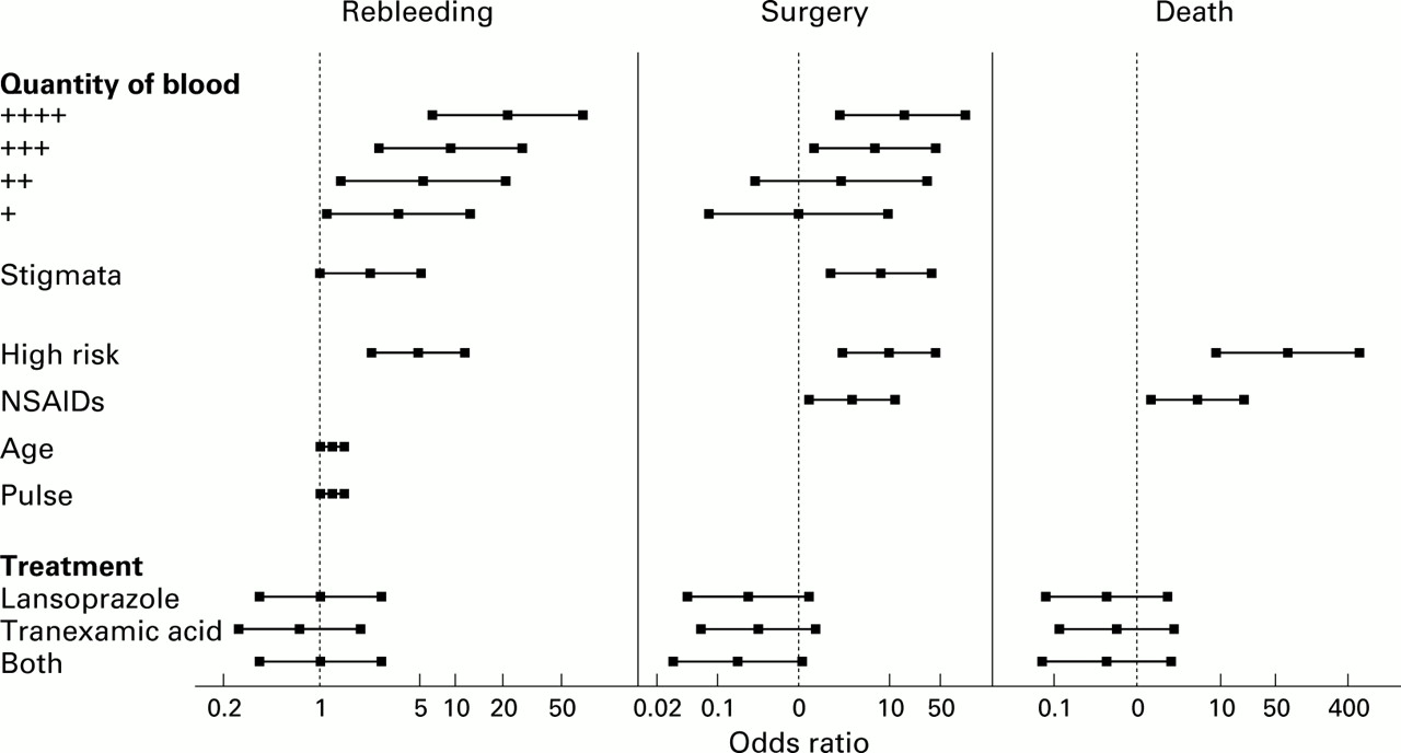
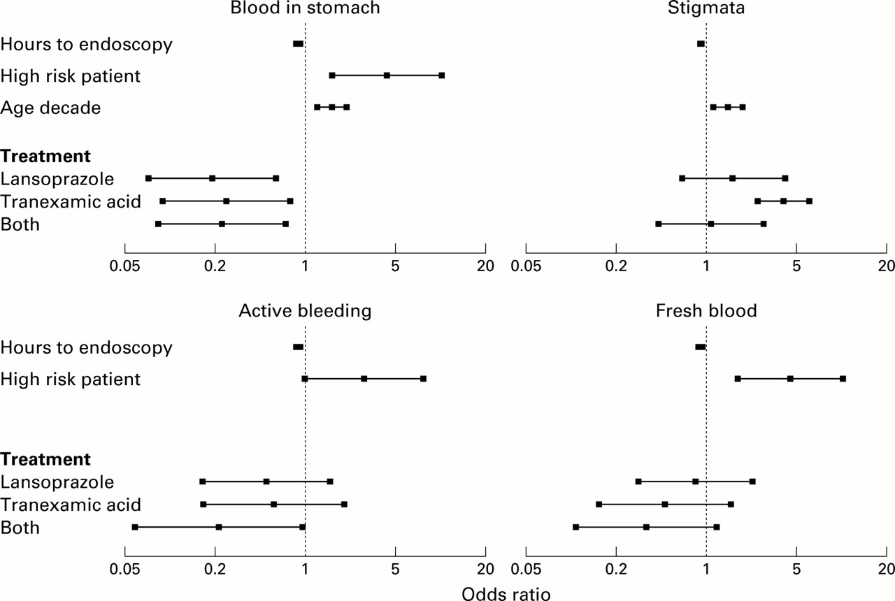
As shown in fig 2, patient risk status, age, and time to endoscopy were significant determinants of finding blood at endoscopy. High risk patients were 3.7 (97% confidence intervals 1.5–9.4) times more likely to have blood in the stomach and the odds increased by 1.5 (1.1–1.9) for each decade of age. The chances of finding blood at endoscopy diminished significantly as the time to endoscopy increased (p=0.001). Initial pulse rate was also a significant influence with an odds ratio of 1.02 (1.00–1.04) for each beat per minute (p=0.035).

Effect of significant factors and of trial treatments on endoscopic findings.
Stigmata of recent haemorrhage
Being a high risk patient was the only factor significantly associated with finding any endoscopic stigma of recent haemorrhage (odds ratio 2.5, 1.1–5.5; p=0.023) (fig 2). The odds of finding stigmata tended to diminish with the number of hours between admission and endoscopy (odds ratio 0.98, 0.96–1.00; p=0.01).
Secondary endoscopic end points
Active bleeding tended to be more likely to be found if there was a tachycardia (odds ratio 1.02, 1.00–1.05 for every 10 beats per minute; p=0.12) and there was a trend to association with a high risk categorisation (odds ratio 2.6, 0.93–7.0; p=0.07). Fresh blood was more likely to be found in high risk patients (odds ratio 3.9, 1.6–9.5; p=0.003). The odds of finding active bleeding (odds ratio 0.94, 0.89–0.98 per hour; p=0.008) or fresh blood (odds ratio 0.95, 0.92–0.99 per hour; p=0.017) diminished with the number of hours to endoscopy.
NON-DRUG DETERMINANTS OF CLINICAL OUTCOMES
Rebleeding
Thirty nine patients (12.3% of all those assessed as having a genuine gastrointestinal bleeding episode) rebled. The factors significantly associated with rebleeding were quantity of blood at endoscopy (p<0.001) and whether the patient was categorised as a high risk patient (odds ratio 3.1, 1.2–7.9; p=0.017) (fig 3). Other endoscopic stigmata were not significantly predictive for rebleeding. Risks increased progressively according to the amount of blood at endoscopy (fig 3) so that the odds of rebleeding in patients with extensive blood at endoscopy were 21.2 (6.2–72.6; p<0.001) compared with patients with no blood. Overall, finding more than a trace of blood was associated with an odds ratio for rebleeding of 9.2 (4.5–18.7) compared with finding none or a trace. Rebleeding occurred in nine of 270 patients with no blood in the stomach at endoscopy (3.3%), in 10 of 63 with a trace or small pool (15.9%), and in 17 of 44 with a large pool or widespread blood (27.3%; p<0.001). Sixteen of 40 patients with active bleeding at endoscopy (40%) rebled compared with 20 of 337 (5.93%) without (p<0.001).
Effect of significant factors and of trial treatments on clinically significant outcomes.
Surgery
Sixteen patients (5.1%) underwent surgery within 30 days of admission. Quantity of blood at endoscopy (p=0.008) and being a high risk patient (odds ratio 8.2, 2.0–33.9; p=0.004) were again significant determinants of the need for surgery (fig 3) but there was also an association with NSAID use (odds ratio 8.2, 2.0–33.9; p=0.021) and endoscopic stigmata (odds ratio 9.8, 2.2–83.8; p=0.003). Extensive blood at endoscopy was associated with an odds ratio for surgery of 18.9 (3.3–30.5) compared with patients with no blood (p=0.0009). Overall, finding more than a trace of blood was associated with an odds ratio for surgery of 8.2 (2.9–22.9) compared with finding none or a trace. Surgery was required in three of 272 patients with no blood at endoscopy (1.1%), in two of 63 with a trace or small pool (3.2%), and in eight of 44 with a large pool or widespread blood (18.2%; p<0.001).
Death
Sixteen patients (5.1%) died during the index admission. As shown in fig 3, high risk patients were 69.9 (8.6–566) times more likely to die compared with lower risk patients (p<0.001). Patients using NSAIDs were also at increased risk, with an odds ratio of 3.9 (1.02–14.9; p=0.049). In the main analysis, none of the endoscopic measurements was significantly associated with subsequent death. However, when analysed separately, finding more than a trace of blood was associated with an odds ratio for death of 3.6 (1.3–10.3) compared with finding none or a trace. Five of 271 patients with no blood at initial endoscopy (1.8%) compared with two of 63 with a trace or small pool (3.2%), and five of those with a large pool or widespread blood (11.4%) died.
Outcomes in ulcer patients
The rate of rebleeding (13.9%), surgery (6.6%), and death (4.4%) in ulcer patients did not differ from the whole patient population with upper gastrointestinal bleeding, and there were relatively few events. Data on ulcer patients (which were broadly similar to the group as a whole) are therefore not presented separately.
EFFECTS OF TRIAL DRUGS ON ENDOSCOPIC END POINTS
Blood at endoscopy
When the effects of treatment were fitted to the model, each treatment was associated with a highly significant reduction in the amount of blood in the stomach although there was no evidence of synergy between them (fig 2). Among patients who received placebo, 53.7% had blood at endoscopy. This was reduced to 25.9% (odds ratio 0.22, 0.07–0.63) in patients treated with lansoprazole (p=0.005), to 33.3% (odds ratio 0.27, 0.09–0.81) with tranexamic acid (p=0.019), and to 25.9% (odds ratio 0.26, 0.09–0.80) with both drugs (p=0.013). As seen in fig 4, the amount of blood at endoscopy in those patients where it was found was also reduced.

Amount of blood in the stomach of patients receiving the four trial treatments.
Stigmata of recent haemorrhage
There were no differences in the prevalence of stigmata at endoscopy between any of the drug treatment groups, whether all patients or only those with ulcers were considered.
EFFECT OF TRIAL DRUGS ON CLINICAL END POINTS
Similar numbers of patients suffered rebleeding in all treatment groups (table 2) and there was no significant influence on the risk of rebleeding (fig 3). Six patients who received placebo underwent surgery (all bleeding related) compared with five on lansoprazole (two for unrelated reasons), five on tranexamic acid, and two on both drugs (table 2). The numbers of patients on active treatment who underwent bleeding related surgery or who died tended to be less in the actively treated patients (table 2, fig 3) but numbers were too small for formal analysis. The effect of the trial treatments on clinical end points was not statistically significant in any instance. There were too few patients with ulcers who experienced clinical end points for valid analysis of the effect of the trial drugs.
SAFETY
There were no significant differences in the number or pattern of adverse events, of severe adverse events, or of adverse events leading to withdrawal in the four treatment groups. Although a number of patients had thrombotic episodes, there was no evidence that these were more common in those receiving tranexamic acid.
Discussion
In this paper we have shown that the simple measure of finding blood in the stomach at initial endoscopy closely reflects baseline measurements associated with higher risk and clinical evaluation of risk status, and is predictive of clinically important outcomes. These relationships appear to be stronger for blood in the stomach than for other endoscopic stigmata. All treatment combinations investigated markedly reduced the chances of finding blood in the stomach at endoscopy. The study was not designed to be large enough to detect effects of treatment on clinical outcomes and indeed was set up to study endoscopic measures as possible surrogates. Nevertheless, the rates of operation and death were numerically less in treated than untreated patients. These observations suggest that blood in the stomach could be used in medium sized pilot studies to identify treatments worth investigating for their effects on rebleeding, surgery, and death in much larger studies.
Our data leave a number of uncertainties. With acid suppression, the pool size within the stomach could be reduced not because of reduced bleeding but because of gastric secretion. This however would not explain the reduction with tranexamic acid. Alternatively, active treatments could increase the rate of gastric emptying of blood. However, there is no pragmatic evidence in favour of this proposition and the fact that blood in the stomach was predictive of clinical outcomes suggests it is more likely to have been a genuine index of intragastric bleeding. Assessments were by a large number of endoscopists and intraobserver agreement was not investigated in this study. Again, however, the predictive value of blood in the stomach suggests it is robust enough to be of value even with a large number of endoscopic observers.
Our study shows that acid suppression with a proton pump inhibitor such as lansoprazole or use of tranexamic acid reduces the amount of blood at endoscopy, as assessed during routine clinical practice. This is probably a real rather than a spurious finding and raises the possibility that in a trial of sufficient size, use of a proton pump inhibitor, or tranexamic acid could improve clinical outcomes in unselected patients. However, there are few grounds to believe that there would be an advantage from using the two approaches simultaneously.
Acknowledgments
Supported by Lederle Laboratories
Abbreviations used in this paper
- NSAID
- non-steroidal anti-inflammatory drug