Article Text

Abstract
Aims—To evaluate the diagnostic yield and safety of a new push type videoenteroscope (PVE) for diagnosis of small bowel disease.
Methods—Three hundred and thirteen patients were referred for one or two way PVE from December 1993 to June 1996. Indications for PVE were: an unexplained iron deficiency anaemia with or without clinically evident gastrointestinal bleeding; or a complementary investigation for suspected small bowel disease, after a small bowel barium follow through (SBBFT) considered as normal or abnormal, but without a definite diagnosis.
Results—A jejunoscopy and a retrograde ileoscopy were carried out in 306 and 234 patients, respectively. In patients with isolated anaemia (n=131) and those with clinically evident gastrointestinal bleeding associated anaemia (n=72), PVE provided a diagnosis in 26 (19.8%) and 22 (30.5%) cases, respectively. Lesions found were located in the jejunoileum in 30 (14.7%) patients and in the gastroduodenum or the colon in 18 (8.8%) patients—that is, within the reach of the conventional gastroscope/colonoscope. In patients with normal (n=54) or abnormal (n=56) SBBFT, PVE provided a diagnosis in 17 (31%) and 27 (48%) cases, respectively. In 25% of cases, the abnormal appearance of SBBFT was not confirmed. The site of the radiological abnormality was not reached in 27% of cases. Lesions were located at the jejunum and the ileum in 59 (64%) and 33 (36%) cases, respectively.
Conclusions—PVE is useful in around 30% of cases of unexplained anaemia or after an SBBFT which failed to provide an accurate aetiological diagnosis. Use of retrograde videoenteroscopy increases diagnostic yield by one third.
- enteroscopy
- small intestine
- gastrointestinal bleeding
- anaemia
- chronic diarrhoea
- intestinal tumour
Statistics from Altmetric.com
The traditional way to investigate the small intestine is small bowel barium follow through (SBBFT) but its sensitivity and specificity are low.1 Only the duodenum and the first or second loops of the jejunum can be reached by upper endoscopy using an adult or paediatric colonoscope. Similarly, only the last centimetres of the ileum can be reached by colonoscopy.2 Endoscopic access to the rest of the small intestine requires the use of a purpose designed enteroscope. This organ is however not immediately accessible, and it is mobile and tortuously folded in the peritoneal cavity.
Many techniques and instrument designs for nonoperative enteroscopy have been described3-5 but at the present time, only the sonde and the push enteroscopes are commercially available. The sonde enteroscope, whose progression is facilitated by a balloon inflated after the pylorus, reaches the distal ileum in 70% of cases. This instrument has no tip angulation or biopsy channel, and only an estimated 50–70% of the bowel surface reached by the endoscope can be properly visualised.6 Furthermore, this technique is time consuming, uncomfortable, and potentially dangerous.7 The push enteroscope has sufficient shaft length and rigidity, tip angulation, and optimal optical characteristics for small bowel exploration but cannot reach the totality of the small bowel. However, this instrument can also be used in the same session by the retrograde route for investigation of a variable length of the ileum (two way enteroscopy). Until now, push videoenteroscopy (PVE) has been the subject of a few reports which concerned only small and heterogeneous series of patients explored by the oral route.8-10 We describe here our findings in 313 patients investigated by single or two way PVE in whom the indication was either unexplained iron deficient anaemia or presumed small bowel disease not diagnosed by previous upper and lower endoscopies and SBBFT. Our experience confirms that PVE is a useful investigation for these indications.
Patients and methods
STANDARD PUSH VIDEOENTEROSCOPY TECHNIQUE
The endoscope used was the Olympus SIF-100, with a total length of 2475 mm, a working length of 2175 mm, an external diameter of 11.2 mm, and a biopsy channel of 2.8 mm (Olympus, Rungis, France). The tip deflection and field of view were those of usual endoscopes, allowing exploration of nearly 100% of the mucosa. To limit gastric loop formation, it was used with an overtube, which consisted of a stiff but flexible proximal shaft and a softer distal section with radio-opaque markers; it had a length of 61 cm and an internal diameter of 18 mm. The procedure was carried out under general anaesthesia (propofol and midazolam) with tracheal intubation and artificial ventilation. The proximal small bowel was usually explored first with the patient in the dorsal decubitus position. Once the tip of the instrument was in the distal duodenum, the shaft was pulled to a straight position, and the overtube, previously preloaded onto the endoscope, was pushed so that the flexible portion lay within the duodenum. The progression of the tip of the instrument in the small bowel was helped using continuous abdominal pressure performed by an assistant. Examination was performed during both the progression and the withdrawal of the instrument. The examination was considered a failure if the ligament of Treitz was not passed.
Exploration of the ileum was performed during the same session by retrograde ileoscopy following colonoscopy. Thus, all patients who may have required an ileoscopy had colonic cleansing with polyethylene glycol 4000 before the endoscopy. No fluoroscopy was performed. The final depth of jejunal and ileal intubation was estimated by straightening the instrument to remove the gastric and colonic loops and subtracting 80 cm from the length inserted, corresponding to the distance from the mouth to the ligament of Treitz or from the anus to the caecum. All PVEs were performed by senior endoscopists.
PATIENTS
A total of 313 consecutive adult patients (mean age 50 (SD 17) years, male:female sex ratio 1.3), were referred for single or two way PVE, from December 1993 to June 1996. All had had one or more previous endoscopic examinations of both the upper and lower gastrointestinal tract, which were considered normal, and 218 (70%) had had an SBBFT.
INDICATIONS FOR PVE
As the recruitment of patients corresponded to a heterogeneous population referred from internal medicine or gastroenterological units for suspected small bowel disease, two categories were distinguished: unexplained iron deficiency anaemia; and complementary investigation after an SBBFT.
Unexplained iron deficiency anaemia
In 203 patients, unexplained iron deficiency anaemia was associated with clinically evident gastrointestinal bleeding (n=72) or isolated occult bleeding (n=131). All these patients had had previous negative upper and lower endoscopies, and 108 had had an SBBFT which was considered normal. In the cases of isolated anaemia, all the potential causes of anaemia (of urinary tract, gynaecological, inflammatory, and marrow origins) had been previously ruled out. In patients with clinically evident gastrointestinal bleeding, the haemorrhage was melaena (79%), haematochezia (18%), or haematemesis (3%). The mean concentration of haemoglobin was 7.1 (2.3) g/dl before transfusion.
Complementary investigation after an SBBFT
In 110 patients in which SBBFT was performed, it was considered normal in 54 and abnormal in 56, but without an accurate aetiological diagnosis. The main indications for the SBBFT were chronic diarrhoea (n=50), unexplained malabsorption syndrome (n=36), or bowel obstruction syndrome (n=24). As a majority of patients were referred from other gastroenterological centres, SBBFT examinations were not performed using a standard technique. However, they were always reviewed before PVE examination. When no indices were available to suggest the site of intestinal lesions, upper and then lower PVEs were performed. When an abnormality was identified during the upper PVE, the lower examination was not performed. When the putative small intestinal lesion was located with certainty in the distal ileum (with an SBBFT), the upper PVE was omitted.
Results
TECHNICAL ASPECTS
A jejunoscopy was performed in 306 patients and ileoscopy was performed in 234. Upper and lower enteroscopies were attempted in 236 subjects during the same session. Seventy patients had only jejunoscopy and seven only ileoscopy. The median length of jejunal and ileal progression was evaluated at 120 (30–200) and 50 (5–200) cm, respectively. Procedure time was 20 (8–50) and 30 (10–70) minutes, respectively. No technical failures occurred during jejunoscopy, but there were 59 failures of retrograde ileoscopy—19 due to bad colonic cleansing, and 40 due to technical difficulties, mainly the impossibility of entering the ileocaecal valve (n=32).
AETIOLOGICAL DIAGNOSIS
Tables 1 and 2 show the prevalence of positive diagnosis related to the indication for PVE. Table 3 presents the aetiological diagnoses in patients undergoing PVE.
Prevalence of positive diagnosis in accordance with single or two way push videoenteroscopy in 203 patients referred for unexplained iron deficiency anaemia after upper and lower digestive endoscopies
Prevalence of positive diagnosis in accordance with single or two way push videoenteroscopy in 110 patients referred for complementary investigation after a small bowel barium follow through (SBBFT)
Aetiological diagnosis in 313 patients undergoing single or two way push videoenteroscopy
In patients with anaemia, PVE provided a diagnosis in 48 (23.6%) cases: jejunoileal lesions were found in 30 (14.7%) cases, and lesions located in the gastroduodenum (n=15) or colon (n=3) which were missed at prior endoscopy were found in 18 (8.8%) cases.
In patients with clinically evident gastrointestinal bleeding and anaemia, PVE provided a diagnosis in 22/72 (30.6%) cases: tumour (adenocarcinoma in one, lymphoma in one, ulcerated leiomyoma in one, and adenoma in one); arteriovenous malformation (varices in four and angiodysplasia in three); diverticulum in one; and gastroduodenal or colonic lesions which had been missed by previous endoscopies (duodenal ulcer in four, gastric ulcer in one, duodenal angiodysplasia in two, gastric cancer in one, colonic angiodysplasia in one, and colonic cancer in one). Small bowel lesions were located at the jejunum and the ileum in nine and three patients, respectively.
In patients with isolated anaemia, PVE provided a diagnosis in 26/131 (19.8%) cases: tumour (adenocarcinoma in one and leiomyoma in one); arteriovenous malformation (angiodysplasia in six and varices in one); Crohn’s disease in two; non-steroidal anti-inflammatory drug (NSAID) associated ulceration in one; mucosal diaphragm in one; lymphangiectasia in one; diverticulum in two; radiation enteritis in one; graft versus host disease in one; and gastroduodenal or colonic lesions which had been missed by previous endoscopies (duodenal ulcer in four, gastric ulcer in two, water melon stomach in one, and colonic angiodysplasia in one). Small bowel lesions were located at the jejunum and the ileum in 12 and six cases, respectively.
In patients without anaemia and an SBBFT regarded as normal, PVE provided a diagnosis in 17/54 (31.5%) cases: tumour (lymphoma in three, lymphomatous polyposis in two); Crohn’s disease in four; NSAID associated ulceration in two; lymphangiectasia in three; ischaemic ulceration in one; cytomegalovirus infection in one; and unexplained ulceration in one. Small bowel lesions were exclusively located at the jejunum and the ileum in nine and four cases, respectively, and were diffuse in four cases.
In patients without anaemia and an SBBFT regarded as abnormal, PVE provided a diagnosis in 27/56 (48.2%) cases: tumour (ulcerated lymphoma in five, stenosing lymphoma in one, lymphomatous polyposis in two, carcinoid tumour in one, adenoma in one); Crohn’s disease in six; jejunal coeliac disease in two; lymphangiectasia in one; ulceration associated with NSAIDs in one; stenosis associated with NSAIDs in two; unexplained stenosis in two; ileal ischemia in one; radiation enteritis in one; and amyloidosis in one. Small bowel lesions were exclusively located at the jejunum and the ileum in seven and 17 cases, respectively, and were diffuse in three cases. In 14/56 (25%) cases, the SBBFT appearances were endoscopically shown as an artefact. The site of the radiological abnormality was not reached in 15/56 (26.7%) cases located in the distal jejunum (n=2) and proximal ileum (n=13).
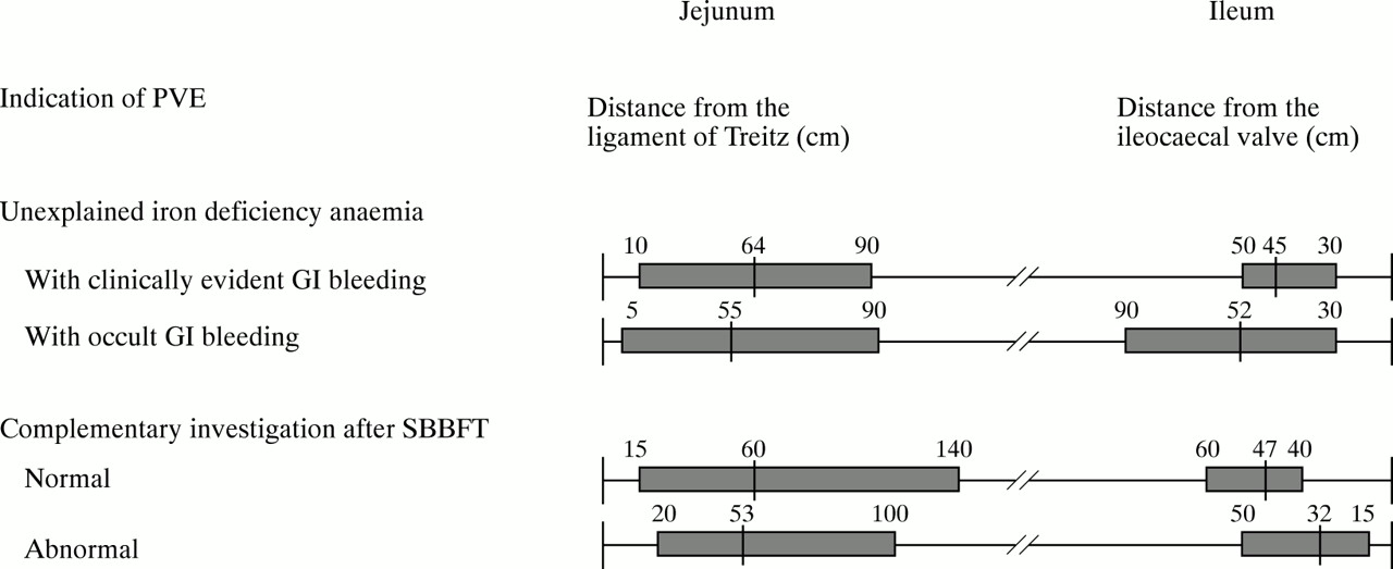
Figure 1 shows the distance of intestinal lesions from the ligament of Treitz and the ileocaecal valve, respectively.

Site of the 74 intestinal lesions found in 313 patients undergoing two way PVE. GI, gastrointestinal; SBBFT, small bowel barium follow through.
TOLERANCE OF PVE
No major complications (haemorrhage, perforation, death) were noted. Minor symptoms (sore throat, transitory dysphagia, etc) were not systematically noted.
Discussion
We found PVE to be a relatively easy and useful technique for the diagnosis of small intestinal diseases not identified by previous routine investigations. Of 313 patients undergoing PVE, a positive diagnosis was made in 29.4% of cases, a percentage similar to that reported by Davies et al in 43 patients (29%),9 but lower than that reported by Rossiniet al in 120 patients studied only by upper way enteroscopy (48%).10 In the experience of some authors,9 ,10 the use of x ray examination or fluoroscopy is not helpful for either the progression or the placement of the overtube. The main purpose of these techniques is probably to estimate accurately the jejunal intubation depth. We evaluated this by guesswork, which is probably a less accurate method than fluoroscopy, but when comparing our estimations on the site of lesions to those measured from an abnormal SBBFT or in patients finally operated on, the accuracy of our estimations was within 10 cm (data not shown). Furthermore, as the entire length of the small intestine is not explored using PVE, only positive results are of value and an accurate knowledge of the site of the lesions does not seem to be essential for patient management.
Approximately 30% of patients with unexplained anaemia will not have an identifiable source of blood loss despite upper and lower endoscopies.11 The usual second line examination is SBBFT but its yield is very low, from 0 to 10%.11-13 In our patients, SBBFT was normal in the 108 cases in which it was performed, although the quality was uncertain; an aetiological diagnosis was made by PVE in 7.4% of cases (arteriovenous malformation in four, tumours in three, and Crohn’s disease in one; data not shown). Most previous published studies of PVE concerned its use in the investigation of clinically evident gastrointestinal bleeding.4 ,13-16 The source of gastrointestinal bleeding was detected in the literature in 17–46% of cases.9 ,17 ,18 Using sonde enteroscopy in similar cases, success rates have been similar, ranging from 26 to 38%.6 ,19 The two methods were equivalent in terms of diagnostic efficiency, but the mean duration of the procedure was six hours with sonde and 35 minutes with push enteroscopy.20
In our study, of 203 patients referred for unexplained anaemia, jejunoileal lesions were found in 30 (14.7%) cases; in addition, 15 gastroduodenal and three colonic lesions missed by previous endoscopies were found (8.8%), with a higher rate in the case of clinically evident gastrointestinal bleeding (13.8%) than in isolated anaemia (6.1%). Interestingly, this rate of missed lesions is similar to that reported in most published studies—from 25% to 60%8 ,10-13 20-22—and raises the following dilemma when investigating unexplained anaemia: whether to repeat gastroduodenal and colonic endoscopies first or to perform an enteroscopy immediately. If the first upper and lower endoscopies had been performed for isolated anaemia in satisfactory conditions by an experienced operator, we think that it is more judicious to perform the PVE directly, to avoid the discomfort and risk of a third endoscopic examination if the second proves also to be negative. In other cases, especially when clinically evident gastrointestinal bleeding is present at the time of first endoscopy, a second upper, but not lower, endoscopy should be performed before PVE.
It must be emphasised that a lower enteroscopy was not performed in all published studies evaluating PVE.8-10 It is claimed that most lesions are located at the jejunum, but in our 48 patients with anaemia and a positive diagnosis provided by PVE, we found nine (19%) lesions located in the ileum and 21 (44%) in the jejunum. This rate of distal lesions is higher than the 10% reported in a long term follow up study after 40 upper PVE examinations in patients with gastrointestinal bleeding of obscure origin.21
The most common cause of anaemia in our patients was an arteriovenous malformation, found in 23% of patients with lesions. In the study of Lewis and Wayne, arteriovenous malformations were also the main cause of gastrointestinal bleeding, accounting for 80% (16/20 cases) of the diagnoses made in 60 patients.18 The second most common cause of anaemia was a small bowel tumour, found in 12% cases: the mean age of these patients was 54.4 years (range 21–71; data not shown). Small bowel tumours account for 5–10% of small bowel bleeding.23 In a study of 258 patients with obscure gastrointestinal bleeding investigated by sonde enteroscopy, small bowel tumours were the most common cause in patients under 50 years of age, found in 13 cases.24
It is noteworthy that in our patients investigated for unexplained anaemia, we failed to find a lesion in 76.3% of cases. Similar proportions have been found in most published studies.9 ,10 ,25 ,26 PVE cannot explore the entire length of the small intestine, and 80–100 cm of the mid-intestine remains inaccessible. The main problem is what to do with these patients. Their follow up has shown contradictory results. Sahay and Scott27 noted spontaneous disappearance of the anaemia in 89% of 75 patients with a mean follow up of six years, but Acosta and Civac noted a rate of death of 23% in relation to comorbid illness in 40 patients with a mean follow up of 26 months.21
The investigation of patients with clinicobiological symptoms suggesting small bowel disease, but without an accurate diagnosis after normal or abnormal SBBFT, was an excellent indication for PVE, with a global diagnostic rate of 40%. PVE permitted us to correct the interpretation of SBBFT and to detect a false positive report in 25% of cases. This high level of false positives is probably partially related to the fact that SBBFT examinations were done in a variety of institutions, by operators of variable skilfulness; it does however reflect the true medical practical and not the ideal conditions which are rarely obtained routinely. This high rate of false positives using SBBFT has already been reported, especially for small intestinal tumours.28 One of the main advantages of enteroscopy when performed to complement an abnormal SBBFT is that PVE allows biopsy examination. In our study, of 110 patients referred for complementary investigation after a normal/abnormal SBBFT, an intestinal tumour was diagnosed in 14% (lymphoma or lymphomatous polyposis in 12), which was malignant in 93% of cases. In most cases, patients could be treated by chemotherapy without requiring surgery.
In spite of the high diagnostic rate of PVE reached in this indication, it does not seem reasonable to perform a PVE before the SBBFT when suspecting small bowel disease on the basis of diarrhoea/malabsorption or suspected small intestinal obstruction, as suggested by others.9 ,29 As the entire length of the small bowel cannot be explored using PVE, a negative result has no value for eliminating a mid-small bowel lesion. The high rate of technical failure we had (26.7%) was explained by the distal site of abnormalities in 13 patients. Lower enteroscopy is harder and more time consuming than upper enteroscopy, because the insertion tube is more flexible than that of the normal colonoscopes, and the passage through the ileocaecal valve can be very difficult, as shown by our high global rate of technical failure (16.4%).
Tolerance of PVE can be considered excellent, as no major complications such as haemorrhage, perforation, or death were noted. However, even minor symptoms, such as sore throat or dysphagia did not occur. Barkinet al 30 reported five complications in 37 enteroscopies: abdominal pain in three, which rapidly resolved, and acute pancreatitis in one and Mallory-Weiss syndrome in one, both of which needed hospitalisation. Thus, the rate of enteroscopy complications does not seem to be much higher than that reported with oesophagogastroduodenoscopy or ileocolonoscopy.
In conclusion, PVE is an additional and useful test in the evaluation of patients with gastrointestinal bleeding or to complement an SBBFT which failed to determine an accurate aetiology. The procedure has an acceptably wide range of uses and reasonable diagnostic success rate to justify its development. Our experience shows that this technique can be performed on an ambulatory basis, is quick to do, not particularly difficult to learn, and relatively safe. In the patient with chronic unexplained anaemia, we advocate its use directly after routine negative oesophagogastroduodenoscopy and ileocolonoscopy, instead of SBBFT, because of the poor performance of the latter. Furthermore, it has been recently shown in a modelling study of diagnostic yields and cost implications that early enteroscopy was the best method for evaluating obscure gastrointestinal bleeding.31 From our results, ileoscopy using the same instrument increases further the yield of these investigations, when upper PVE is negative.
Acknowledgments
We thank Drs M Hautefeuille, JP Therond, B Vernisse, K Vahedi, and Mrs A Beauplet and Mr D Brezephin for their collaboration.