Article Text

Abstract
Objective Cholestatic liver diseases in humans as well as bile acid (BA)-feeding and common bile duct ligation (CBDL) in rodents trigger hyperplasia of cholangiocytes within the portal fields. Furthermore, elevation of BA levels enhances proliferation and invasiveness of cholangiocarcinoma (CCA) cells in animal models, thus promoting tumour progression. TGR5 is a G-protein coupled BA receptor, which is highly expressed in cholangiocytes and postulated to mediate the proliferative effects of BA.
Design BA-dependent cholangiocyte proliferation was examined in TGR5-knockout and wild type mice following cholic acid (CA)-feeding and CBDL. TGR5-dependent proliferation and protection from apoptosis was studied in isolated cholangiocytes and CCA cell lines following stimulation with TGR5 ligands and kinase inhibitors. TGR5 expression was analysed in human CCA tissue.
Results Cholangiocyte proliferation was significantly reduced in TGR5-knockout mice in response to CA-feeding and CBDL. Taurolithocholic acid and TGR5-selective agonists induced cholangiocyte proliferation through elevation of reactive oxygen species and cSrc mediated epidermal growth factor receptor transactivation and subsequent Erk1/2 phosphorylation only in wild type but not in TGR5-knockout-derived cells. In human CCA tissue TGR5 was overexpressed and the pathway of TGR5-dependent proliferation via epidermal growth factor receptor and extracellular signal-regulated kinase (ERK)1/2 activation also translated to CCA cell lines. Furthermore, apoptosis was inhibited by TGR5-dependent CD95 receptor serine phosphorylation.
Conclusions TGR5 is an important mediator of BA-induced cholangiocyte proliferation in vivo and in vitro. Furthermore, TGR5 protects cholangiocytes from death receptor-mediated apoptosis. These mechanisms may protect cholangiocytes from BA toxicity under cholestatic conditions, however, they may trigger proliferation and apoptosis resistance in malignantly transformed cholangiocytes, thus promoting CCA progression.
- BILE ACID
- CHOLANGIOCARCINOMA
- APOPTOSIS
- CELL PROLIFERATION
Statistics from Altmetric.com
Significance of this study
What is already known on this subject
Cholangiocyte proliferation is a hallmark of cholestatic liver diseases in humans. Rodent models for cholangiocyte proliferation include bile acid (BA)-feeding and common bile duct ligation (CBDL). Both models have been associated with elevated levels of cyclic AMP and activation of mitogen-activated protein (MAP)-kinases (eg, Erk1/2) in cholangiocytes.
Cholangiocarcinoma (CCA) is the second most common primary liver cancer with very dismal prognosis. CCA response to conventional chemotherapy is limited, therefore, novel therapeutic targets are needed. BAs have been linked to CCA progression in animal models and cell culture studies.
TGR5 (Gpbar1) is a membrane-bound receptor responsive to primary and secondary BAs and is expressed in cholangiocytes and gallbladder epithelial cells. TGR5 has been linked to proliferation in a cholangiocyte cell line (H69) and cell lines derived from oesophageal and gastric adenocarcinomas. The contribution of TGR5 to BA-dependent cholangiocyte proliferation in vivo as well as the receptor's potential role in CCA progression is unknown.
What are the new findings
TGR5 is required for BA-induced cholangiocyte proliferation in animal models as well as in isolated biliary epithelial cells. Using isolated cholangiocytes we demonstrate that TGR5 promotes cell proliferation through elevation of reactive oxygen species (ROS) and subsequent activation of Rous sarcoma oncogene (cSrc), epidermal growth factor receptor (EGFR) and Erk1/2. Furthermore, this pathway can be activated by TGR5 ligands in CCA cell lines EGI-1 and TFK-1.
Activation of TGR5 induces antiapoptotic effects through serine phosphorylation of the CD95 death receptor in murine cholangiocytes and CCA cell lines.
Under cholestatic conditions TGR5-deficient mice show reduced cholangiocyte hyperplasia and diminished hepatocyte proliferation and increased liver injury, suggesting that TGR5 exerts protective effects against BA toxicity in liver.
TGR5 is highly expressed in the tumour cells of human CCA. In this tissue elevated ERK1/2 phosphorylation and CD95 receptor serine phosphorylation was detected, indicating that the TGR5-dependent pathways identified in cholangiocytes may play a role in CCA cells.
How might it impact on clinical practice in the foreseeable future?
TGR5 mediates proliferative and antiapoptotic and thus protective effects in cholangiocytes. TGR5 agonists may therefore be a valuable treatment of cholestatic liver disease affecting the biliary tree, such as primary biliary cirrhosis.
On the other hand, in CCA tissue, overexpression of TGR5 could be associated to tumour cell proliferation and apoptosis resistance. Hence, development of TGR5 inhibitors could represent a novel therapeutic approach in CCA.
Introduction
Bile acids (BAs) are signalling molecules with diverse endocrine and paracrine functions.1–5 BAs play an important role for BA, glucose, lipid and energy homoeostasis as well as for the immune response.3 ,5 ,6 Thus, BAs and BA receptors have emerged as targets for the treatment of metabolic diseases, such as obesity, diabetes, steatohepatitis and atherosclerosis.3 ,5–12
Furthermore, various BAs exert proliferative effects on different cell types including hepatocytes, cholangiocytes and intestinal epithelial cells.13–17 In rodents, cholic acid (CA)-feeding triggers proliferation in hepatocytes and BA depletion by cholestyramine slows liver regeneration after partial hepatectomy or after acetaminophen overdose.17–19 While hydrophilic BAs such as taurocholic (TC) acid and tauroursodeoxycholic acid promote protective and antiapoptotic effects in hepatocytes, hydrophobic BAs such as taurolithocholic (TLC) acid and glycochenodeoxycholic acid trigger hepatocyte apoptosis and liver damage.2 ,18 ,20–24 In contrast to hepatocytes, TLC acid induces proliferation of cholangiocytes in vitro and in vivo.13 ,14 Whether BAs exert protective or toxic effects depends on their biochemical properties and concentration, and on the BA receptors expressed in the respective cell type.9 In liver several different BA receptors are expressed, which comprise nuclear BA receptors, such as the farnesoid-X-receptor, as well as plasma membrane-bound, G-protein coupled receptors (GPCRs) like the sphingosine-1-phosphate receptor-2 (S1PR2) or TGR5 (Gpbar1).5 ,9 ,25–29
TGR5 is localised in different non-parenchymal cells of the liver, including Kupffer cells (KCs), sinusoidal endothelial cells, cholangiocytes and gallbladder epithelial cells.9 ,30–37 TGR5 is coupled to a stimulatory G-protein and is responsive to different progesterone derivatives and unconjugated and conjugated BAs with TLC acid (half maximal effective concentration (EC50)=0.29 µM) being the most potent agonist.25 ,27 ,38 ,39 Stimulation of TGR5 by TLC acid in gallbladder tissue led to a significant rise in intracellular cyclic AMP (cAMP) and incubation of isolated gallbladder epithelial cells with a TGR5 agonist triggered chloride secretion.30 ,31 Furthermore, treatment with the TGR5 agonist INT-777 (6α-ethyl-23(S)-methyl-CA) significantly enhanced bile flow in rodents.32 ,40 ,41 Additionally, TGR5 may confer protection from BA toxicity by preventing death receptor-mediated apoptosis.32 Stimulation of TGR5 in isolated murine cholangiocytes with INT-777 inhibited CD95 ligand-induced cell death.32 Moreover, cholangiocyte proliferation is enhanced by TC acid and TLC acid, which are potent TGR5 ligands.14 ,32 ,39 Treatment of the human cholangiocyte cell line H69 with TLC acid or INT-777 triggered proliferation in non-confluent, non-ciliated cells, indicating a role of TGR5 for cholangiocyte proliferation.31 ,32 ,34 ,39 However, the underlying signalling mechanisms are incompletely understood. The aim of this study was to investigate whether TGR5 promotes BA-dependent cholangiocyte proliferation in vivo and to elucidate the underlying signalling pathway in vitro. Since BAs also enhance cholangiocarcinoma (CCA) proliferation, progression and invasiveness,42–44 we investigated the expression of TGR5 in human CCA tissue as well as the receptor's role for proliferation and resistance to apoptosis in CCA cell lines.
Material and methods
TGR5 agonists
TGR5-Ago-T(4-((3,5-bis(Trifluoromethyl)phenyl)methyl)-6-(2-fluorophenyl)-4,5-dihydropyrido-(3,2-f)-1,4-oxazepin-3(2H)-one) (CAS 877052-79-4; patent JP2006056881) and TGR5-Ago-R RO5527239 ((R,E)-1-(4-(3-(hydroxyimino)-3-(2-methylpyridin-4-yl)-1-o-tolylpropyl)phenyl)piperidine-4-carboxylic acid)45 were kindly provided by F. Hoffmann-La Roche, (Basel, Switzerland).30 ,45 For all other chemicals see online supplementary material.
Animal studies
TGR5-knockout (KO) mice were a gift from Schering-Plough Research Institute (Kenilworth, New Jersey, USA).35 Male mice, 8–10 weeks old, were either fed standard chow diet or the same diet supplemented with 0.5% (weight per weight) CA (Ssniff, Soest Germany) for 7 days. Common bile duct ligation (CBDL) was carried out under anaesthesia with isoflurane.46 Cholangiocytes were isolated from 6–8-week old mice33 ,47 (see online supplementary material).
For proliferation-assay (BrdU), epidermal growth factor (EGF) ELISA, reactive nitrogen and oxygen species (RNOS) measurements, 3-(4,5-dimethylthiazol-2-yl)-2,5-diphenyltetrazolium bromide (MTT)-assay, lactate dehydrogenase (LDH) release, terminal deoxynucleotidyl transferase dUTP nick end labelling (TUNEL) assay, western blotting, CD95R immunoprecipitation, immunohistochemistry, immunofluorescence staining, quantitative real-time PCR, BA measurements see online supplementary material.
Human liver tissue
The study was performed according to the guidelines of the declaration of Helsinki and informed written consent was obtained from all patients. Liver tissue was collected immediately after resection and shock-frozen in liquid nitrogen (see online supplementary material).
Statistics
Data are given as means±SEM. Results were analysed using the two-sided Student's t test and/or the Mann-Whitney U test. Application of the Mann-Whitney U test is indicated in the respective figure legends. A p value <0.05 was considered statistically significant.
Results
BA feeding induces cholangiocyte proliferation in wild type but not in TGR5 KO mice
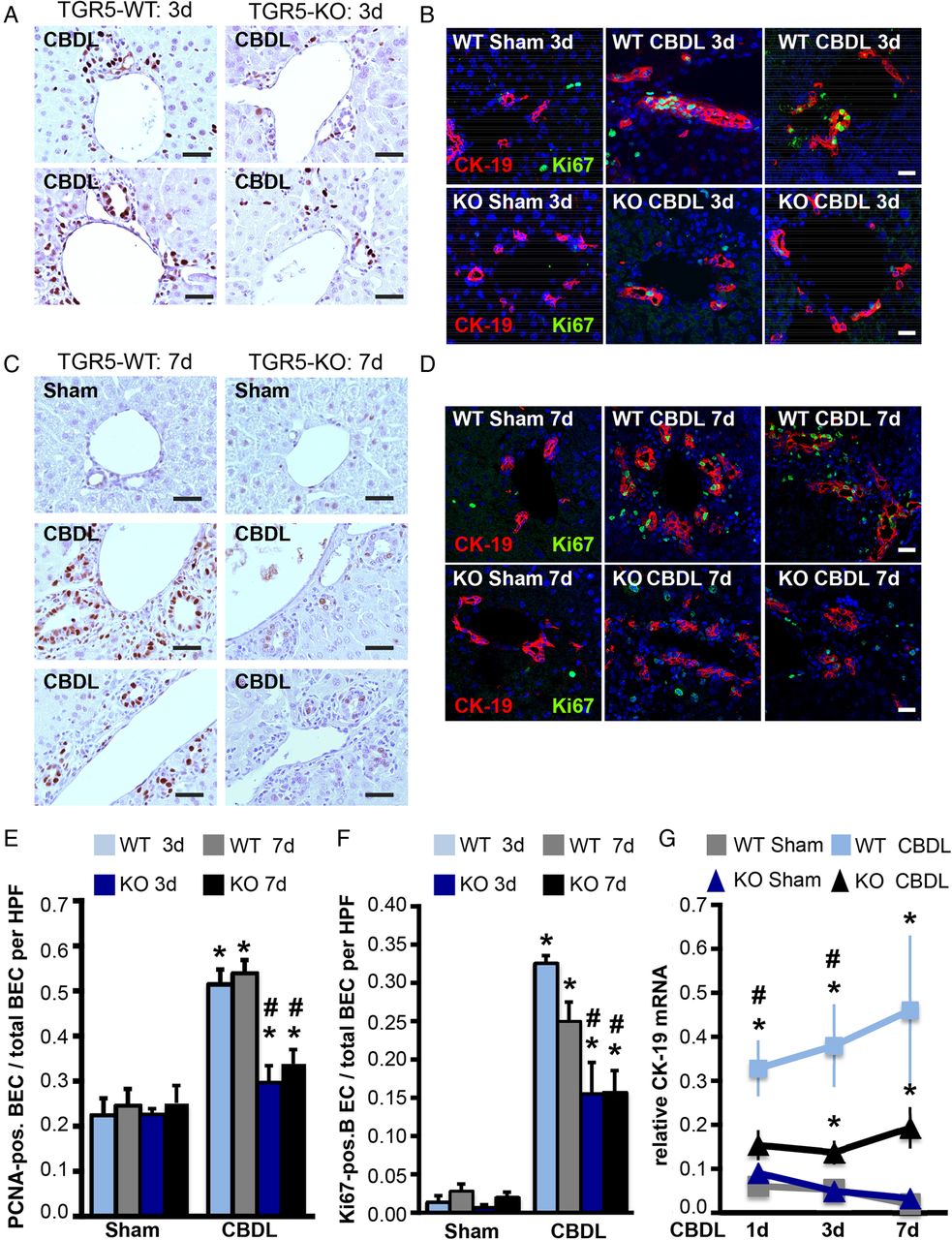
Feeding of CA (0.1–1.0%) triggers hepatocyte and cholangiocyte proliferation in rodents.14 ,17 Since TGR5 is highly expressed in cholangiocytes but not hepatocytes,9 ,36 the role of TGR5 for cholangiocyte proliferation was investigated in wild type (WT) and TGR5-KO mice fed with a diet containing 0.5% CA for 7 days. To quantify cholangiocyte proliferation, liver sections were stained with antibodies against the proliferation marker proteins Ki67 or the proliferating cell nuclear antigen (PCNA) (figure 1A, B). In chow-fed animals of both genotypes only few cytokeratin-19 (CK-19)-positive cholangiocytes also stained positive for Ki67 (1.9±1.2% in WT; 1.5±0.8% in TGR5-KO, n=3 each, p=0.82). CA-feeding significantly increased the number of Ki67-positive CK-19-expressing cholangiocytes in liver cryosections of both genotypes, however, this rise was significantly more pronounced in WT animals (23±2% Ki67-positive cholangiocytes) as compared with TGR5-KO mice (12±1%, n=5 each, p<0.01) (figure 1A, C). Analysis of PCNA staining also demonstrated an elevation of PCNA-positive cholangiocytes after CA-feeding in both genotypes. Again, the increase was significantly more pronounced in WT (54±5% PCNA-positive cholangiocytes) as compared with TGR5-KO mice (38±2%, n=10 each, p<0.01) (figure 1 B, D). In chow-fed animals of both genotypes similar amounts of cholangiocytes stained positive for PCNA (33±1% in WT; 31±1% in TGR5-KO, n=8–10, p=0.26). Furthermore, mRNA levels and the immunofluorescence staining for the cholangiocyte marker protein CK-19 increased significantly in livers of CA-fed WT as compared with TGR5-KO animals (p<0.01), indicating that CA-induced cholangiocyte proliferation is reduced in the absence of TGR5 (figure 1 E–G). There was no difference in liver CK-19 mRNA levels or CK-19 immunofluorescence staining between genotypes on chow diet (figure 1 E–G).

Cholic acid (CA)-feeding promotes cholangiocyte proliferation in TGR5-wild type (TGR5-WT) but not in TGR5-knockout (TGR5-KO) mice. (A and B). Male TGR5-WT and TGR5-KO (6–8 weeks old) mice were either fed with chow or the same diet supplemented with 0.5% (weight per weight) CA for 7 days. Cell proliferation was detected with antibodies against Ki67 (A) or proliferating cell nuclear antigen (PCNA) (B). Representative images of one chow-fed and two CA-fed animals per genotype are shown. (A) Cholangiocytes were stained with an antibody against cytokeratin-19 (CK-19) and nuclei were made visible using Hoechst. (B) Haematoxylin was used to stain nuclei. Bars=25 μm. (C) Cells positive for Ki67 and CK-19 were counted and divided by the total number of CK-19-positive cholangiocytes per high power field (HPF). At least five images of portal fields for each animal were analysed (n=3 animals on chow; n=5 animals on CA diet per genotype each). (D) PCNA-positive cholangiocytes and total number of cholangiocytes were counted in each HPF. At least 10 images per animal were analysed (n=8–10 animals/group). (E) CK-19 mRNA was measured in relation to hypoxanthine-phosphoribosyltransferase 1 (HPRT1) mRNA in livers from chow-fed (n=5 animals/genotype) and CA-fed animals (n=5–6 animals/genotype). CK-19 mRNA expression was significantly elevated in CA-fed TGR5-WT animals. Data are means±SEM. *significant difference from chow (p<0.05); #significant difference from WT of the same condition (p<0.05). (F) Cryosections were stained with an antibody against CK-19 (in red) to visualise cholangiocytes. Nuclei were stained with Hoechst (blue). Representative images of one chow-fed and two CA-fed animals per genotype are shown. Bars=50 µm. (G) Quantitative analysis of relative CK-19 fluorescence staining (number of pixels × average fluorescence intensity per pixel) in livers from TGR5-WT and TGR5-KO mice. No difference in CK-19 staining was observed between chow-fed mice (squares). However, the CK-19 fluorescence signal was significantly increased in livers from TGR5-WT mice fed with CA as compared with the TGR5-WT animals on chow diet. This increase in CK-19 staining following CA feeding was not observed in TGR5-KO littermates (triangles). Thus, CA-fed TGR5-KO showed significantly less CK-19 staining as compared with the CA-fed TGR5-WT animals. Ten images per animal were analysed from four different animals per condition. Statistical analysis was performed with the Mann-Whitney U-test.
CA-feeding induced a significant rise in serum BA concentrations and a mild liver injury as indicated by a moderate increase in aspartate aminotransferase (AST), alanine aminotransferase (ALT) and bilirubin levels in WT and TGR5-KO mice. Alkaline phosphatase levels were unchanged by this treatment. Only AST levels were significantly different between genotypes following CA feeding with a 6.9-fold rise in AST in TGR5-KO mice as compared with a 2.3-fold increase in WT littermates, supporting the role of TGR5 in the protection of the liver from BA toxicity.48 CA-feeding triggered a more pronounced hepatocyte proliferation as determined by Ki67-staining and cyclin D1 mRNA levels in TGR5-WT livers, resulting in a significant increase in liver size by about 35% in WT and only 15% in TGR5-KO mice (see online supplementary table S1 and figure S1C, D).
Bile duct ligation triggers a pronounced cholangiocyte proliferative response only in TGR5-WT mice
CBDL also induces cholangiocyte proliferation,49 therefore, TGR5-WT and TGR5-KO mice were subjected to CBDL for 3 days or 7 days. CBDL resulted in a significant increase in AST, ALT, alkaline phosphatase, bilirubin and BA serum levels in WT and TGR5-KO animals as compared with the sham-operated littermates. TGR5-KO mice showed a tendency towards higher AST, ALT and BA levels, however, this difference did not reach statistical significance (see online supplementary table S2). Hematoxylin eosin (HE) staining demonstrated significantly more necrotic areas 3 days after CBDL in livers from TGR5-KO mice. This difference was no longer observed 7 days after CBDL (see online supplementary figure S2E). PCNA staining of CBDL livers showed a significant increase in PCNA-positive cholangiocytes in WT animals after 3 days (52±3%, p<0.01, n=6) and 7 days (54±3%, p<0.01, n=6) as compared with sham-operated WT controls (3 days: 23±4%, 7 days: 25±4%, n=4–5). In contrast, only a mild cholangiocyte proliferative response was observed in CBDL livers of TGR5-KO mice after either 3 days (30±4%, n=6, p=0.12 vs sham) or 7 days (34±3%, n=5, p=0.12 vs sham) as compared with the sham-operated TGR5-KO mice (3 days: 23±1%, 7 days: 25±4%, n=5 each) (figure 2A, C, E). In sham-operated animals of both genotypes less than 3% of CK-19-positive cholangiocytes also stained positive for the proliferation marker Ki67 after 3 days or 7 days (0.7–2.9%, n=4–5/group) (figure 2B, D, F). CBDL increased the number of Ki67-positive CK-19-expressing cholangiocytes in livers of both genotypes, however, this rise was significantly more pronounced in WT animals (TGR5-WT: 33±1% and 25±3% Ki67-positive cholangiocytes after 3 days or 7 days, respectively, n=4–6/group) (TGR5-KO: 16±4% and 16±3%, n=4–6/group, p<0.05 vs WT-CBDL) (figure 2F). Furthermore, CBDL triggered a significant elevation of CK-19 mRNA expression over time, which was more prominent in livers from WT animals after 1 day and 3 days (figure 2G). Since proliferation of cholangiocytes can also be promoted by cytokines and growth regulators including tumour necrosis factor α (TNFα), interleukin-6 (IL-6) or TNF-like weak inducer of apoptosis (TWEAK),50 serum levels of cytokines as well as liver mRNA expression of cytokines, TWEAK and its receptor Fn14 were determined after CBDL (see online supplementary figure S3). CBDL for 1–7 days led to a significant increase in IL-1β, TNFα, IL-6 and monocyte chemoattractant protein-1 (MCP-1) serum levels in both genotypes. Only MCP-1 serum concentrations were significantly higher in the TGR5-deficient animals after 3–7 days of CBDL. On the mRNA level no significant differences in IL-1β, TNFα, IL-6 or MCP-1 expression were detected between genotypes after 1 day or 7 days of CBDL. In contrast, IL-1β, TNFα, MCP-1, but not IL-6 mRNA expression was significantly elevated in livers from TGR5-KO mice 3 days after CBDL. TWEAK mRNA expression was unchanged in CBDL livers after 7 days in either genotype, while mRNA levels of the TWEAK receptor Fn14 significantly increased in CBDL livers of both genotypes (see online supplementary figure S3). Similar to CA-feeding, CBDL also promoted hepatocyte proliferation, which again was significantly increased in TGR5-WT animals as compared with TGR5-KO mice 7 days after CBDL as measured by Ki67 staining (see online supplementary figure S2F).

Common bile duct ligation (CBDL) triggers a more pronounced cholangiocyte proliferation in TGR5-wild type (TGR5-WT) as compared with TGR5-knockout (TGR5-KO) mice. (A–D) Male mice (8–10 weeks old) were subjected to CBDL-operation or sham-operation for 3 days or 7 days (D). Cell proliferation was detected with antibodies against proliferating cell nuclear antigen (PCNA) (A and C) or Ki67 (B and D). A. Images of two different animals after 3 days of CBDL stained for PCNA per genotype are shown. Nuclei were visualised with haematoxylin. (B) Cholangiocytes were stained with an antibody against cytokeratin-19 (CK-19). Nuclei were visualised with Hoechst (blue). Representative images of one sham and two CBDL animals 3 days after CBDL per genotype are shown. (C) Representative images of one sham-operated and two 7-day-CBDL-operated animals per genotype were stained with PCNA and haematoxylin. (D) Co-staining of liver cryosections 7 days after sham or CBDL with antibodies against CK-19 and Ki67. Nuclei are stained with Hoechst. Bars=25 μm. (E) PCNA-positive cholangiocytes and total number of cholangiocytes were counted in each high power field (HPF). At least 10 images per animal from 4–6 different animals were analysed for each condition. (F) Cholangiocytes positive for Ki67 and CK-19 as well as the total amount of CK-19-positive cells per HPF were determined. At least five images per animal were analysed for each condition (n=4–6 animals per group). (G) CK-19 mRNA was measured in relation to HPRT1 mRNA in livers of CBDL-operated or sham-operated animals after 1 day, 3 days or 7 days. CK-19 mRNA expression was significantly elevated in CBDL TGR5-WT as compared with TGR5-KO animals 1 day and 3 days after CBDL (n=4–7 animals/group). Data are means±SEM; *significant difference from sham (p<0.05); #significant difference from wild type of the same condition (p<0.05).
Activation of TGR5 promotes cholangiocyte proliferation in vitro through Erk1/2 activation
To elucidate the role of TGR5 for BA-induced cholangiocyte proliferation, primary cholangiocytes isolated from TGR5-WT and TGR5-KO mice were incubated with growth medium, which contained EGF as well as forskolin,33 and proliferation was measured by BrdU-incorporation. Growth medium triggered a similar 2.0±0.1-fold and 1.9±0.2-fold increase in proliferation in TGR5-WT- and TGR5-KO-derived cholangiocytes, respectively (n=12–15). A 1.7-fold increase in BrdU-incorporation was observed in WT and TGR5-deficient cholangiocytes (n=7–10) following stimulation with forskolin (10 μM) (figure 3A). However, treatment with TC acid (10 μM, 25 μM), TLC acid (10 μM, 25 μM) or the TGR5 agonist (Ago-R; 1 μM, 10 μM) led to a dose-dependent increase in proliferation only in WT cells (n=6–13), but not in cells from TGR5-KO mice (n=4–10) (figure 3A; see online supplementary figure S6A, B). Stimulation with a second TGR5 agonist (Ago-T, 25 μM, n=5–8) triggered a significant 1.4±0.1-fold BrdU-incorporation only in TGR5-WT cells.
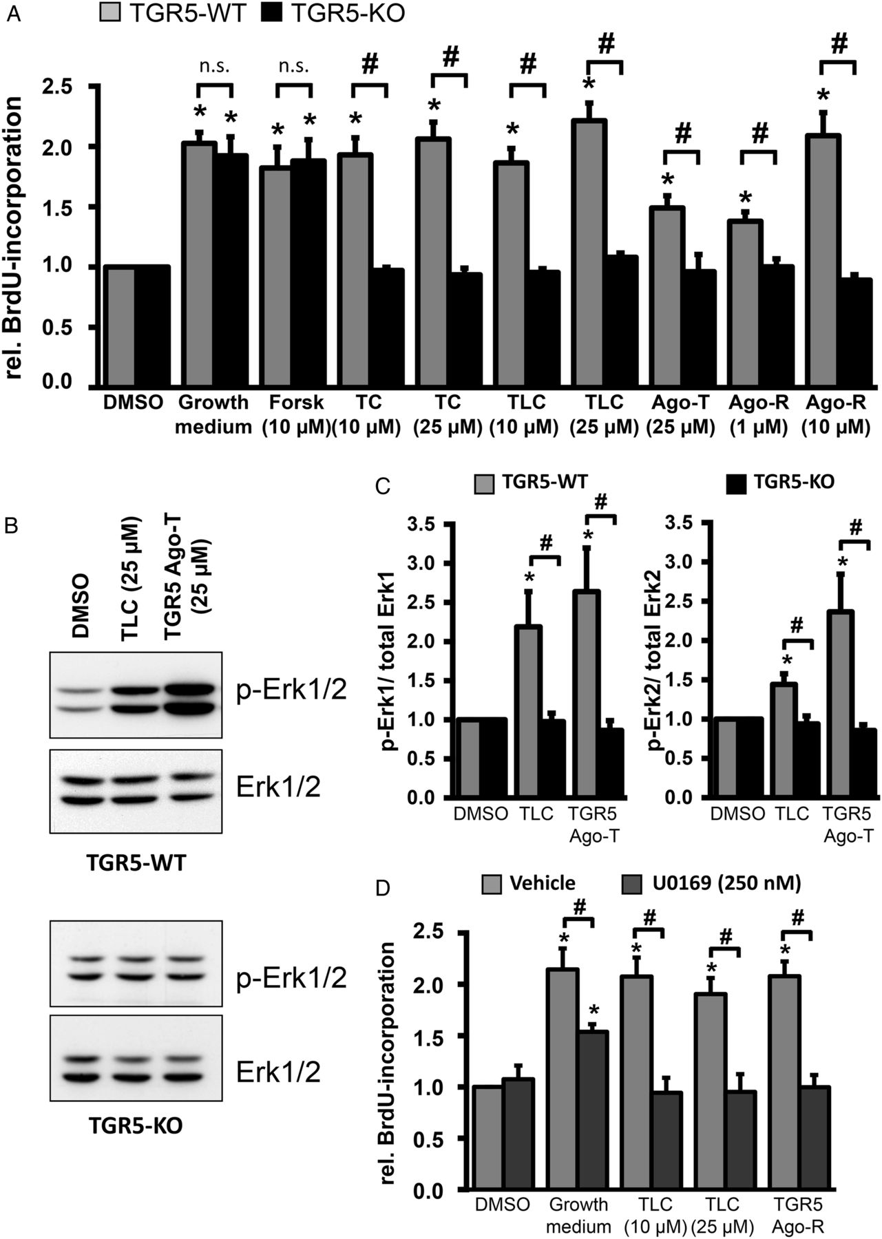
Taurolithocholic (TLC) acid-induced cholangiocyte proliferation is TGR5 dependent and Erk1/2 dependent. (A) Cholangiocytes were isolated from TGR5-wild type (TGR5-WT, grey) and TGR5-knockout mice (TGR5-KO, black). Cell proliferation was measured by BrdU-incorporation in the presence of growth medium (positive control), forskolin (Forsk), taurocholic (TC) acid, TLC acid or specific TGR5 agonists. Incubation with growth factor-containing medium and forskolin induced a significant increase in BrdU-incorporation in cholangiocytes from TGR5-WT (n=10–15) and TGR5-KO (n=7–12) mice. In contrast TC acid, TLC acid and the two different TGR5-specific agonists significantly raised BrdU incorporation only in the WT-derived (n=6–13) cells but not in cells from TGR5-KO (n=4–10) mice. (B) Western blot analysis of Erk1 and Erk2 phosphorylation in isolated cholangiocytes. Cells were stimulated with TLC acid or a TGR5 agonist (25 µM each) for 30 min. Incubation with both substances induced a significant increase in Erk1 and Erk2 phosphorylation in TGR5-WT cells (n=15) but not in cholangiocytes from TGR5-KO mice (n=5). (C) Erk1 and Erk2 phosphorylation versus total Erk1 and Erk2 was determined respectively by densitometric analysis. The values for dimethyl sulfoxide (DMSO)-treated cells for each n were set to 1.0. (D) Cholangiocytes from TGR5-WT mice were cultivated with the MEK1-inhibitior U0169 (250 nM, n=5) 30 min prior to stimulation with growth medium, TLC acid or the TGR5 agonist. Treatment with U0169 completely abolished TLC acid-induced and TGR5 agonist-induced cell proliferation and significantly reduced BrdU-incorporation in the presence of growth medium. Data are expressed as means±SEM; *significant difference from control conditions (p<0.05); #significant difference from WT cells (A and C) or vehicle treated cells (D) (p<0.05). n.s., not significant.
Western blot analysis of WT cholangiocytes treated for 30 min with either TLC acid (25 μM, n=15) or a TGR5 agonist (Ago-T; 25 μM, n=15) showed a significant 2.2±0.4-fold and 2.6±0.6-fold increase in Erk1 phosphorylation and a 1.4±0.1-fold and 2.4±0.5-fold rise in Erk2 phosphorylation as compared with dimethyl sulfoxide (DMSO)-treated WT cells (p≤0.01), which was absent in TGR5-KO cholangiocytes (n=5, figure 3B, C). Incubation of WT cholangiocytes with the mitogen-activated protein kinase kinase-1 (MEK1) inhibitor U0169 (250 nM) 30 min prior to stimulation with growth medium, TLC acid or a TGR5 agonist completely abolished TLC acid-induced and TGR5 agonist-induced BrdU incorporation, while the growth medium-dependent rise in proliferation was significantly reduced from 2.1±0.2-fold to 1.5±0.1-fold (n=5, p≤0.01, figure 3D).
TGR5-mediated cholangiocyte proliferation involves EGFR activation
Stimulation of TGR5-WT cholangiocytes with TLC acid and a TGR5 agonist led to an increase in tyrosine phosphorylation of the epidermal growth factor receptor (EGFR) at positions 845, 1045 and 1173 within 30 min (n=7–8, p<0.05, figure 4A, B). Inhibition of the EGFR kinase by AG1478 (10 nM) completely suppressed the TGR5 ligand-dependent BrdU-incorporation of TGR5-WT cholangiocytes, while the growth medium-induced proliferation was only partially inhibited by AG1478 (figure 4C). To test, whether EGFR activation is mediated through ADAM-dependent or matrix metalloproteinase (MMP)-dependent EGF shedding, proliferation of TGR5-WT cholangiocytes was measured after incubation with the ADAM/MMP inhibitor batimastat (60 nM).51 Pretreatment of the cells with batimastat abolished TLC acid-dependent and TGR5 agonist-dependent BrdU-incorporation, while the response to the EGF-containing growth medium was unaffected (figure 4D). Furthermore, treatment of TGR5-WT cholangiocytes with a TGR5 agonist (Ago-R, n=14) and TLC acid (25 μM, n=19) induced a significant 1.55±0.18-fold and 1.62±0.16-fold increase in soluble EGF in the cell culture supernatant (p≤0.01), respectively, within 20 min. No increase in EGF shedding was measured in the supernatant of TGR5-KO cells treated with different TGR5 ligands (n=7–19) (figure 4E). Preincubation of cholangiocytes with either batimastat (60 nM), 4-amino-5-(4-chlorophenyl)-7-(t-butyl)pyrazolo[3,4-d]-pyrimidine (PP2) (0.5 μM) or N-acteylcysteine (3 mM) inhibited TLC acid-mediated EGF shedding in WT-derived cholangiocytes (figure 4E).

Stimulation of TGR5 leads to epidermal growth factor receptor (EGFR) activation. (A and B) Western blot analysis of EGFR phosphorylation in isolated wild type (WT) cholangiocytes. Cells were stimulated with taurolithocholic (TLC) acid or a TGR5 agonist (25 µM each) for 30 min. Incubation with both substances induced a significant increase in EGFR tyrosine phosphorylation at positions 845, 1045 and 1173. (B) EGFR Y845, Y1045 and Y1173 phosphorylation versus total EGFR was determined by densitometric analysis (n=7–8). The value for dimethyl sulfoxide (DMSO)-treated cells for each n was set to 1.0. (C) Cholangiocytes from WT mice were cultured with the EGFR kinase inhibitor AG1478 (10 nM) 30 min prior to stimulation. Treatment with AG1478 abolished TLC acid-dependent and TGR5 agonist-dependent cell proliferation and significantly reduced BrdU-incorporation in the presence of growth medium (n=6). (D) Cholangiocytes from WT mice were cultured with a matrix metalloproteinase/ADAMs inhibitor batimastat (60 nM) 30 min prior to stimulation. Treatment with batimastat abolished TLC acid-dependent and TGR5 agonist-dependent cell proliferation (n=5). (E) Shedding of epidermal growth factor (EGF) was determined in the cell supernatant with the quantikine mouse EGF ELISA after treatment of TGR5-WT and TGR5-knockout (KO) cholangiocytes with TLC acid (25 µM)(n=19) or a TGR5 agonist (10 µM)(n=7–14). EGF in the supernatant was normalised to the total protein amount of the cells in each vial. Preincubation (30 min) with batimastat (bati, 60 nM, n=6–7), the Src-kinase inhibitor 4-amino-5-(4-chlorophenyl)-7-(t-butyl)pyrazolo[3,4-d]-pyrimidine (PP2) (0.5 μM, n=5–6) or N-acetylcysteine (NAC, 3 mM, n=5–6) prevented TLC acid-induced EGF-shedding in cholangiocytes from WT animals. Data are expressed as means±SEM; *significant difference from control conditions (p<0.05); #significant difference from vehicle treated cells (p<0.05). n.s., not significant.
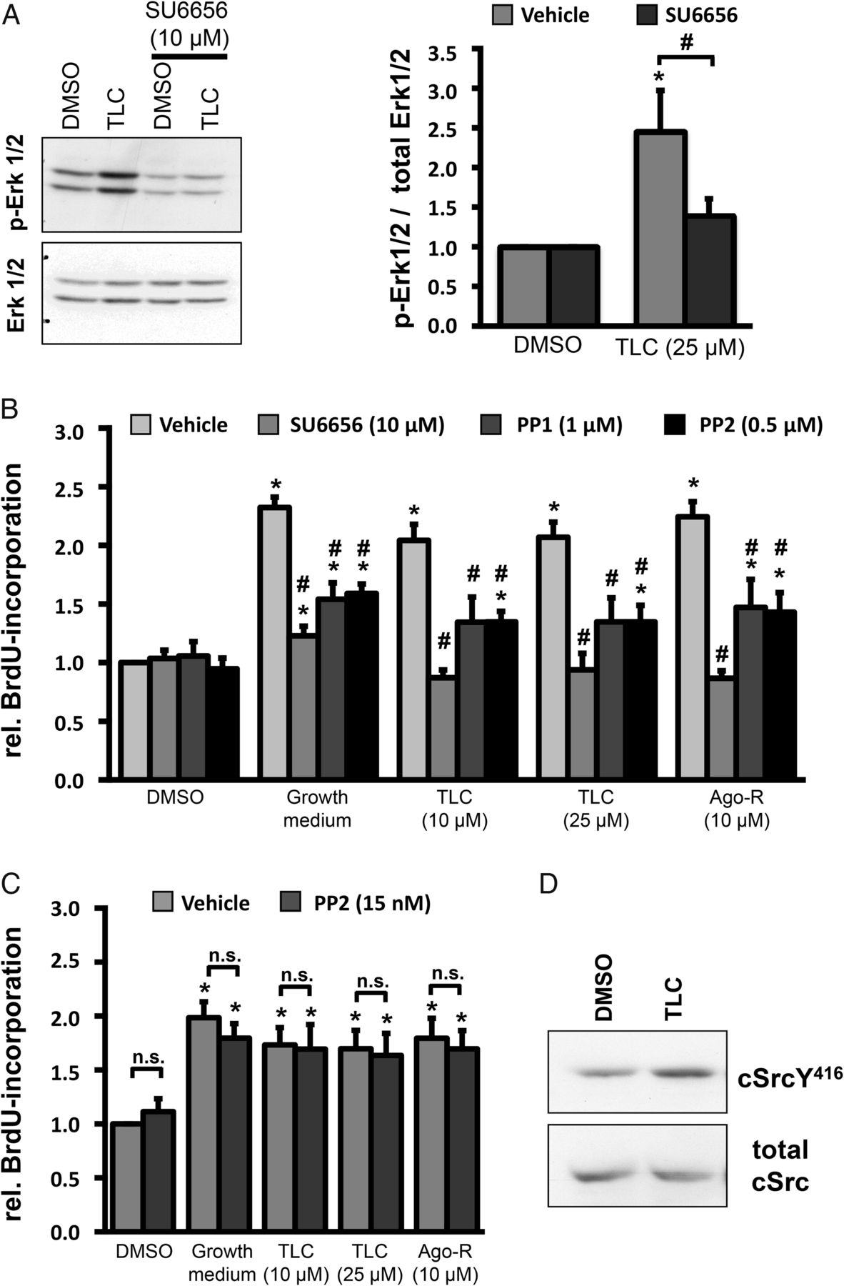
Src kinases and RNOS, but not adenylate cyclase, mediate the TGR5-induced cholangiocyte proliferation
Since Src kinases can promote EGF shedding as well as direct activation of the EGFR at tyrosine-845,52 ,53 we tested whether inhibition of Src kinases could suppress TLC acid-induced Erk1/2 phosphorylation and TGR5 ligand-mediated proliferation in WT cholangiocytes. Preincubation of cells with SU6656 (10 μM), an inhibitor of Src kinases, prevented TLC acid-dependent Erk1/2 phosphorylation (n=7, p<0.05 vs TLC acid) (figure 5A). Furthermore, treatment of WT cholangiocytes with different Src kinase inhibitors (SU6656, PP1 (1 μM), PP2 (0.5 μM)) prior to stimulation with growth medium, TLC acid or the TGR5 agonists significantly lowered BrdU-incorporation (n=6–8) (figure 5B). The effect was most pronounced with SU6656 and was dose-dependent for PP2, where 15 nM (n=8) did not show any inhibitory effect, while 0.5 μM significantly reduced growth medium- and TGR5 ligand-dependent proliferation (figure 5C), indicating that the effects were mediated via Rous sarcoma oncogene (cSrc). This is supported by the finding that TLC acid (25 μM) triggered phosphorylation of cSrc at tyrosine-416 in WT cholangiocytes (n=7, p<0.05) (figure 5D).
Activation of a Src kinase is essential for cholangiocyte proliferation. (A) TGR5-wild type (WT) cholangiocytes were stimulated with taurolithocholic (TLC) acid (25 µM) and Erk1/2 phosphorylation was detected by western blotting. Preincubation of the cells with the Src kinase inhibitor SU6656 inhibited TLC acid-mediated Erk1/2 phosphorylation. Densitometric analysis of Erk1/2 phosphorylation versus total Erk1/2 (n=7). (B) Cholangiocytes from WT mice were stimulated with different Src kinase inhibitors (SU6656, PP1, 4-amino-5-(4-chlorophenyl)-7-(t-butyl)pyrazolo[3,4-d]-pyrimidine (PP2)) 30 min prior to stimulation with growth medium, TLC acid or the TGR5 agonist. Treatment with SU6656 (10 µM), PP1 (1 µM) and PP2 (0.5 µM) significantly reduced TLC acid-mediated and TGR5 agonist-mediated cholangiocyte proliferation (n=6–8). (C) In contrast lower PP2 concentrations (15 nM) had no effect on growth medium-induced, TLC acid-induced or TGR5 agonist-induced BrdU-incorporation (n=8). (D) Stimulation of WT cholangiocytes with TLC acid (25 µM) significantly enhanced phosphorylation of Rous sarcoma oncogene (cSrc) at tyrosine-416. Data are expressed as means±SEM; *significant difference from control conditions (p<0.05); #=significant difference from vehicle treated cells (p<0.05).
Stimulation of TGR5 can promote the formation of RNOS,38 which in turn can activate Src kinases.54 Incubation of TGR5-WT and TGR5-KO cholangiocytes with TC acid, TLC acid and the TGR5 agonist (Ago-R) induced a significant increase in RNOS formation only in WT-derived but not in TGR5-KO-derived cells (n=4–9, figure 6A, B). Using N-acteylcysteine (3 mM) and apocynin (300 μM), we could demonstrate that both compounds significantly inhibited growth medium-dependent, TLC acid-dependent and TGR5 agonist-dependent cholangiocyte proliferation (figure 6C).

Reactive nitrogen and oxygen species (RNOS) are important for bile acid (BA)-mediated cholangiocyte proliferation. (A) Cholangiocytes from TGR5-wild type (WT) and TGR5-knockout (KO) mice were incubated with the RNOS-sensitive dye 2',7'-dichlorofluorescein (DCF). Stimulation with taurocholic (TC) acid and taurolithocholic (TLC) acid (10 µM and 25 µM each) for 1 h led to a significant increase in DCF-fluorescence in TGR5-WT but not in TGR5-KO-derived cells (n=6–9). H2O2 (10 μM) was used as a positive control. (B) TGR5-WT and TGR5-KO cholangiocytes were incubated with the reactive oxygen species (ROS)-sensitive dye dihydroethidium (DHE) for 30 min. Application of the TGR5 agonist (Ago-R, 10 μM) for 1 h led to a significant increase in DHE fluorescence only in TGR5-expressing WT cells (n=4–5). (C) WT cholangiocytes were incubated with the antioxidant N-acetylcysteine (NAC, 3 mM, n=6) or the NADPH-oxidase inhibitor apocynin (300 µM, n=7) 30 min prior to stimulation with growth medium, TLC acid or the TGR5 agonist. (D) WT cholangiocytes were treated with the adenylate cyclase inhibitors SQ22 536 (60 µM, n=6–13) and dideoxyadenosine (DDA, 10 µM, n=6–8) 30 min before stimulation with growth medium, TLC acid or the TGR5 agonist. Data are expressed as means±SEM; * significant difference from control conditions (p<0.05); # significant difference from vehicle treated cells (p<0.05).
Since TGR5 is coupled to a stimulatory G-protein25 ,27 and elevation of cAMP has been shown to promote cholangiocyte proliferation (see figure 3A), the contribution of adenylate cyclase (AC) to TGR5-dependent cholangiocyte proliferation was assessed with two different inhibitors. SQ22 536 (60 μM, p<0.01, n=6) and 2′,5′-dideoxyadenosine (10 μM, p<0.01, n=6) significantly suppressed forskolin-dependent BrdU-incorporation (figure 6D). In contrast, neither AC-inhibitor (SQ22,536, n=13; 2′,5′-dideoxyadenosine n=8) had any effect on growth medium-induced, TLC acid-induced or TGR5 agonist-induced cholangiocyte proliferation, indicating AC-independent signalling pathways for EGF-containing growth medium as well as the TGR5 ligands (figure 6D).
To determine whether TGR5 deficiency modulates the response of cholangiocytes towards other growth stimuli, cells from TGR5-WT and TGR5-KO mice were incubated with EGF (50 ng/mL), H2O2 (10 nM) or cytokines such as IL-1β, TNFα, IL-6. Each of these factors triggered a similar and significant increase in BrdU-incorporation in cholangiocytes from both genotypes, suggesting that the proliferative response towards growth regulators other than BA is independent of TGR5 (see online supplementary figure S6C).
TGR5 is highly expressed in human CCA
Liver tissue was collected from patients with CCA at the time of surgery. From each liver two specimens were obtained: one from the macroscopically visible tumour lesion (CCA) and one from the non-cancerous liver resection margin. Twenty patients were diagnosed with intrahepatic CCA, while three patients received liver surgery for intrahepatic spread of a hilar/central or a distal extrahepatic CCA (see online supplementary table S3). Immunofluorescence staining of the non-cancerous liver samples showed a strong TGR5 signal in the bile ducts of the portal fields as demonstrated by the double labelling with antibodies specific for TGR5 and CK-19 (figure 7A). TGR5 was mainly localised in the apical membrane domain of cholangiocytes, which is in line with previous data from human livers and gallbladder epithelial cells.30 ,33 Furthermore, TGR5 was detected in CD163-positive KCs and in cells lining the liver sinusoids (see online supplementary figure S9). Immunofluorescence staining of all 23 CCA samples with the anti-TGR5 antibody resulted in a strong fluorescence signal in CCA cells, which were also positive for CK-19 and CK-7. No TGR5 immunoreactivity was visible in stromal cells between the tumour cells (figure 7B–D, see online supplementary figures S7 and S8). Within the tumour cells TGR5 was detected near or in the plasma membrane as well as in intracellular vesicular structures, which is in line with the western blot data showing the fully glycosylated as well as core-glycosylated and unglycosylated forms of the protein (see online supplementary figure S10A). The mean TGR5 fluorescence intensity per cell was significantly higher in CCA cells as compared with TGR5-positive cholangiocytes in the non-cancerous liver tissue (124.5±1.6 arbitrary units in CCA cells; 108.8±2.2 arbitrary units in cholangiocytes, p<0.05). Furthermore, the amount of TGR5-positive pixels per cell were significantly increased in CCA cells (10 885±775 TGR5-positive pixels/cell) as compared with the TGR5-positive cholangiocytes in non-cancerous liver tissue (3343±434 TGR5-positive pixels/cell; p<0.05). In contrast, the amount of CK-7-positive pixels per cell were similar in CCA cells and TGR5-positive cholangiocytes, thus when normalised to CK-7, TGR5 expression as measured by positive pixels per cell was 1.6±0.2 in CCA cells and 0.5±0.0 in non-cancerous cholangiocytes (p<0.01). Accordingly, the tumour cells showed a 3.6-fold higher relative TGR5 expression as measured by TGR5-positive pixels per cell or a 3.3-fold higher TGR5 expression as normalised to CK-7 in comparison to non-cancerous cholangiocytes.

Immunolocalisation of TGR5 in cholangiocarcinoma (CCA) tissue. Cryosections were double-labelled for TGR5 (red) and cytokeratin-19 (CK-19, green), a marker protein of cholangiocytes. (A) In non-cancerous liver tissue the TGR5 staining was localised in the apical membrane domain of CK-19-positive cholangiocytes. (B–D). Immunolocalisation of TGR5 in CCA tissue. A strong immunofluorescence signal was observed in the CCA samples with the anti-TGR5 antibody. TGR5 staining was detected in tumour cells intracellularly as well as in a submembranous compartment (arrowheads), but was absent in the stromal cells between the tumour cell clusters. Nuclei were stained with Hoechst (blue). Bars=10 µm.
Stimulation of TGR5 in CCA cell lines promotes proliferation through EGFR and ERK activation
To determine whether activation of TGR5 also promotes cell proliferation of CCA cells, BrdU-incorporation was measured in cholangiocarcinoma cell line EGI-1 and TFK-1 cells. EGI-1 and TFK-1 cells are derived from a human intrahepatic, low-differentiated and an extrahepatic, well-differentiated CCA, respectively.55 Immunofluorescence staining localised TGR5 in the primary cilia of TFK-1 cells as demonstrated by colocalisation of TGR5 with the ciliary marker protein acetylated α-tubulin. In EGI-1 cells TGR5 was detected intracellularly as well as in or near the plasma membrane (see online supplementary figure S11). Incubation of EGI-1 cells with TLC acid (10 μM; 25 μM) and the TGR5 agonist Ago-R (10 μM) led to a significant 1.6±0.2-fold, 1.6±0.1-fold and 1.6±0.2-fold increase in cell proliferation, respectively (n=9–12, p<0.05, figure 8A). Stimulation of TFK-1 cells with TLC acid (10 μM; 25 μM) and a TGR5 agonist (Ago-R, 10 μM) triggered a significant 1.5±0.1-fold, 1.6±0.1-fold and 1.6±0.1-fold rise in cell proliferation, respectively (n=13–14, p<0.01, figure 8A). Growth factor medium, EGF (50 ng/mL) and forskolin (10 μM) promoted a 1.5±0.1-fold, 1.4±0.1-fold and 1.5±0.1-fold rise in BrdU-incorporation in EGI-1 cells (n=8–12) and a 1.6±0.1-fold, 1.5±0.0-fold and 1.7±0.1-fold increase in TFK-1 cells (n=12–15). Knockdown of TGR5 with siRNA significantly reduced TGR5 agonist-mediated and TLC acid-mediated BrdU incorporation in TFK-1 cells as compared with cells transfected with scrambled siRNA (see online supplementary figure S11C,D). TLC acid (25 μM) as well as the different TGR5 agonists (Ago-T: 25 μM; Ago-R: 10 μM) triggered ERK1/2 phosphorylation in EGI-1 and TFK-1 cells within 30 min as determined by densitometric analysis (n=17–18, p<0.01, figure 8B, see online supplementary figure S12A). Furthermore, stimulation of CCA cells with TGR5 ligands also induced phosphorylation of the EGFR at tyrosine residues 1045 and 1173 (n=10–16, p<0.05, figure 8C, see online supplementary figure S12B–D). These data suggest that activation of TGR5 in CCA cells promotes cell proliferation through similar mechanisms as in mouse cholangiocytes. We therefore analysed ERK1/2 phosphorylation in human CCA tissue by western blotting. Densitometric analysis demonstrated a significant 1.7-fold increase in phosphorylated ERK1/2 versus total ERK1/2 in CCA as compared with the non-cancerous liver tissue (n=10 pairs, p<0.05, figure 8D). Furthermore, immunofluorescence staining of the EGFR in human CCA samples revealed a strong signal in the tumour cells mainly in the plasma membrane (figure 8E).

Activation of TGR5 induces proliferation and ERK1/2 phosphorylation in cholangiocarcinoma (CCA) cell lines. (A) CCA cell lines, cholangiocarcinoma cell line EGI-1 (n=7–11) and TFK-1 (n=12–14) were treated with growth factor-containing media, forskolin (10 μM), epidermal growth factor (EGF) (50 ng/mL), taurolithocholic (TLC) acid (25 μM) or a TGR5 specific agonist (Ago-R, 10 μM). Cell proliferation was measured by 5-bromo-2'-deoxyuridin (BrdU)-incorporation. (B) CCA cell lines were stimulated with TLC acid or the two different TGR5 agonists for 30 min. Phosphorylated ERK1/2 and total ERK1/2 was determined by western blotting (n=14–18). (C) Simulation of TFK-1 cells with TLC and the two TGR5 agonists for 30 min induced an increase in phosphorylation of the epidermal growth factor receptor (EGFR) at tyrosine residues 1045 and 1173. (D) Western blot analysis of ERK1/2 phosphorylation in non-cancerous (liver) and CCA tissues. Densitometric analysis of phosphorylated ERK1/2 versus total ERK1/2 (n=10). (E) A strong immunofluorescence signal was observed in the CCA samples with the anti-EGFR antibody (in red). Bar=10 µm. *significantly different from (DMSO)-treated controls or non-cancerous liver tissue (p<0.05).
TGR5 has antiapoptotic effects in cholangiocytes and CCA cell lines
Activation of TGR5 by TLC acid and subsequent elevation of cAMP can promote CD95 receptor (CD95R) serine phosphorylation in sinusoidal endothelial cells.37 Phosphorylation of the CD95R at serine/threonine residues leads to retention of the death receptor inside the cell, thereby preventing apoptosis.56 To determine whether TGR5 confers a protective role for cell survival, cholangiocytes from TGR5-WT and TGR5-KO mice were incubated for 18 h with different BAs. Cell viability was analysed by MTT-assay. TC acid, taurochenodeoxycholic acid, glycochenodeoxycholic acid and TLC acid significantly reduced cell survival in TGR5-deficient cholangiocytes (n=4–6) but not in WT cholangiocytes (figure 9A, n=3–8, p<0.05). Staurosporine and CD95 ligand (CD95L, 100 ng/mL) served as positive controls. While TGR5-KO-derived and WT-derived cells showed a similar response to staurosporine treatment, CD95L triggered a significantly stronger reduction in cell viability in TGR5-deficient cells. To analyse the role of TGR5 for death receptor-mediated apoptosis, cholangiocytes from TGR5-WT mice were treated with CD95L (18 h) and LDH release was measured. CD95L significantly increased LDH release, which was inhibited by pretreatment of the cells with either a TGR5 agonist (Ago-R) or TLC acid (n=4–6 per condition and genotype, p<0.05, figure 9B). DNA fragmentation is a hallmark of apoptosis and was determined using TUNEL assay. Under control conditions no difference in the number of TUNEL-positive cells was observed between TGR5-WT and TGR5-deficient cells. However, treatment with CD95L resulted in a significantly higher apoptosis rate of 43.0±2.9% (n=3) in TGR5-KO cholangiocytes as compared with WT cells (25.3±4.7%; n=7, p<0.05) (figure 9C). Incubation of TGR5-WT cholangiocytes with TLC acid or a TGR5 agonist (Ago-T, 25 µM each) for 30 min increased CD95R serine phosphorylation significantly by 1.5±0.2-fold (n=7, p<0.05) and 1.4±0.2-fold (n=11, p<0.05), respectively, as compared with DMSO-treated cells, which was not observed in cells derived from TGR5-KO mice (n=5) (figure 9D).

Role of TGR5 for cell viability and apoptosis. Cholangiocytes were isolated from TGR5-wild type (TGR5-WT, grey bars) and TGR5-knockout (KO) mice (TGR5-KO, black bars). (A) Cell viability was measured using an 3-(4,5-dimethylthiazol-2-yl)-2,5-diphenyltetrazolium bromide (MTT)-assay. Incubation of WT cholangiocytes with different bile acids (BAs) (250 μM each) for 18 h did not reduce cell viability. In contrast, TGR5-KO-derived cholangiocytes showed reduced cell viability after treatment with taurocholic (TC) acid, taurochenodeoxycholic (TCDC) acid, glycochenodeoxycholic (GCDC) acid or taurolithocholic (TLC) acid (n=3–8, p<0.05). CD95 ligand (CD95L 100 ng/mL) and staurosporine (Stauro, 2.5 µM) were used as positive controls. While no difference in cell viability was observed between genotypes after Stauro treatment, incubation with CD95L significantly reduced cell viability in TGR5-deficient cholangiocytes as compared with WT-derived cells. (B) Lactate dehydrogenase (LDH) release (U/L) was measured in WT cholangiocytes following stimulation with CD95L. Pretreatment of cells with either a TGR5 agonist (Ago-R, 10 μM) or TLC acid (25 μM) completely abolished the CD95L-induced LDH release. Staurosporine (2.5 µM) was used as positive control (n=4–6). (C) Cholangiocytes were treated with soluble CD95 ligand (CD95L, 100 ng/mL, 18 h). Apoptosis was measured by TUNEL assay. Incubation with CD95L resulted in a significant increase in TUNEL-positive cells (n=3–7). However, TGR5-KO cholangiocytes were more susceptible to CD95L-induced apoptosis as demonstrated by the significantly higher percentage of TUNEL-positive cells compared with cells from WT mice. (D) Cholangiocytes were stimulated with (DMSO), the TGR5 agonist (Ago-T) and TLC acid (25 µM each) for 30 min. Immunoprecipitation of CD95R was performed and the amount of serine phosphorylation versus total CD95R protein determined by densitometric analysis. Stimulation with the TGR5 specific agonist (Ago-T, n=7) and TLC acid (n=11) led to a significant increase in CD95R serine phosphorylation. (E) Stimulation of TFK-1 cells with TLC acid and the TGR5 agonist (Ago-T, 25 µM each) triggered an increase in CD95R serine phosphorylation (n=7–11, p<0.05). (F) Tissue lysates of tumour samples (cholangiocarcinoma, CCA) and adjacent non-cancerous liver samples (liver) were used for immunoprecipitation of the CD95 receptor. Serine phosphorylation versus total CD95R was determined by densitometric analysis. The values for non-cancerous liver tissue of each paired sample were set to 1.0. CD95R serine phosphorylation was significantly higher in the tumour samples as compared with the respective liver samples (n=10 pairs). Data are expressed as means±SEM; *significant difference from control conditions (p<0.05); #significant difference from WT cells (p<0.05).
Treatment of TFK-1 cells with TLC acid (25 µM), TGR5 agonist Ago-T (25 µM) and TGR5 agonist Ago-R (10 µM) for 30 min triggered a 1.4±0.1-fold (n=7, p<0.01), 1.1±0.0-fold (n=7, p<0.05) and 1.3±0.1-fold (n=11, p<0.01) elevation of CD95R serine phosphorylation, respectively (figure 9E). Immunoprecipitation of the CD95R using tissue lysates of CCA and non-cancerous liver samples showed a 2.1-fold increase in serine phosphorylation of the CD95R in the CCA as compared with the non-cancerous liver tissue (n=10 pairs, p<0.05) (figure 9F). Thus, activation of TGR5 may mediate antiapoptotic effects in cholangiocytes and in CCA cells.
Discussion
Proliferation of hepatocytes and cholangiocytes occurs as a direct effect of BAs on the cells and also as an adaptive response to liver injury caused by BA overload in cholestatic diseases.57–60 In the present study, induction of cholestasis by BA-feeding or CBDL resulted in more liver damage as assessed by transaminase levels and liver histology in TGR5-KO mice as compared with WT littermates, indicating that TGR5 exerts protective effects in models of BA overload. This is in line with previous data, which showed a slowed hepatic regeneration capacity and more pronounced liver injury in TGR5-deficient mice following partial hepatectomy.48 Proliferation of cholangiocytes is a hallmark of various cholestatic liver diseases57–60 and can be induced in rodents by BA-feeding (CA, TC acid, TLC acid), CBDL and models of liver damage, such as carbon tetrachloride administration or partial hepatectomy.58 BA-feeding and CBDL promote biliary hyperplasia within the portal tracts in WT animals,13 ,14 ,58 ,61 which was significantly decreased in livers from TGR5-KO mice in our study. Compared with WT littermates, TGR5-KO mice showed reduced CK-19-positive bile ducts and lower numbers of PCNA-positive and Ki67-positive cholangiocytes in response to CA-feeding and CBDL. Besides BA, cholangiocyte proliferation can be triggered in vivo and in vitro by different growth factors such as EGF and vascular endothelial growth factor as well as by cytokines including IL-6, IL-1, TNFα and TWEAK.50 ,62 ,63 Thus, bone marrow-derived macrophages and liver-resident KCs play an important role for cholangiocyte, hepatocyte and hepatic progenitor cell proliferation as well as for hepatic progenitor cell differentiation.50 ,64 ,65 Since TGR5 is expressed in KCs and CD14-positive monocytes,36 ,25 changes in cytokine levels may contribute to the reduced cholangiocyte and hepatocyte proliferation in TGR5-deficient mice. However, there was no genotype-specific difference in serum cytokine levels following CBDL, albeit cytokine mRNA levels were increased in livers from TGR5-KO mice. Additionally, no change in TWEAK mRNA expression was observed in CBDL livers (7 days) from both genotypes. Incubation of isolated cholangiocytes with IL-6, IL-1β, TNFα, H2O2 or EGF showed a comparable proliferative response in TGR5-expressing and TGR5-deficient cells. In contrast, stimulation of cholangiocytes with BA triggered a proliferative response only in WT-derived cells. In rats, cholangiocyte proliferation after BA feeding or CBDL has been linked to elevated cAMP levels and activation of a protein kinase A-Src-MEK-ERK1/2-dependent signalling cascade.58 ,66 This is in line with our data, demonstrating that elevation of cAMP by forskolin promotes a significant rise in 5-bromo-2'-deoxyuridin (BrdU)-incorporation in cholangiocytes from WT and TGR5-KO mice. However, stimulation with TC acid, TLC acid or different TGR5 agonists only induced proliferation in WT-derived but not in TGR5-KO-derived cholangiocytes, underscoring the role of TGR5 for BA-induced cholangiocyte proliferation. Furthermore, inhibition of AC did not abolish the TGR5 ligand-dependent BrdU-incorporation, indicating that TGR5 triggers proliferation through an AC-independent mechanism. Stimulation of TGR5 promotes RNOS formation and subsequent activation of cSrc, which in turn triggers direct phosphorylation of the EGFR at tyrosine-845,53 as well as ligand-induced EGFR activation through EGF-shedding. These mechanisms were sensitive to inhibitors of RNOS formation, Src and MMP/ADAMs activation (figure 10).

Proposed TGR5-dependent signalling in cholangiocytes and cholangiocarcinoma (CCA) cells. Activation of TGR5 in cholangiocytes promotes cell proliferation through a ROS-Rous sarcoma oncogene (cSrc)- epidermal growth factor receptor (EGFR)-MEK-ERK pathway, which is independent of adenylate cyclase activation. Stimulation of adenylate cyclase via TGR5, however, induces CD95R serine phosphorylation thereby preventing apoptosis. BA, bile acid; cAMP, cyclic AMP; EGF, epidermal growth factor; MMP, matrix metalloproteinase.
Stimulation of GPCRs, such as the β2-adrenergic receptor can induce RNOS formation through nicotinamide adenine dinucleotide phosphate hydrogen (NADPH) oxidases.67–69 Inhibition of RNOS generation prevented ligand-induced elevation of the β2-adrenergic receptor second messenger cAMP and activation of the downstream targets protein kinase A (PKA) and ERK1/2.68 ,69 Stimulation of TGR5 has been shown to trigger RNOS formation in cholangiocytes (this study), astrocytes as well as in an oesophageal adenocarcinoma cell line.38 ,70 Oxidative stress in turn promotes phosphorylation of tyrosine kinases, such as members of the Src family, through inhibition of protein phosphatases.54 ,71 ,72 Activated cSrc can stimulate the metalloproteinase ADAM17, thereby increasing shedding of EGFR ligands.73 Thus, GPCR ligands, including BA, through elevation of RNOS promote Src phosphorylation and subsequent MMP/ADAM-mediated EGF shedding resulting in transactivation of the EGFR and mitogen-activated protein (MAP)-kinase phosphorylation.23 ,74–77
BA-dependent TGR5 activation has been linked to EGFR-induced and ERK1/2-induced cell proliferation in cholangiocytes in our study and in a cell line derived from gastric adenocarcinoma.78 However, the signalling from TGR5 to the MMPs/ADAMs was not addressed in that study, which also did not provide data from human tumour tissue.78 Conjugated BA as well as bile duct obstruction have been implicated in CCA growth and progression.42 ,43 ,79 Recently, overexpression of the BA receptor S1PR2 was demonstrated in human CCA and CCA cell lines, where its activation enhanced cell proliferation.42 The present study demonstrates that TGR5 is also highly expressed in human CCA, where the receptor was exclusively detected in CK-7-positive and CK-19-positive tumour cells. Confocal laser scanning microscopy revealed that the amount of TGR5-positive pixels, the mean TGR5-fluorescence intensity and the relative TGR5-fluorescence intensity per cell were significantly higher in the tumour cells as compared with cholangiocytes from non-cancerous liver tissue, indicating that the receptor is overexpressed in CCA cells. Using CCA cell lines (EGI-1, TFK-1) we could demonstrate that activation of TGR5 induced EGFR and ERK1/2 phosphorylation leading to increased cell proliferation. This suggests that the TGR5-ROS-cSrc-EGFR-ERK1/2 proliferation pathway identified in murine cholangiocytes in our study may also play a role in CCA cells. Furthermore, we detected a high level of EGFR staining and of ERK1/2 phosphorylation in CCA tissue, which is in line with published data.80 ,81
CCA tissue also showed an increased amount of serine-phosphorylated CD95R. Serine/threonine phosphorylation of the CD95R can be triggered by BA via TGR5 and leads to internalisation of the CD95R thereby preventing death-inducing signalling complex (DISC) formation and apoptosis execution.37 ,56 This mechanism may protect cholangiocytes from BA toxicity and also contribute to the resistance of CCA cells against apoptosis induction by an agonistic anti-CD95 antibody.82 Therefore, this antiapoptotic pathway complements previously described mechanisms by which CCA cells can disable CD95R signalling towards cell death, including the upregulation of antiapoptotic proteins.81–83
Therefore, BA via TGR5 can trigger cholangiocyte proliferation as well as cell protective and antiapoptotic effects, which may represent important mechanisms to protect cholangiocytes, which are exposed to millimolar BA concentrations, from BA-induced cell injury and may as a consequence of this alleviate liver damage under cholestatic conditions. In turn, reduced TGR5 expression may contribute to a higher susceptibility for cholestatic injury as seen in the TGR5-KO mice.48 In humans, a common single nucleotide polymorphism (SNP) has been identified in the non-coding exon 1 of the TGR5 gene and is associated with lower TGR5 mRNA expression.84 Interestingly, this single nucleotide polymorphisms (SNP) is significantly more common in patients with primary sclerosing cholangitis.84 In Mdr2-KO mice, which serve as a model for primary sclerosing cholangitis, treatment with a TGR5 agonist did not alleviate liver injury, albeit administration of a dual FXR/TGR5 agonist significantly improved liver damage.85 However, TGR5 expression has not been analysed and may be altered in Mdr2-KO mice, thus affecting efficacy of TGR5 agonist treatment.32 While TGR5 exerts cytoprotective effects in non-cancerous cholangiocytes, the receptor may confer apoptosis resistance and enhance proliferation in malignant transformed biliary epithelial cells and may thus modulate disease progression.
Acknowledgments
The authors thank Stefanie Lindner, Nicole Eichhorst and Ursula Kristek for expert technical assistance. TGR5 knockout mice were a generous gift from Dr Vassileva from Schering-Plough Research Institute (Kenilworth, New Jersey, USA). Zeta-Software was developed at the Fraunhofer Institute (Sankt Augustin, Germany) by Thomas Berlage and Olga Domanova.
References
Supplementary materials
Supplementary Data
This web only file has been produced by the BMJ Publishing Group from an electronic file supplied by the author(s) and has not been edited for content.
- Data supplement 1 - Online supplement
Footnotes
Contributors MR, KD, AS, SK, DHe, CK and VK performed experiments and analysed results. DHe and EM developed methods for bile acid measurements used in the study. SK and RK developed methods to quantify immunofluorescence staining for this study. VK and DHä designed the study, critically analysed the data and wrote the manuscript. CU and WTK helped with data interpretation and contributed important intellectual content. All authors critically revised the manuscript.
Funding This study was supported by the Deutsche Forschungsgemeinschaft through Sonderforschungsbereich 974 Düsseldorf and Klinische Forschergruppe 217 as well as through Düsseldorf School of Oncology (DSO).
Competing interests CU is an employee of F. Hoffman-La Roche AG.
Provenance and peer review Not commissioned; externally peer reviewed.
Data sharing statement We agree to share all primary data and protocols used in this study.