Article Text

Abstract
Objective Recent data supports a significant role for immune checkpoint inhibitors in the treatment of solid tumours. Here, we evaluate gastric and gastro-oesophageal junction (G/GEJ) adenocarcinomas for their expression of programmed death-ligand 1 (PD-L1), infiltration by CD8+ T cells and the relationship of both factors to patient survival.
Design Thirty-four resections of primary invasive G/GEJ were stained by immunohistochemistry for PD-L1 and CD8 and by DNA in situ hybridisation for Epstein–Barr virus (EBV). CD8+ T cell densities both within tumours and at the tumour–stromal interface were analysed using whole slide digital imaging. Patient survival was evaluated according to PD-L1 status and CD8 density.
Results 12% of resections showed tumour cell membranous PD-L1 expression and 44% showed expression within the immune stroma. Two cases (6%) were EBV positive, with one showing membranous PD-L1 positivity. Increasing CD8+ densities both within tumours and immune stroma was associated with increasing percentage of tumour (p=0.027) and stromal (p=0.005) PD-L1 expression. Both tumour and immune stromal PD-L1 expression and high intratumoral or stromal CD8+ T cell density (>500/mm2) were associated with worse progression-free survival (PFS) and overall survival (OS).
Conclusions PD-L1 is expressed on both tumour cells and in the immune stroma across all stages and histologies of G/GEJ. Surprisingly, we demonstrate that increasing CD8 infiltration is correlated with impaired PFS and OS. Patients with higher CD8+ T cell densities also have higher PD-L1 expression, indicating an adaptive immune resistance mechanism may be occurring. Further characterisation of the G/GEJ immune microenvironment may highlight targets for immune-based therapy.
- CANCER IMMUNOBIOLOGY
- GASTRIC CANCER
- IMMUNE RESPONSE
- IMMUNOTHERAPY
Statistics from Altmetric.com
Significance of this study
What is already known on this subject?
Advanced gastric cancer is inherently resistant to systemic chemotherapy as a result of histological, molecular and aetiological heterogeneity.
The link between infection and chronic inflammation has long been recognised in upper GI tumours. An improved understanding of the immune microenvironment that contributes to the development of gastro-oesophageal cancer is of critical importance if we are to improve on existing therapeutics.
Preliminary data indicates that a subset of patients with gastric cancer may have a significant response to checkpoint inhibitors targeting the programmed death-1/programmed death-ligand 1 axis.
What are the new findings?
A total of 12% of gastric cancers showed tumour cell membranous programmed death-ligand 1 (PD-L1) expression and 44% showed PD-L1 expression within the immune stroma.
Increasing CD8+ densities both within tumours and immune stroma was associated with increasing percentage of tumour and stromal PD-L1 expression indicating an adaptive immune resistance pattern.
Tumour and immune stromal PD-L1 expression are associated with worse progression free and overall survival.
Introduction
With the exception of trastuzumab and ramucirumab, clinical trials using targeted agents have been disappointing in gastric cancer.1–3 The Cancer Genome Atlas project recently completed a comprehensive molecular characterisation of gastric adenocarcinomas and has proposed subdividing tumours into four subtypes.4 It is unclear if improved tumour stratification based on this molecular classification system will lead to meaningful clinical benefits in future targeted trials given the lack of identifiable oncogenic driver mutations. Interestingly, the majority of gastric cancers are associated with infectious agents most notably Helicobacter pylori and Epstein–Barr virus (EBV).5 ,6 The link between infection, chronic inflammation and malignancy has long been recognised in gastric cancer and suggests that targeting the immune system may lead to improved outcomes in a tumour type inherently resistant to systemic treatments as a result of histological, molecular and aetiological heterogeneity.7–9
Binding of programmed death-ligand 1 (PD-L1) to its receptors PD-1 and B7.1 (CD80) suppresses T cell migration, proliferation and secretion of cytotoxic mediators, ultimately restricting tumour killing by diminishing effector T cell functioning.10 ,11 PD-1 is expressed on activated T cell and B cell whereas PD-L1 is typically expressed on many types of immune cells such as macrophages and dendritic cells, but can be induced by inflammatory cytokines in a variety of tissue types. Multiple tumour types most notably melanoma, non-small cell lung cancer and renal cell carcinomas have been shown to express PD-L1 effectively co-opting a native tolerance mechanism.12 ,13 In general at least two mechanisms for the regulation of PD-L1 by tumour cells have emerged: intrinsic immune resistance and adaptive resistance. Adaptive immune resistance occurs when PD-L1 is upregulated on tumour cells in response to interferon gamma secreting CD8+ T cells.14–16 Intrinsic resistance results in increased PD-L1 expression on tumour cells secondary to oncogenic signalling.17 ,18 Furthermore, it has been suggested that checkpoint inhibition is most effective in patients in which pre-existing immunity is suppressed by PD-L1 which is then reinvigorated via antibody therapy.12 ,15 ,19 ,20 Releasing the PD-1/PD-L1 checkpoint in pre-existing tumour-antigen specific T cells can lead to T cell proliferation, intratumoral infiltration and increased effector function.15
The immune response appears to be important to gastric cancer development. Given the promise of PD-L1/PD-1-based therapies, in the current study we sought to evaluate PD-L1 expression in gastric tumours. We examined the invasive tumour interface (stromal–tumour edge) and the cancer microenvironment (stroma). We also determined if CD8+ T cell density and PD-L1 expression impacts patient survival.
Materials and methods
The primary objective of this study was to estimate the proportion of patients with gastric cancer who are considered positive for PD-L1 on primary gastric cancer cells and on tumour immune stroma.
Patients
This retrospective analysis includes 34 patients with resected primary gastric and gastro-oesophageal junction (G/GEJ) who were treated at Johns Hopkins from 2000 to 2013. Tumours were assigned a pathological tumour, node, metastases stage as defined by the American Joint Committee on Cancer 7th edition. For each case the diagnosis was confirmed by review of the H&E stained slides, and a representative block from each specimen was chosen for immunohistochemical (IHC) analysis. Patient characteristics were collected from the Johns Hopkins electronic medical records (table 1).
Relationship of PD-L1 expression by tumour and immune stroma to clinical parameters and CD8 infiltration in gastric adenocarcinomas
Staining and evaluation
Serial 5 μm thick sections from the formalin-fixed paraffin-embedded samples of primary gastric tumours were cut onto glass slides and advanced for IHC staining. Immunohistochemistry for PD-L1 (5H1 clone) was performed as previously described.21 The percentage of malignant cells demonstrating cell surface (membranous) PD-L1 was scored at 5% increments. Cases demonstrating ≥5% tumour cell expression were considered positive for this compartment. Tumour infiltrating and stromal immune cells including tumour-infiltrating lymphocytes (TILs) and tumour-associated macrophages (TAMs) were scored separately. Any expression (>1%) of PD-L1 on TIL and TAM (intratumoral and immune stroma) was considered positive. The tumours were labelled by IHC for CD8 and by DNA in situ hybridisation (ISH) for EBV using standard automated methods in a Clinical Laboratory Improvement Amendments laboratory using batch controls. Specific IgG controls were additionally used for PD-L1 staining. Slides stained for CD8 were annotated for areas of tumour, tumour–stromal interface and non-tumour. CD8+ density was quantified at each location using digital image analysis on a PIP platform using scanned whole slides as previously described (see online supplementary methods for detailed description of digital image analysis).22 ,23 All scoring was performed by pathologists (RAA and EDT), who were blinded to clinical outcomes.
Statistical analysis
The Cochran–Armitage test for trend was used to determine if the probability of positive PD-L1 expression increased with increasing stage of disease and increasing levels of CD8 expression. Other associations were assessed with χ2 or Fisher’s exact tests as appropriate. Means were compared with two-sample Student's t test. Probabilities of overall and event-free survival were estimated using the Kaplan–Meier (KM) method and compared using the log-rank statistic or the Cox proportional hazards regression model. When assessing tumour and stromal CD8 expression for associations with survival outcomes, markers were first analysed as continuous. If significant, CD8 markers were categorised for plotting KM curves and interpretation. For assessment of tumour and stromal PD-L1 expression association with survival, markers were analysed as categorical. In all cases, the statistics reported are for categorical analyses.
When creating categories of expression from continuous data, the upper quartile was used as the cutpoint for stroma CD8 and the approximate lower and upper quartiles were used for tumour CD8. Statistical analyses were performed using SAS V.9.2 (SAS Institute, Cary, North Carolina, USA) and R V.2.14.1.
Results
Clinicopathological features of G/GEJ adenocarcinomas
The clinicopathological features of 34 patients with newly diagnosed primary G/GEJ are described in detail in table 1. The cases were distributed evenly between males and females with a median patient age of 67 (range 21–92). Intestinal and diffuse pathological patterns were equally represented. The majority of the tumours were located in the fundus or body of the stomach, with a small minority (5%) located in the GEJ. Patients presented with a range of stages with 26% diagnosed with stage I, 41% with stage II, 24% with stage III and 9% with stage IV disease. Approximately half of the patients (44%) had received neoadjuvant chemotherapy and/or radiation prior to surgical resection. Over a median follow-up time of 40 months (3.3 years), 13 patients (38%) died due to disease with a 5 year overall survival (OS) probability of 62% (95% CI 44% to 86%).
Expression of PD-L1 by gastric adenocarcinomas and associated immune stroma
Primary G/GEJ was evaluated for membranous PD-L1 staining. A subset of tumours (12%) (4/34, 95% CI 3.3% to 27.5%) showed strong, diffuse staining by neoplastic cells in a membranous pattern (table 1 and figure 1). Certain gastric adenocarcinomas are associated with EBV infection, which has been associated with amplification of PD-L1.4 We next evaluated our gastric adenocarcinoma cohort for EBV using ISH. Only two (6%) tumours were positive for EBV (table 1 and figure 2). Of the EBV positive tumours, only one was also PD-LIpos. The remainder of the PD-L1pos tumours was negative for EBV, demonstrating that membranous PD-L1 expression in G/GEJ is not only a phenomenon associated with EBV infection (table 1 and figure 2).

Expression of PD-L1 by gastric adenocarcinomas. H&E (A, B, E and F) and programmed death-ligand 1 (PD-L1) (C, D, G and H) immunohistochemical staining in representative examples of PD-L1pos (panel 1) and PD-L1neg (panel 2) intestinal-type gastric adenocarcinoma. PD-L1 staining is observed on both tumour cells and infiltrating immune cells. Panel 1 (×100 original magnification) and panel 2 (×400 original magnification).

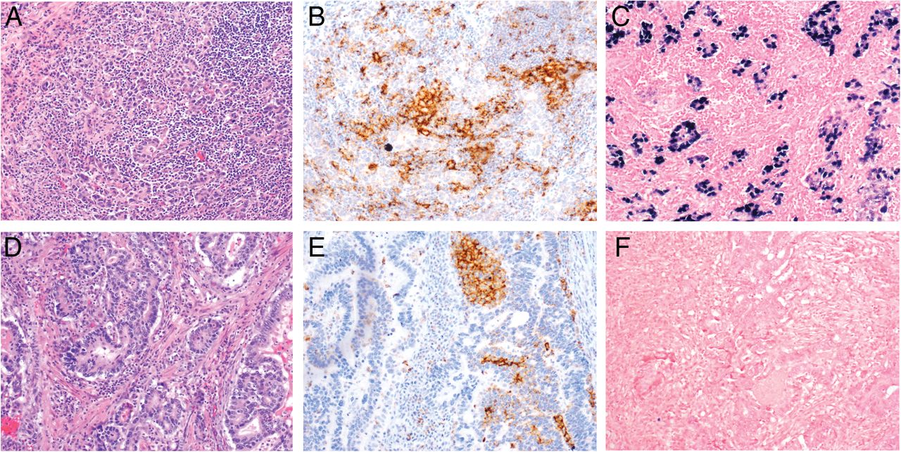
Epstein–Barr virus (EBV) status in programmed death-ligand 1 (PD-L1pos) gastric adenocarcinomas. H&E (A and D) and PD-L1 (B and E) staining and in situ hybridisation (ISH) for EBV (C and F) in two PD-L1pos gastric adenocarcinomas. Evidence of EBV infection by ISH in shown in C, while the tumour in F is negative for EBV. Shown at ×100 original magnification.
Our cohort of G/GEJ was associated with a mixed immune infiltration, which was present in both a peritumoral and ‘interface’ pattern at the interface between tumour and surrounding stroma and within the tumour (intratumoral). Overall, 44% (15/33, 95% CI 28.2% to 63.7%) of G/GEJ showed expression of PD-L1 within the infiltrating and interface immune stroma (table 1 and figure 3). Expression of PD-L1 in the immune stroma was also present almost exclusively in a peritumoral pattern, consistent with the predominant pattern of immune infiltration (figure 3).

Expression of programmed death-ligand 1 (PD-L1) by immune stroma associated with gastric adenocarcinomas. H&E (A–C) and PD-L1 staining (D–F) in two gastric adenocarcinomas with PD-L1pos (A, B, D and E) or PD-L1neg immune stroma (C and F). PD-L1 expression by immune stroma is seen in a peritumoral ‘interface’ pattern. Shown at ×100 (A, C, D and F) and ×400 (B and E) original magnification.
Association of PD-L1 expression with clinical parameters in gastric adenocarcinomas
The associations between tumour and immune stromal PD-L1 expression with clinical features of G/GEJ patients are detailed in table 1. PD-L1 expression in the both tumour and immune stroma was associated with increased age at diagnosis (p=0.14 and 0.04, respectively) with a median age of 74 in PD-L1pos tumours and 65.5 in PD-L1neg tumours and 73 in tumours with PD-L1pos stroma and 59 in tumours with PD-L1neg stroma. More tumours with intestinal-type histology (15%) showed membranous expression of PD-L1 than those with diffuse histology (6%), while there was little difference seen in PD-L1 expression in the immune stroma between intestinal and diffuse-type histology. No GEJ tumours showed membranous PD-L1 staining and only one showed PD-L1 expression in the immune stroma. In contrast, 14% of tumours located in the fundus/body showed membranous PD-L1 staining on tumour cells and 48% had PD-L1pos stroma. Finally, 20% of G/GEJ treated with neoadjuvant therapy prior to resection showed membranous tumour cell PD-L1 staining, while only 5% of treatment-naïve tumours showed expression. In contrast, similar numbers of treated and treatment-naïve tumours showed expression of PD-L1 by immune stroma. However, the above differences were not statistically significant. Overall, only age was significantly associated with increased PD-L1 expression by immune stroma (p=0.04). No significant associations were found between either tumour or immune stroma PD-L1 expression and patient gender (p=0.36 and 0.38), tumour histology (p=0.43 and 0.57), tumour location (p=0.24 for immune stroma PD-L1), stage at presentation (p=0.43 and 1.0), prior neoadjuvant therapy (p=0.22 and 0.90) or EBV status (p=0.14 and 0.89), though the sample size is too small to make conclusions regarding the latter factor. Tumour cells and immune cells may demonstrate a differential susceptibility to locally secreted factors driving PD-L1 expression,16 though the relative contributions of these different factors as well as the impact of previous treatment and the contribution of the native tissue stroma are areas of active investigation.
CD8+ T cell infiltration in G/GEJ and association with PD-L1 expression
As CD8+ T cells are central to adaptive immune resistance associated with PD-L1 expression in tumours, we quantified CD8+ infiltration in G/GEJ and associated peritumoral stroma using whole slide image analysis. Density of CD8+ T cells was measured in representative intratumoral, tumour–stromal interface and non-tumour regions of each case in our G/GEJ cohort. Average CD8+ T cell densities in each area are shown in table 1. We next compared average CD8+ densities in gastric adenocarcinomas with or without expression of PD-L1 on the tumour cells or in the immune stroma. As shown in table 1, the density of CD8+ T cells was 16-fold higher within PD-L1pos tumours and fourfold higher in the peritumoral/interface region. Furthermore, the density of CD8+ T cells was sixfold higher within tumours with PD-L1pos immune stroma and 3.5-fold higher in the peritumoral/interface regions of those tumours. Increasing CD8+ density within tumours was positively correlated with PD-L1 expression by tumour cells and the immune stroma (figure 4). Intratumoral and stromal CD8+ densities were broken into levels by quartiles: low (<147/mm2), mid (147–500/mm2) and high (>500/mm2). The correlation between CD8+ density and PD-L1 expression by tumour cells and immune stroma was then determined. Increasing CD8+ density was associated with increasing PD-L1 expression by both tumour cells and immune stroma. No PD-L1pos tumours showed low intratumoral CD8+ density, while 7% and 38% of tumours with mid or high CD8+ densities, respectively, were PD-L1pos (p=0.027). Similarly, while 23% and 36% of tumours with low or mid stromal CD8+ densities, respectively, were PD-L1pos, 89% of tumours with high stromal CD8+ densities were PD-L1pos (p=0.005). As chemotherapy and radiation have the potential to alter the tumour microenvironment, we also evaluated average CD8 densities in patients who did and did not receive neoadjuvant therapy. There were no statistically significant differences in infiltration. The mean intratumoral CD8 densities were 712.4 in the non-neoadjuvant group and 1075.6 in the neoadjuvant group (p=0.3), while the mean stromal CD8 densities were 458.2 in the non-neoadjuvant group and 263.6 in the neoadjuvant group (p=0.13).

Programmed death-ligand 1 (PD-L1) expression by tumour cells and immune stroma increases with increasing CD8 density in each location. CD8 density within gastric and gastro-oesophageal junction was determined by digital image analysis and densities were divided by quartiles into low (<147/mm2), mid (147–500/mm2) and high (>500/mm2). Correlation between CD8 density and PD-L1 expression by location were determined using the exact version of the Cochran–Armitage trend test. PD-L1 expression by both tumour cells (p=0.0027) and immune stroma (p=0.005) increased with increasing CD8 density in each location.
Association of PD-L1 expression and CD8+ infiltration with progression free and OS in gastric adenocarcinomas
Both progression-free survival (PFS) and OS were evaluated according to the PD-L1 status of the tumour and immune stroma as well as the density of CD8+ T cells at the tumour–stromal interface. PD-L1 expression on both tumour cells and in the immune stroma was associated with worse PFS and OS in patients with G/GEJ, but only the association with tumour expression of PD-L1 reached the level of statistical significance (PFS: HR=6.0 (95% CI 1.46 to 24.65), p=0.01 and OS: HR=7.81 (95% CI 1.71 to 35.68), p=0.01) (figure 5). PFS and OS were also evaluated according to CD8+ T cell density. Intratumoral CD8+ T cell density was divided into categories designated as: low (<147/mm2), mid (147–500/mm2) and high (>500/mm2). Immune stroma CD8+ density was divided into categories designated as high (>500/mm2) and low (<500/mm2). Tumours with either high intratumoral CD8+ T cell density or high stromal CD8+ T cell density had worse PFS and OS compared with tumours with low intratumoral or low stromal CD8+ densities (intratumoral CD8 PFS: (HR=11.22 (95% CI 1.31 to 96.32), p=0.03); intratumoral CD8 OS: (HR=10.48 (95% CI 1.22 to 89.93), p=0.03) and immune stroma CD8 PFS: (HR=3.91 (95% CI 1.32 to 11.59), p=0.01); immune stroma CD8 OS: (HR=3.46 (95% CI 1.09 to 10.96), p=0.03)) (figure 6). The reported HRs for intratumoral CD8 density reflect the survival comparison between tumours with high density (>500 CD8 T cells/mm2) and tumours with low density (<147 CD8 T cells/mm2).

Tumour programmed death-ligand 1 (PD-L1) expression in gastric and gastro-oesophageal adenocarcinomas is correlated with worse progression-free survival (PFS) and overall survival (OS). PFS and OS associations with PD-L1 expression by the tumour (A) and in the immune stroma (B). Probabilities of overall and event-free survival were estimated using the Kaplan–Meier method and compared using the log-rank statistic or the Cox proportional hazards regression model.

Increasing intratumoral and stromal CD8+ T cell density in gastric and gastro-oesophageal adenocarcinomas is correlated with worse progression-free survival (PFS) and overall survival (OS). PFS and OS associations with CD8+ T cell density within tumours (A) and in the immune stroma (B). Probabilities of overall and event-free survival were estimated using the Kaplan–Meier method and compared using the log-rank statistic or the Cox proportional hazards regression model. The upper quartile was used as the breakpoint for stroma CD8 and the approximate lower and upper quartiles were used for tumour CD8. The reported HRs for intratumoral CD8 density reflect the survival comparison between tumours with high density (>500 CD8 T cells/mm2) and tumours with low density (<147 CD8 T cells/mm2).
We also stratified our model to account for neoadjuvant therapy status in our patient cohort (see online supplementary table S1). Small numbers precluded evaluation of the association between OS, tumour PD-L1 and neoadjuvant therapy. However, in all evaluable models, neoadjuvant therapy status did not impact the survival trends or statistical significance of tumour PD-L1, stromal PD-L1, intratumoral CD8 T cell density or stromal CD8 T cell density. In addition, we also stratified our model by stage to account for the impact of extent of disease on our survival analyses. For intratumoral CD8 density, small numbers precluded evaluation of the association between OS, intratumoral CD8 density and stage. However, for the vast majority of variables analysed, stage, like neoadjuvant status, did not impact the survival trends or statistical significance of tumour PD-L1, stromal PD-L1, intratumoral CD8 T cell density or stromal CD8 T cell density. When stratified by stage, the statistical impact of stromal CD8 density on worsening OS showed a change in p value from 0.03 to 0.07 (see online supplementary table S1).
These results contradict findings that high density of TILs are associated with better disease outcomes in gastric cancer.24–27 Previous studies have used a variety of modalities to measure TIL. Our focus on absolute CD8+ T cell numbers and analysis by compartment location may partially explain the differences in our findings. These discrepancies highlight the need for further investigation of specific immune cell subtypes and localisation within the tumour microenvironment in order to better understand the complicated interactions present and to better predict outcome in individual tumour types.
Discussion
Gastric cancer has limited treatment options in the locally advanced and metastatic setting with chemotherapy resistance limiting efficacy beyond the first-line setting. Tumours escape immune surveillance by a number of mechanisms of which four groups have now been proposed on the basis of their PD-L1 status and the presence or absence of TILs. These include type I (PD-L1pos with TILs driving adaptive immune resistance), type II (PD-L1 negative with no TIL indicating immune ignorance), type III (PD-L1pos with no TIL indicating intrinsic induction) and type IV (PD-L1 negative with TIL indicating the role of other suppressor(s) in promoting immune tolerance).28 Malignant melanoma has been extensively studied and a high proportion of type I and type II microenvironments are seen21 whereas this information is yet to be defined in gastric cancers. In two of the four proposed immune resistance mechanisms, PD-L1 expression is hypothesised to play a key role. It is currently unclear what the early events are that leads to the aberrant expression of PD-1/PD-L1 by tumour cells and/or host immune cells in gastric cancer. Genomic aberrations in tumour cells that lead to aberrant PD-L1 expression have been proposed and microsatellite instability may have a predictive role as may EBV status.4 ,22 Emerging data in other tumour types suggest that negative immune checkpoint proteins are usually upregulated in tumour tissues with a ‘T cell inflamed phenotype’ and that infiltration of tumours by effector T cells is necessary to drive upregulation of immune checkpoints.14 This finding suggests that targeting the PD-1/PD-L1 axis in gastric cancer may only be clinically effective for the subgroup of tumours that contain tumour-infiltrating immune cells.
Here, we evaluated the expression of PD-L1 on tumour cells and tumour-associated immune stromal cells in patients with operable gastric cancer. We determined the CD8+ T cell density within the tumour and at the invasive tumour margin (immune stroma). Finally, we correlated PD-L1 expression and CD8+ T cell density with PFS and OS. We found 12% of G/GEJ showed membranous PD-L1 expression on tumour cells. In contrast, 44% of G/GEJ showed expression of PD-L1 in the immune stroma, most notably at the invasive tumour–stromal margin. While it is quite clear PD-L1 expression on tumour cells has predictive and prognostic features,12 ,13 ,15 ,19–21 ,29 it is increasingly recognised that PD-L1 expression on inflammatory cells, particularly at the invasive front, may also be an immunosuppressive mechanism.30 In this study, the expression of PD-L1 in tumour cells and immune stroma was associated with a worse PFS and OS in patients with gastric cancer, however, only the association with tumour cell expression was statistically significant. The association of PD-L1 expression with poor clinical outcome has been reported in a diverse set of tumour types, including gastric cancer.29 ,31–36
We found more CD8+ T cell infiltration in the tumours and peritumoral interfaces of patients that were also PD-L1pos compared with those that were PD-L1neg. When CD8+ T cell densities were broken into low, mid and high categories we found 89% of stroma PD-L1pos tumours had high CD8 densities. This underscores the importance of the linkage between CD8+ T cells, mechanistically thought to be a source of cytokines such as interferon gamma and expression of PD-L1.21 Depending on the type of cancer, the presence of TILs can be seen as a favourable or unfavourable prognostic feature and the interplay between CD8+ cells (as well as other immune cells) and resistance via enhanced PD-L1 expression needs further exploration.37–43 Our study highlights the complicated interactions within the tumour microenvironment and emphasises that impact of individual types of immune cells may be highly dependent on tumour type.
The importance of tumour and stromal PD-L1 expression was apparent in preliminary data from the KEYNOTE-012 trial. The investigators showed the anti-PD-1 antibody pembrolizumab has significant efficacy in patients with advanced gastric cancer (NCT01848834).44 Only patients with distinctive stromal or ≥1% tumour cell PD-L1 staining (22C3 antibody) were eligible. Of the 162 patients screened, 65 (40%) were considered PD-L1+. In our study, a total of 44% of patients had either tumour and/or stromal PD-L1 expression, similar to the rate seen in the above trial. Ultimately, PD-L1 expression level was associated with overall response rate of 22% to pembrolizumab (95% CI 10% to 39%) by central review and 33% (95% CI 19% to 50%) by investigator review. Median time to response was 8 weeks (range 7–16), with a median response duration of 24 weeks (range 8+ to 33+). PD-L1 expression level was associated with overall response rate (ORR) (one-sided p=0.10). The 6-month PFS rate was 24%. The 6-month OS rate was 69%, which is impressive in a highly treated patient population.
Our study has several important findings that may have implications for targeting the PD-1/PD-L1 axis in gastric cancer. Here, we show for the first time that CD8+ T cell infiltrated gastric tumours contain high presence of PD-L1 expression arguing that an adaptive immune resistance-type mechanism is active in these tumours, which may be overcome by the administration of anti-PD-1/PD-L1. We did not see a significant difference in PD-L1 expression between stages of disease at presentation, indicating that checkpoint inhibition may have activity in early stage as well as late stage disease. Our findings also suggest that the diversity of clinical outcomes seen in patients with similar stages of gastric cancer may be explained in part by differences in TIL density and PD-L1 expression. Interestingly, we did not detect evidence that EBV positivity was necessary for PD-L1 expression in gastric cancer. The clinical relevance of other factors such as the potential contributions of myeloid-derived suppressor cells, CD4+CD25+FoxP3+ Tregs and indoleamine 2,3-dioxygenase needs to be elucidated. Such additional characterisation of the G/GEJ tumour immune microenvironment may yield a more nuanced understanding of the interactions between CD8+ T cells and immunosuppression and provide further targets for immune-based therapy. Overall, our results support continued clinical development of PD-1/PD-L1 checkpoint inhibitors for patients with gastro-oesophageal cancers.
References
Supplementary materials
Supplementary Data
This web only file has been produced by the BMJ Publishing Group from an electronic file supplied by the author(s) and has not been edited for content.
- Data supplement 1 - Online supplement
- Data supplement 2 - Online table
Footnotes
Contributors EDT, MZ, RAA and RJK were responsible for study design, data collection, data analysis and manuscript writing. AM, TC, NC, EA and JMT were responsible for data collection, data analysis and manuscript writing. SY, MD and NA were responsible for data collection.
Funding This study was supported by a GI SPORE CDA awarded to RJK (NIH grant CA62924), the COLE Foundation and by SKCCC core grant NCI CCSG P30 CA006973.
Competing interests None declared.
Ethics approval Johns Hopkins IRB.
Provenance and peer review Not commissioned; externally peer reviewed.