Article Text

Abstract
The cholinergic anti-inflammatory pathway (CAIP) has been proposed as a key mechanism by which the brain, through the vagus nerve, modulates the immune system in the spleen. Vagus nerve stimulation (VNS) reduces intestinal inflammation and improves postoperative ileus. We investigated the neural pathway involved and the cells mediating the anti-inflammatory effect of VNS in the gut. The effect of VNS on intestinal inflammation and transit was investigated in wild-type, splenic denervated and Rag-1 knockout mice. To define the possible role of α7 nicotinic acetylcholine receptor (α7nAChR), we used knockout and bone marrow chimaera mice. Anterograde tracing of vagal efferents, cell sorting and Ca2+ imaging were used to reveal the intestinal cells targeted by the vagus nerve. VNS attenuates surgery-induced intestinal inflammation and improves postoperative intestinal transit in wild-type, splenic denervated and T-cell-deficient mice. In contrast, VNS is ineffective in α7nAChR knockout mice and α7nAChR-deficient bone marrow chimaera mice. Anterograde labelling fails to detect vagal efferents contacting resident macrophages, but shows close contacts between cholinergic myenteric neurons and resident macrophages expressing α7nAChR. Finally, α7nAChR activation modulates ATP-induced Ca2+ response in small intestine resident macrophages. We show that the anti-inflammatory effect of the VNS in the intestine is independent of the spleen and T cells. Instead, the vagus nerve interacts with cholinergic myenteric neurons in close contact with the muscularis macrophages. Our data suggest that intestinal muscularis resident macrophages expressing α7nAChR are most likely the ultimate target of the gastrointestinal CAIP.
- NERVE-GUT INTERACTIONS
- NEURAL-IMMUNE INTERACTIONS
- MACROPHAGES
- ENTERIC NERVOUS SYSTEM
Statistics from Altmetric.com
Significance of this study
What is already known about this subject?
-
In models of systemic inflammation, VNS induces the release of ACh from splenic T cells leading to a reduced cytokine production by macrophages and improved survival.
-
The cytokine-suppressing mechanism of the CAIP in the spleen requires α7nAChR expression on splenic macrophages.
-
VNS, systemic administration of selective α7nACh agonists and central stimulation of the vagus nerve reduce intestinal inflammation and consequently POI.
What are the new findings?
-
Activation of the vagus nerve prevents POI through a circuit independent of the spleen and T cells.
-
In the murine small bowel, vagal efferent terminals mainly contact cholinergic myenteric neurons and do not directly innervate intestinal muscularis resident macrophages.
-
α7nAChR expression on muscularis externa resident macrophages, but not on non-immune cells, is essential for the CAIP in the gut.
-
α7nAChR activation modulates ATP-induced Ca2+ response in small intestinal muscularis externa resident macrophages.
How might it impact on clinical practice in the foreseeable future?
-
Electrical stimulation of the vagus nerve is currently evaluated as therapy in patients undergoing abdominal surgery and in patients with Crohn's disease.
-
Our study provides further insight into the vagal anti-inflammatory pathway of the intestine, which is crucial to understand its mechanism of action and the receptors and cells involved.
-
This will ultimately lead to improved treatment of immune-mediated diseases of the gastrointestinal tract, such as POI and potentially inflammatory bowel disease.
Introduction
In 2000, Tracey and co-workers introduced the concept of the ‘cholinergic anti-inflammatory pathway’ (CAIP).1 In a model of systemic inflammation such as sepsis, these investigators demonstrated that electrical vagus nerve stimulation (VNS) improved survival by inhibiting cytokine production by splenic macrophages in an α7 nicotinic receptor (α7nAChR)-dependent fashion.1 ,2 In contrast to the initial hypothesis proposing direct contacts between vagal nerve fibres and splenic macrophages,3 ,4 it is now clear that the vagus nerve rather indirectly modulates the innate immune response in the spleen by activating the adrenergic neurons in the prevertebral ganglia. In line with this hypothesis, only in mice with an intact and innervated spleen, VNS is able to exert its anti-inflammatory effect.5 Recent studies suggest that acetylcholine (ACh) released by the vagus nerve in the coeliac mesenteric ganglia activates noradrenergic neurons of the splenic nerve, leading to the release of norepinephrine in the spleen.6 At this site, these adrenergic nerve fibres stimulate ACh synthesis in splenic memory T cells in a β2-adrenoceptor-dependent manner.7
In 2005, we extended the concept of the vagal anti-inflammatory pathway in the gastrointestinal tract. We demonstrated that VNS prevented surgery-induced intestinal inflammation and improved the course of postoperative ileus (POI),8 an immune-mediated condition characterised by localised inflammatory reactions in the muscularis externa evoked by intestinal handling during surgery.9–12 Initiation of this inflammatory response is evidently dependent on activation of a macrophage population that resides between the circular and longitudinal muscle layers of the intestinal wall (muscularis externa)13 making resident macrophages a potential target to prevent POI. Surgical handling of the intestine triggers the activation of these normally quiescent macrophages, which results in the production of pro-inflammatory cytokines,13 upregulation of endothelial adhesion molecules, and subsequent influx of leucocytes into the muscularis externa.11 This inflammatory response is associated with prolonged dysmotility of the bowel due to the release of nitric oxide and prostaglandins.14–17
POI is associated with localised or subtle inflammatory processes in the gut and does not trigger a systemic response that will activate splenic immune cells. Therefore, taking into account that the gastrointestinal tract is densely innervated by the vagus nerve,18–21 we hypothesise that the vagus might directly modulate the intestinal muscularis resident macrophages independent of the spleen. Moreover, it still remains unclear whether α7nAChR on intestinal muscularis macrophages may represent the final target of the CAIP in the gut. Indeed, current data suggesting the involvement of α7nAChR are rather indirect because in vitro data evaluating the anti-inflammatory effect of nicotine were obtained using macrophage cell lines, splenic, peritoneal or bone marrow-derived macrophages (but not intestinal resident muscularis macrophages),1 ,8 ,22 whereas in vivo studies reported the effect of systemic administration of an α7nAChR agonist.23 The expression of α7nAChR has, however, not been demonstrated in intestinal muscularis macrophages and the effect of pharmacological blockade or the absence of α7nAChR on VNS has not yet been investigated. Therefore, to what extent the anti-inflammatory effect of VNS in the gut is mediated by dampening of splenic macrophages or by direct interaction with the intestinal macrophages through α7nAChR activation remains unclear. Notably, insight into the nature of the pathway involved in the anti-inflammatory effect of VNS is crucial ultimately to develop new therapeutic approaches, not only for POI, but also for other immune-mediated disorders of the gut.
Here, we provide evidence that the vagus nerve reduces local intestinal inflammation through a circuit that differs from that previously reported in models of systemic inflammation, that is, independent of the spleen and T cells. This process requires α7nAChR expression on resident muscularis macrophages and it most likely involves cholinergic enteric neurons as the source of ACh. Our findings support the idea that the vagus nerve, through the enteric nervous system (ENS), may play a crucial role in maintaining intestinal immune homeostasis, potentially opening new avenues for therapy.
Materials and methods
Please see supplementary materials and methods (available online only) for an expanded version of this section.
Animals
Mice deficient for the α7 nicotinic receptor (α7nAChR−/−; B6.129S7-Chrna7tm1Bay),24 Rag-1 knockout mice (Rag-1−/−; B6.129S7-Rag1tm1Mom/J)25 and B6.SJL-Ptprca Pepcb/BoyJ mice were purchased from the Jackson Laboratory (USA). Wild-type mice (C57BL/6JOlaHsd) were purchased from Harlan (The Netherlands). All mice were maintained at the University of Leuven animal facility under specific pathogen-free conditions on a 12 : 12-h light–dark cycle and provided with commercially available chow (ssniff R/M-H; ssniff Spezialdiäten GmbH) and freely available tap water. Gastrointestinal transit and food intake from wild-type, α7nAChR−/− and Rag-1−/− mice was assessed and no differences were found between groups for both parameters (see supplementary figure S1, available online only). All experimental procedures were approved by the Animal Care and Animal Experiments Committee of the University of Leuven (Leuven, Belgium).
Surgical procedure to induce POI
Mice were anaesthetised by intraperitoneal injection of a mixture of ketamine (Ketalar 100 mg/kg; Pfizer) and xylazine (Rompun 10 mg/kg; Bayer). Anaesthetised mice underwent a laparotomy alone or a laparotomy followed by intestinal manipulation (IM).8 ,10 ,26 ,27 Surgery was performed under sterile conditions using a sterile moist cotton applicator attached to a device enabling the application of a constant pressure of 90 mN. The small intestine was manipulated three times from the caecum to the distal duodenum. The total duration of the surgery was between 10 and 15 min, while the duration of the IM was between 5 and 7 min. After surgery, mice were allowed to recover on a heating pad for 3 h and then provided with food and freely available water.
Electrical VNS
VNS was applied for 5 min before IM. The right cervical vagus nerve was isolated from the carotid artery and stimulated electrically using a bipolar platinum electrode (Bilaney). Electrical stimuli consisted of square pulses (5 V, 1 ms duration) at 5 Hz (Astro-Med Inc).8 For sham stimulation, the right cervical vagus nerve was isolated but not stimulated.
Statistics
Unpaired Student's t test was used to evaluate differences between two experimental groups. To compare multiple groups, one-way analysis of variance followed by the Bonferroni post-hoc test was performed. Wilcoxon signed-rank or one-way analysis of variance followed by Dunn's multiple comparison tests were used to analyse calcium imaging experiments. Statistical analysis was performed using GraphPad Prism software (Graphpad Software Inc.).
Results
VNS modulates intestinal muscularis inflammation independently of splenic innervation and of T cells
As previously reported,11 ,28 IM resulted in a delay in gastrointestinal transit 24 h after abdominal surgery compared to laparotomy without IM (see supplementary figure S2A,B, available online only). In addition, a significant increase of pro-inflammatory cytokine gene expression (il6, il1β, il1α and ccl2; see supplementary figure S2E, available online only) and recruitment of myeloperoxidase-positive cells were detected in the muscularis externa (see supplementary figure S2D, available online only). Interestingly, VNS before IM significantly improved gastrointestinal transit compared with mice subjected to IM alone (see supplementary figure S2A,B, available online only). In addition, VNS reduced surgery-induced inflammation as shown by reduced influx of myeloperoxidase-positive cells (see supplementary figure S2C,D, available online only), decreased pro-inflammatory cytokine gene expression (il6, il1β, il1α and ccl2; see supplementary figure S2E, available online only), as well as reduction of CD45-positive cells, neutrophils and monocytes in the muscularis externa (see supplementary figure S2F, available online only).
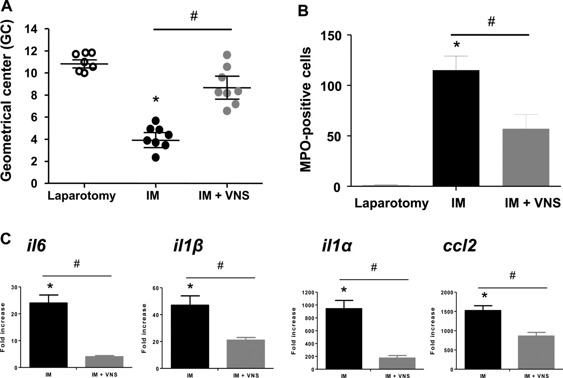
Knowing that the spleen mediates the vagal anti-inflammatory reflex during systemic inflammation such as sepsis,29 we determined whether this organ was also mediating the beneficial effect of VNS in the gut. To this end, mice subjected to splenic denervation were used. As previously reported,29 VNS in splenic denervated mice failed to lower lipopolysaccharide-induced tumour necrosis factor α (TNFα) levels in serum, proving proper disruption of the splenic innervation (see supplementary figure S3A, available online only). Interestingly, VNS was still able to improve gastrointestinal transit after surgery in splenic denervated mice (figure 1A), and reduced muscularis inflammation as revealed by decreased il6, il1β, il1α, and ccl2 gene expression (figure 1C) and reduced the number of myeloperoxidase-positive cells in the muscularis externa (figure 1B).

Anti-inflammatory effect of vagus nerve stimulation (VNS) in the gut is independent of the spleen. (A–C) Wild-type mice with splenic denervation were subjected to intestinal manipulation (IM) with or without VNS. (A) Geometric centre (GC) values of dextran distribution through the intestinal segments. (B) Histogram represents mean±SEM of myeloperoxidase-positive cells in the muscularis externa from an area of 0.5 mm2. (C) il6, il1β, il1α and ccl2 mRNA expression in the muscularis externa 24 h after IM. Data are expressed as fold increase over laparotomy mice. *p<0.01 compared with the laparotomy group and #p<0.01 compared with the IM group (one-way analysis of variance followed by Bonferroni post-hoc test and unpaired t test).
The ability of splenic T cells to secrete ACh upon VNS has been proved to be critical for dampening systemic inflammation.7 Therefore, to address the involvement of T cells in the vagal control of intestinal inflammation, we evaluated the effect of VNS in T-cell-deficient Rag-1−/− mice subjected to IM.25 Remarkably, in Rag-1−/− mice, as in wild-type control mice, VNS before IM significantly improved gastrointestinal transit (figure 2A). In addition, VNS in Rag-1−/− mice resulted in a significant reduction of myeloperoxidase-positive cells (figure 2B) and decreased il6, il1β, il1α, and ccl2 gene expression in the muscularis compared to Rag-1−/− mice subjected to IM only (figure 2C). Taken together, our data demonstrate that the vagal anti-inflammatory effect in the intestinal muscularis is independent of splenic T cells.

Vagus nerve stimulation (VNS) reduces intestinal inflammation in the gut independently of T cells. (A–C) Rag1−/− were subjected to laparotomy, intestinal manipulation (IM) alone or IM with VNS. (A) Geometric centre (GC) values of dextran distribution through the intestinal segments. (B) Histogram represents mean±SEM of myeloperoxidase-positive cells in the muscularis externa from an area of 0.5 mm2. (C) il6, il1β, il1α and ccl2 mRNA expression in the muscularis externa 24 h after surgery. Data are expressed as fold increase over laparotomy mice. Histograms represent mean±SEM. *p<0.01 compared with the laparotomy group and #p<0.01 compared with the IM group (one-way analysis of variance followed by Bonferroni post-hoc test and unpaired t test).
Distribution of efferent vagal terminals in the small bowel
As we showed that the anti-inflammatory effect of VNS is independent of the spleen, we anticipated that the vagus nerve interacts directly with the intestinal immune system. To unravel the neural circuit involved, we performed anterograde tracing of vagal efferents to the gut by stereotactic injection of biotin-conjugated dextran amine into the dorsal motor nucleus of the vagus (DMV).30 The presence and the distribution of biotin-labelled vagal efferent nerve fibres were later identified using fluorescence-labelled streptavidin. As shown in figure 3A, efferent vagal terminals were mainly localised at the level of the myenteric plexus region within the interganglionic nerve bundles and ganglia. In addition, vagal efferent terminals were strongly immunoreactive for choline acetyltransferase and closely associated with cholinergic enteric neurons (figure 3B). Within the myenteric ganglia, the vagal efferents showed varicosities in the vicinity of a subgroup of enteric neurons revealing putative sites of transmitter release (white arrows in figure 3C). In contrast, direct contacts between efferent vagal terminals and muscularis resident macrophages (F4/80-positive cells) were not observed (figure 3C). Notably, cholinergic enteric nerve fibres form a dense network in close proximity to muscularis F4/80-positive macrophages suggesting possible neuromodulation of these intestinal immune cells by the ENS (figure 3C).

The efferent vagus nerve contacts cholinergic enteric neurons that are in close contact with muscularis resident macrophages. Efferent vagus nerve terminals (green) were anterograde traced by injection of biotin-dextran amine in the dorsal motor nucleus of the vagus. Enteric neurons were labelled with anti-PGP 9.5 (red) or with anti-choline acetyltransferase (ChAT) (red) while intestinal muscularis resident macrophages were labelled with anti-F4/80 (blue). (A) Confocal images show that efferent vagus nerve terminals are located within enteric neurons of the myenteric plexus region. (B) High power magnification image of a myenteric ganglion. Note vagus nerve efferent fibres and terminals have cholinergic nature (ChAT-positive) and target ChAT immunoreactive cell bodies of the enteric neurons. (C) Maximum intensity projection of confocal stacks shows that intestinal muscularis resident macrophages are not directly innervated by the vagus nerve. Notably, cholinergic enteric nerve fibres are located in close proximity to muscularis F4/80-positive macrophages. Top inset shows close proximity between macrophages (blue) and ChAT-positive enteric neurons red while the bottom inset shows contacts between efferent vagus nerve terminals and cell bodies of ChAT-positive enteric neurons. White arrows point at possible contact sites between efferent vagus nerve terminals and ChAT neurons.
α7nAChR expression on immune cells is essential for the vagal anti-inflammatory effect in the gut
Previous studies in several preclinical models of systemic inflammation have convincingly demonstrated that the CAIP requires the presence of α7nAChR;2 ,31 however, the role of α7nAChR and the cells expressing this receptor in the gut have not been established. Therefore, to evaluate the contribution of this receptor to the vagal anti-inflammatory effect in the intestine, α7nAChR−/− knockout mice24 were subjected to IM with or without VNS, as described above. Of note, in α7nAChR−/− mice VNS before IM failed to improve gastrointestinal transit (figure 4A) and to reduce the inflammatory response in the muscularis externa. VNS before IM in α7nAChR−/− mice did not decrease the number of myeloperoxidase-positive cells (figure 4B) or gene expression of the pro-inflammatory cytokines (il6, il1β, il1α and ccl2) compared to α7nAChR−/− mice only subjected to IM (figure 4C). In addition, VNS before IM in α7nAChR−/− mice was not able to reduce the recruitment of CD45-positive cells, neutrophils and monocytes in the muscularis externa (figure 4D).

α7nAChR is required to reduce intestinal inflammation upon vagus nerve stimulation. (A) Graph shows geometric centre (GC) values from α7nAChR−/− mice subjected to laparotomy, intestinal manipulation (IM) alone and IM with vagus nerve stimulation (VNS). (B) Histogram represents myeloperoxidase-positive cells from an area of 0.5 mm2, in α7nAChR−/− mice. (C) il6, il1β, il1α and ccl2 mRNA expression in the muscularis externa 24 h after surgery. Data are expressed as fold increase over laparotomy mice. (D) Immune cell subsets in the muscularis externa 24 h after surgery. Results are expressed as mean±SEM. *p<0.05 compared to laparotomy (analysis of variance followed by Bonferroni's multiple comparison test and unpaired t test).
Although our data imply a crucial role for α7nAChR in the intestinal vagal anti-inflammatory pathway, the cell type (neurons6 vs immune cells) carrying the functional α7nAChR remained to be elucidated. To address this question, bone marrow chimaera mice were generated by transplanting either α7nAChR+/+ bone marrow cells into irradiated recipient α7nAChR−/− mice, or α7nAChR−/− bone marrow into irradiated α7nAChR+/+ recipient mice (figure 5). Following VNS, we observed that only mice carrying α7nAChR+/+ bone marrow displayed significantly improved intestinal transit, reduced recruitment of inflammatory cells and decreased pro-inflammatory gene expression (il6, il1β, il1α and ccl2) (figure 5A–C), as well as reduced CD45-positive cells, neutrophils and monocytes in the muscularis (see supplementary figure S4A, available online only). In contrast, the vagal anti-inflammatory effect was absent in α7nAChR+/+ mice reconstituted with α7nAChR−/− bone marrow cells (figure 5D–E and see supplementary figure S4B, available online only). As expected, VNS effectively reduced inflammation and normalised gastrointestinal transit in wild-type mice reconstituted with wild-type bone marrow cells, whereas VNS was not effective in α7nAChR−/− mice reconstituted with α7nAChR−/− bone marrow cells (see supplementary figure S5, available online only). In view of our bone marrow chimaera experiments it is clear that intestinal immune cells expressing α7nAChR may represent the final target of the vagal anti-inflammatory pathway.

Anti-inflammatory effect of vagus nerve stimulation (VNS) requires α7nAChR expression in bone marrow (BM)-derived cells. Bone marrow chimaera mice with α7nAChR deficiency in BM-derived cells (α7nAChR−/−→α7nAChR+/+ mice) or in non-BM-derived cells (α7nAChR+/+→α7nAChR−/− mice) were generated by adoptive transfer of α7nAChR+/+ or α7nAChR−/− BM cells into irradiated α7nAChR−/− or α7nAChR+/+ mice, respectively. (A and D) Geometric centre (GC) values of dextran distribution through the intestinal segments in chimaera mice. (B and E) Recruitment of myeloperoxidase-positive cells in the muscularis externa of the small intestine. Results are expressed as myeloperoxidase-positive cells from an area of 0.5 mm2. (C and F) il6, il1β, il1α and ccl2 mRNA expression 24 h after surgery in the muscularis externa of the indicated BM-chimaera mice. Data are expressed as fold increase over the same chimaera laparotomy mice. Histograms represent mean±SEM. *p<0.01 compared with the laparotomy group and #p<0.01 compared with the intestinal manipulation (IM) group (one-way analysis of variance followed by Bonferroni post-hoc test and unpaired t test).
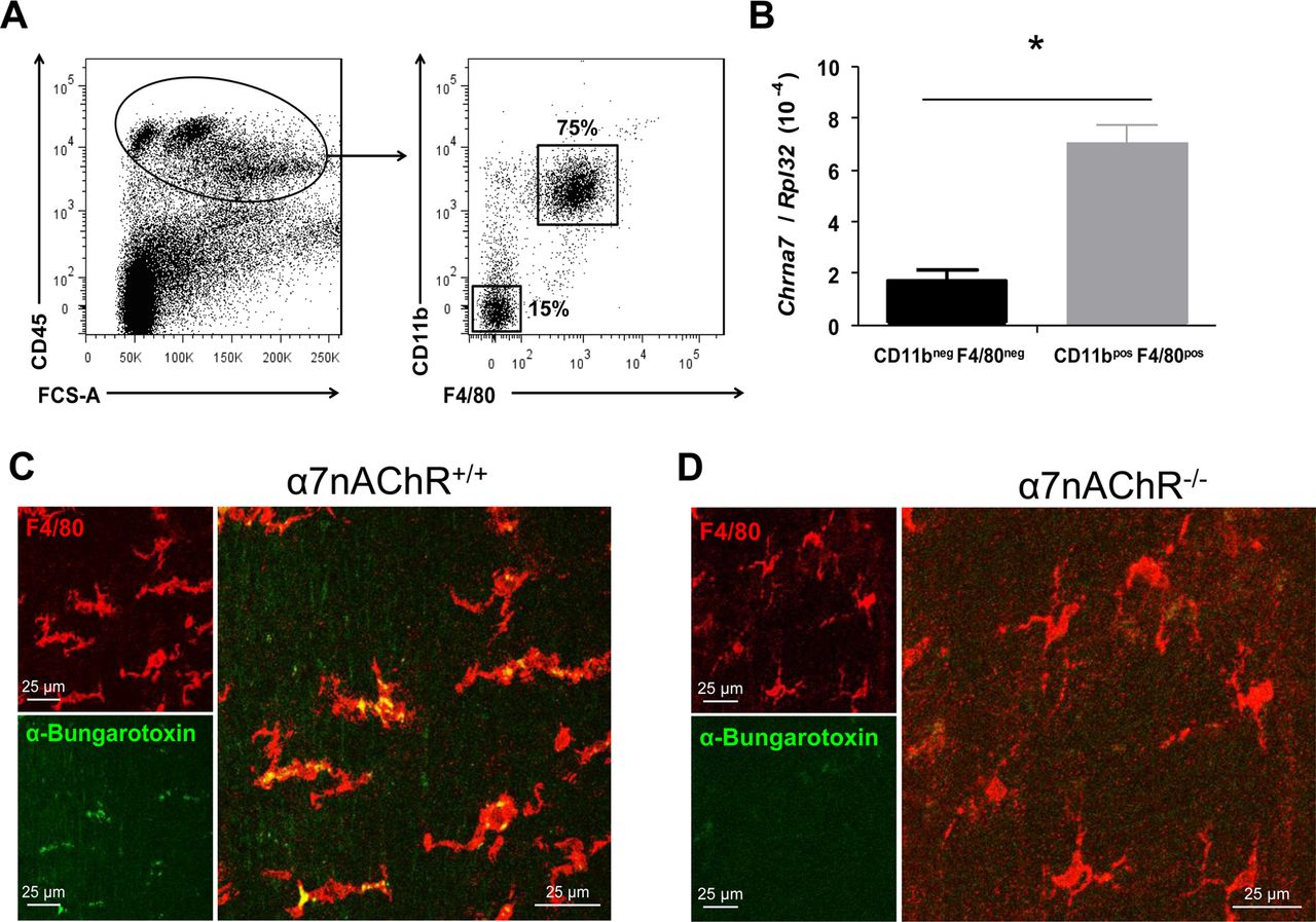
To identify the immune cells carrying α7nAChR, muscularis externa from the small intestine was enzymatically digested and immune cells were separated by sorting as shown in figure 6A. Of note, intestinal muscularis resident macrophages (CD45+CD11b+F4/80+) are the cells expressing preferentially α7nAChR messenger RNA whereas other immune cells (CD45+CD11b−F4/80−) express significantly lower α7nAChR levels (figure 6B). To confirm this finding we performed staining of the muscularis externa using fluorescently labelled α-bungarotoxin. Figure 6C shows that, in α7nAChR wild-type mice, muscularis externa resident macrophages (F4/80 positive, red) are positive for FITC-labelled α-bungarotoxin (green). Of note, similar experiments performed in α7nAChR−/− mice showed no binding of α-bungarotoxin to F4/80-positive cells suggesting that muscularis resident macrophages indeed express α7nAChR at the protein level (figure 6D).

Intestinal muscularis resident macrophages express α7nAChR that is specifically recognised by α-bungarotoxin. Intestinal muscularis immune cells isolated from C57BL/6 wild-type mice were sorted in two different populations; F4/80− CD11b− and F4/80+ CD11b+ cells. (A) Sorting strategy, CD45-positive cells were further separated in F4/80− CD11b− and F4/80+ CD11b+ cells. (B) mRNA expression of α7nAChR (Chrna7) was determined by reverse transcriptase quantitative PCR in CD45+ F4/80− CD11b− and CD45+ F4/80+ CD11b+ cells. Histogram represents mean±SEM from three independent sorting experiments. *p<0.01 (unpaired t test). (C) Muscularis externa whole mount from the jejunum of α7nAChR+/+ mouse was stained with FITC-labelled α-bungarotoxin and with anti-mouseF4/80. The images shown are maximum intensity projections of stacks of confocal optical sections. Note, merged panel shows co-localisation of the FITC-bungarotoxin (green) with F4/80-positive (red) resident macrophages. (D) Confocal images of the muscularis externa from α7nAChR−/− mice. Note the absence of α-bungarotoxin signal in α7nAChR−/− mice providing evidence supporting the specificity of α-bungarotoxin staining for α7nAChR.
ATP response of intestinal muscularis resident macrophages is modulated by activation of α7nAChR
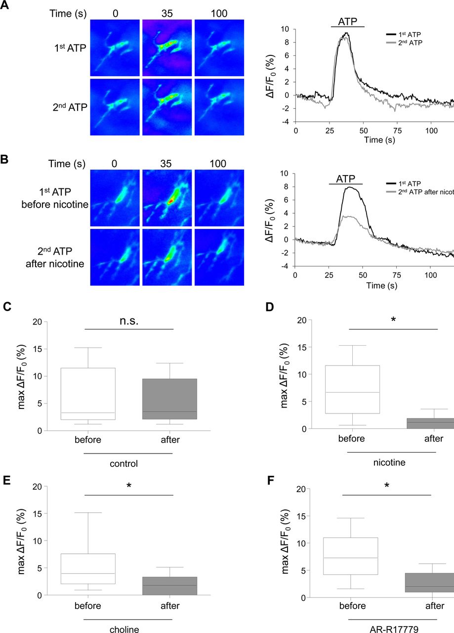
To demonstrate finally that muscularis resident macrophages can indeed be modulated by the CAIP via the activation of α7nAChR, we evaluated the effect of nicotine and two α7nAChR agonists (choline and AR-R17779) ‘in situ’ in an intact mouse jejunum muscularis preparation using [Ca2+]i imaging. The jejunum muscularis isolated from Cx3cr1GFP/+ mice was used to allow clear and specific identification of the macrophages. Indeed, 96–98% of the muscularis externa resident macrophages were green fluorescent protein positive (see supplementary figure S6, available online only). To visualise [Ca2+]i changes in resident macrophages, intestinal muscularis externa preparations were loaded with the Ca2+ indicator Fluo-4 AM. Due to the high dynamic range of the camera used, Fluo-4 changes could be recorded even in the green fluorescent protein cells (see supplementary figure S7, available online only). Using this experimental set-up, resident macrophages were stimulated with ATP, a typical tissue damage associated molecule,32 before and after treatment with one of the α7nAChR agonists: nicotine (10 μM), choline (50 mM) or AR-R17779 (10 μM). Notably, nicotine, choline and AR-R17779 significantly decreased ATP-induced [Ca2+]i transients in muscularis resident macrophages (figure 7D–F). The effect of these α7nAChR agonists on ATP-induced [Ca2+]i transients was not due to desensitisation because two consecutive ATP stimuli without α7nAChR activation resulted in reproducible responses (figure 7A,C). In addition, as described in supplementary figure S8 (available online only) treatment with nicotine, choline or AR-R17779 also induced [Ca2+]i transients in muscularis resident macrophages, albeit of smaller magnitude compared to the response evoked by ATP. These findings confirm that resident macrophages express functional α7nAChR and that the activation status of intestinal muscularis resident macrophages can be modulated by local release of ACh.
The adenosine-5′-triphosphate (ATP) response of intestinal muscularis resident macrophages is modulated by activation of α7nAChR. Jejunum muscularis externa preparations from Cx3CR1GFP/+ mice loaded with Fluo-4 were exposed to a first challenge of 100 μM of ATP. Ten minutes later, to induce pharmacological activation of α7nAChR, nicotine (10 μM), choline (50 mM) or AR-R17779 (10 μM) were applied on the tissue surface for 1 min, followed after 10 min by a second ATP administration. (A) Representative images of a Fluo-4 loaded resident macrophage in the jejunum muscularis externa of Cx3CR1GFP/+ mouse in response to two consecutive (20 min delay) ATP applications interspersed with a vehicle application. Right panel shows Ca2+ transients of the same resident macrophage in response to two ATP applications (first: black; second: grey). (B) Representative images from a Fluo-4 loaded resident macrophage in the jejunum muscularis externa of Cx3CR1GFP/+ mouse in response to ATP before and after nicotine application. Right panel shows Ca2+ transients of the same resident macrophage in response to ATP before (black) and after (grey) nicotine application. (C–F) Box plots showing maximum fluorescence changes (% ΔF/F0) in muscularis resident macrophages responding to two consecutive ATP challenges in control (C) or with intermediate application of nicotine (10 μM) (D), choline (50 mM) (E) or AR-R17779 (10 μM) (F). Results are expressed as median (10th percentile/90th percentile) of at least 50 macrophages from three to five preparations isolated from three individual mice. *p<0.001 compared ATP responses before and after application of nicotine, choline or AR-R17779 (Wilcoxon signed rank test).
Discussion
In this study, we have revealed the neural circuit and the cellular target involved in the ‘gastrointestinal CAIP’. First, we showed that the vagus nerve dampens intestinal inflammation by directly interacting with the intestine without the involvement of the spleen. Second, within the intestinal wall, resident macrophages are in close proximity to cholinergic nerve fibres arising from enteric neurons innervated by the vagus nerve. Third, the expression of α7nAChR on intestinal muscularis resident macrophages but not on non-immune cells is crucial for the protective effects of the vagus nerve. Finally, activation of α7nAChR modulates intestinal muscularis resident macrophages. Based on these results, we conclude that in contrast to the cholinergic modulation of the immune system in the spleen, the vagal anti-inflammatory input is direct to the gut and is independent of the spleen. Instead, we speculate that the intestinal immune system is directly modulated by the vagus nerve through the ENS and provide evidence that resident macrophages are the ultimate target of the vagal anti-inflammatory pathway in the gut.
In sepsis, the spleen is considered the main source of pro-inflammatory cytokines contributing to disseminated tissue injury and multi-organ failure.33 ,34 Following injection of lipopolysaccharide, splenic macrophages become activated and secrete inflammatory cytokines into the systemic circulation,34 ,35 leading to an exacerbated immune response and disseminated disease. In addition to the classic compensatory mechanisms (ie, synthesis of anti-inflammatory cytokines and activation of the hypothalamic–pituitary–adrenal axis), Tracey and co-workers recently proposed that the brain through the vagus nerve influences this immune response contributing to recovery of immune homeostasis.4 Using an animal model of sepsis, it was elegantly demonstrated that stimulation of the vagus nerve suppresses cytokine production and increases survival.5 In contrast to the earlier hypotheses proposing a direct effect of ACh released by vagal efferents,2 recent evidence suggests that the vagus indirectly interacts with the spleen through adrenergic neurons located in the coeliac mesenteric ganglia, leading to the release of norepinephrine via the splenic nerve.6 Recently, memory T cells (in a β2-adrenoceptor-dependent fashion rather than vagal efferents), were proposed as a final source of ACh in the spleen, driving lower cytokine production by splenic macrophages.7 ,36 ,37 Clearly, as the spleen plays a central role in sepsis, this organ should be the main target of immune modulation.
Previously, we showed that electrical stimulation of the vagus nerve8 or systemic administration of a nicotinic agonist23 in mice with surgery-induced ileus significantly improved gastrointestinal transit and reduced cytokine production in the gut. However, we reasoned that under this condition of local inflammation, a more direct input of the CAIP would be involved. Here, we aimed to unravel the anatomical and functional mechanisms responsible for the vagus nerve modulation of the intestinal immune system. To this end, surgery-induced intestinal inflammation was provoked in mice that underwent splenic denervation. One striking finding was that unlike sepsis, stimulation of the vagus nerve in animals devoid of splenic innervation was still capable of reducing intestinal inflammation and preventing ileus. In addition, VNS in T-cell-lacking Rag-1−/− mice was still effective in preventing surgery-induced muscularis inflammation. Therefore, our experiments suggest that the vagal anti-inflammatory effect in the gut is independent of splenic T cells. This is in line with recent findings obtained in a model of burn injury in which the beneficial effect of VNS on intestinal barrier function was preserved in splenectomised mice.38 ,39 Altogether, our data imply that the gastrointestinal tract receives direct vagal input, capable of modulating the immune response in the gut in a spleen and T-cell-independent fashion.
The coeliac branch of the vagus nerve densely innervates the gastrointestinal tract with efferent fibres originating mainly from the DMV, with a typical rostro-caudal gradient with the highest density of innervation observed in the stomach followed by a subsequent decrease in the small bowel and colon.20 ,40 By means of anterograde tracer injected into the DMV, we confirmed that efferent vagal nerve terminals are in close apposition to post-ganglionic neurons located in the ENS as previously proposed.40 ,41 Indeed, vagal nerve varicosities were in close contact with cholinergic neurons located at the level of the myenteric plexus region. Of note, no vagus nerve fibres were detected directly in contact with muscularis resident macrophages. However, resident macrophages were in close proximity with cholinergic nerve fibres. Based on these findings, one could speculate that vagal signals are amplified by the ENS and consequently induce a substantial release of ACh in the intestinal microenvironment that leads to modulation of the immune response.
Even though the exact source of ACh (vagal terminals, enteric neurons) remains to be identified, studies using α7nAChR knockout mice and pharmacological α7nAChR agonists/antagonists have shown the importance of this receptor as an end target of the CAIP.2 ,23 ,31 In order to investigate the involvement of this receptor in the gastrointestinal CAIP, we made use of α7nAChR−/− mice.24 As in sepsis, VNS in α7nAChR−/− mice could not reduce intestinal inflammation and did not improve gastrointestinal transit. This supports the notion that in the intestine, α7nAChR represents the final target of the CAIP. Nevertheless, the exact location of α7nAChR is still a matter of debate. In sepsis, the inflammatory reflex requires an intact splenic nerve and α7nAChR expression in the coeliac mesenteric ganglia.5 ,29 However, two reports have recently provided evidence against the neural location of α7nAChR, describing that α7nAChR are instead expressed on splenic macrophages.7 ,31 In agreement with these reports, using bone marrow chimaera mice, we have found that the expression of α7nAChR in bone marrow-derived cells is essential for the beneficial effects of VNS in the gut, while this effect was abolished when the receptor was expressed only in non-bone marrow-derived cells. Using cell sorting and confocal microscopy, we identified resident macrophages as the main immune cells expressing α7nAChR in the muscularis externa. Moreover, we found that resident macrophage activation can be modulated in situ by nicotine and by specific α7nAChR agonists (choline, AR-R17779). Application of these compounds reduced the ATP-induced Ca2+ signal in resident muscularis macrophages, further corroborating the concept that these cells indeed represent the final target of the CAIP.
The fact that other types of immune cells (CD45+ CD11b− F4/80−) also express α7nAChR, even if less than resident macrophages, raises the possibility that these cells could also potentially respond to ACh and play a role in the anti-inflammatory effect of VNS in the gut, albeit inferior to resident macrophages. Clearly, this may be elucidated by future research using targeted transgenic mice with α7nAChR deficiency specifically in macrophages.
Although our morphological data indicate relatively close contact between cholinergic nerve fibres and resident macrophages, it should be emphasised that once released, ACh is rapidly and efficiently broken down by acetylcholinesterase. Therefore, one may question to what extent ACh can modulate macrophages ‘in situ’. In this context, we acknowledge that the breakdown product of ACh, which is choline, is a relatively selective agonist of α7nAChR.42 ,43 Although hypothetical, this could imply that α7nAChR will be preferentially targeted, which is in line with our data. Nevertheless, it is possible that other nicotinic or even muscarinic receptors may be involved. Similarly, as cholinergic neurons release various neurotransmitters (eg, vasoactive intestinal peptide, glutamate, and even nitric oxide) with well-documented immune modulatory properties,44–46 their involvement in the anti-inflammatory effect of VNS cannot be excluded. However, we re-emphasise that VNS has no effect in α7nAChR−/− mice, suggesting that ACh is the major component mediating the gastrointestinal CAIP.
Our data provide new insights about CAIP in the gut and how activation of the vagus nerve does alleviate POI-related inflammatory processes and associated gut dysfunction. From a physiological perspective, one may argue that the neural pathway described here is more involved in the modulation of local inflammatory processes with no or limited systemic influence, representing the first line of neuromodulation. In more disseminated inflammatory conditions, such as sepsis, in which the spleen becomes an important player, the brain will activate a second level of neuromodulatory control through vagal input to the spleen. Furthermore, indirect evidence for a role of the ENS in immunomodulation is provided by the observation that abnormalities in the myenteric plexus have been reported in patients affected by inflammatory bowel disease. Of note, myenteric plexitis has been associated with an increased risk of developing an early recurrence of Crohn's disease following surgery,47–49 suggesting a possible implication of the ENS in the pathogenesis of this illness. Conversely, our data indicate that activation of myenteric cholinergic neurons may represent a new anti-inflammatory strategy to control immune-mediated intestinal disorders. In line with our findings, in vivo activation of enteric neurons using 5-hydroxytryptamine receptor four agonists was recently proposed to be involved in their beneficial effect in surgery-induced intestinal inflammation.50 Finally, electrical stimulation of the vagus nerve, a current treatment of intractable epilepsy,51 may be considered as a new therapy for immune-mediated diseases of the gut. To date, a clinical trial evaluating VNS in patients with inflammatory bowel disease is ongoing in France (NCT01569503), whereas we are currently studying its efficacy in POI (NCT01572155).
References
Supplementary materials
Supplementary Data
This web only file has been produced by the BMJ Publishing Group from an electronic file supplied by the author(s) and has not been edited for content.
Files in this Data Supplement:
- Data supplement 1 - Online supplement
Footnotes
-
Correction notice This article has been corrected since it was published Online First. Figure 1A has been amended.
-
Acknowledgements The authors would like to thank K Lambaerts and R De Keyser for their excellent technical assistance.
-
Contributors GM, PJG-P, MS, KJT, PVB and GEB planned and designed experiments. GM, PJG-P, AN, MDG, CC, SHvB, KM and WB performed experiments. GM, PJG-P and GEB reviewed data and wrote the manuscript. GM and PJG-P contributed equally.
-
Funding This study was supported by grants from Research Foundation—Flanders (FWO) (Odysseus and Hercules program to GEB and FWO grant to PVB), by a FWO postdoctoral research fellowship (to GM, PJG and WB) and by FWO PhD fellowship (to MDG). This work was partly supported by a grant from the European Union 7th Framework Program (to AN)
-
Competing interests None.
-
Provenance and peer review Not commissioned; externally peer reviewed.