Article Text

Abstract
Objective: Our previous coeliac disease genome-wide association study (GWAS) implicated risk variants in the human leucocyte antigen (HLA) region and eight novel risk regions. To identify more coeliac disease loci, we selected 458 single nucleotide polymorphisms (SNPs) that showed more modest association in the GWAS for genotyping and analysis in four independent cohorts.
Design: 458 SNPs were assayed in 1682 cases and 3258 controls from three populations (UK, Irish and Dutch). We combined the results with the original GWAS cohort (767 UK cases and 1422 controls); six SNPs showed association with p<1×10−04 and were then genotyped in an independent Italian coeliac cohort (538 cases and 593 controls).
Results: We identified two novel coeliac disease risk regions: 6q23.3 (OLIG3-TNFAIP3) and 2p16.1 (REL), both of which reached genome-wide significance in the combined analysis of all 2987 cases and 5273 controls (rs2327832 p = 1.3×10−08, and rs842647 p = 5.2×10−07). We investigated the expression of these genes in the RNA isolated from biopsies and from whole blood RNA. We did not observe any changes in gene expression, nor in the correlation of genotype with gene expression.
Conclusions: Both TNFAIP3 (A20, at the protein level) and REL are key mediators in the nuclear factor kappa B (NF-κB) inflammatory signalling pathway. For the first time, a role for primary heritable variation in this important biological pathway predisposing to coeliac disease has been identified. Currently, the HLA risk factors and the 10 established non-HLA risk factors explain ∼40% of the heritability of coeliac disease.
Statistics from Altmetric.com
Coeliac disease is a common intestinal inflammatory disorder, characterised by intolerance to dietary gluten protein from wheat, and related proteins from barley and rye. It is the best understood human leucocyte antigen (HLA)-associated disorder. Coeliac disease is rather special because it shares its pathogenesis with other autoimmune diseases (such as type 1 diabetes (T1D) and rheumatoid arthritis (RA)) and with intestinal inflammatory diseases (such as Crohn’s disease). Shared genetic risk factors for both coeliac disease and Crohn’s disease, as well as for coeliac disease and autoimmunity have been reported.1–4 To search for genetic risk factors, we recently performed a genome-wide association study (GWAS) in coeliac disease, followed by replication of the 1020 most strongly associated single nucleotide polymorphisms (SNPs) in case–control cohorts from three populations. These studies led to the discovery of eight new non-HLA loci.1 5 The results led to three observations:1
only three of the confirmed loci were presented by SNPs located in the top-100 associated signals in the GWAS, whereas three more associated SNPs ranked below the top-500, and one SNP ranked as low as 1004 in the initial GWAS2
seven of the eight new loci contained immune-related genes, four of which are cytokines or cytokine receptors (IL2/IL21, IL18RAP, IL12A, and the CCR1/CCR3 cluster locus)3
four of the new loci (IL2-IL21, IL18RAP, CCR3 and SH2B3) are shared by other autoimmune and inflammatory disorders1–3 5 6
These three observations prompted us to start a second replication study in which we followed up even lower ranking SNPs from our coeliac disease GWAS, and focused on the involvement of the immune pathways in the pathogenesis of coeliac disease (fig 1). We therefore enriched our SNP set with SNPs that showed association to coeliac disease in our GWAS and that mapped to the immune-related genes (mostly interleukins and their receptors). In addition, we sought shared autoimmune and inflammatory genes by investigating an overlap between SNPs associated in our coeliac disease GWAS and to either T1D, RA or Crohn’s disease in the Wellcome Trust Case Control Consortium (WTCCC) GWAS data.7

Scheme representing the single nucleotide polymorphisms (SNPs) selected for the second coeliac genome-wide association study (GWAS) follow-up study. WTCCC, Wellcome Trust Case Control Consortium.
With such a study design we successfully identified two novel, genome-wide significant, coeliac disease loci: the intergenic region on 6q23.3 located in the proximity of the TNFAIP3 gene, and 2p16.1, mapping to the second intron of the REL gene. Both loci indicate an as yet unrecognised role for the nuclear factor kappa B (NF-κB) signalling pathway in the pathogenesis of coeliac disease.
MATERIALS AND METHODS
Subject DNA
DNA was extracted from whole blood, except for the 1958 cohort control samples, which were lymphoblastoid cell line DNA, and 374 cases and 176 controls from the UK2 collection, which were Oragene saliva DNA. Whole-genome amplified (WGA) blood DNA was used for 194 Irish cases and 18 Dutch cases. Genotype cluster theta values for WGA DNA were similar to blood DNA, for a small fraction of markers the intensity (R) was lower.
Detailed characteristics of UKGWAS, UK2, Irish, Dutch and Italian samples are provided in table 1 and previously published studies.1 5 8 Informed consent was obtained from all subjects.
SNP selection
Three groups of SNPs were selected for the genotyping:
To follow up our GWAS SNPs were selected with p values between p>0.000275 and p<0.004 (indicated as SNP-group 1 in table 2 and supplementary data 1; also indicated as category “WGAtop2000_noWTCCCassoc” in supplementary data 1) (n = 300).
SNPs from immune-related genes associated in GWAS with p<0.05 (indicated as SNP-group 2 in table 2 and supplementary data 1; also indicated as category “WGArepli_ImmuneGenes” in supplementary data 1) (n = 55).
SNPs within the top-3000 coeliac–GWAS ranking (p<0.009), which showed also association to either type 1 diabetes (T1D), rheumatoid arthritis (RA) or Crohn’s disease in the WTCCC study with p<0.05 (n = 103). For calculation of association in the WTCCC cohort we used imputed genotypes (WTCCC data accessed on 21 November 2007). T1D, RA and Crohn’s cases were compared to the blood donor WTCCC control cohort (n = 1500). The 1958 birth cohort was excluded from the WTCCC analysis, as the majority of these samples overlapped with the coeliac disease–GWAS control cohort. This SNP category is indicated as SNP-group 3 in table 2 and supplementary data 1; also indicated as category “WGAtop3000associatedWTCCC” in supplementary data 1).
Golden Gate genotyping
Genotyping of all samples was performed following the manufacturer’s protocol. Genotyping data and clustering was performed in BeadStudio. Clustering clouds were manually investigated and adjusted if necessary. Four hundred and fifty-eight SNPs were included in the genotype analysis. Ten SNPs with <95% call rate, because of poor amplification or poor genotype cloud clustering, were excluded. For the top-6 associated SNPs we investigated clustering in subgroups of blood, saliva and lymphoblastoid cell line DNAs. All groups showed similar patterns and comparable theta values. All plates included one duplicate sample to control for plate swaps. Six SNPs were out of Hardy–Weinberg equilibrium (p>0.001) and were excluded from further analysis. In total, 1682 cases and 3258 controls from the three populations (replication cohort 1; R1) were successfully genotyped for 442 SNPs.
Genotype concordance
A single control DNA sample was included in each 96-well plate. Genotype concordance for this sample was 99.9% for 45 replicates of 442 SNPs.
Additional genotype quality control
Pairwise comparisons of identity-by-descent were made for all samples (UK2, Irish and Dutch) using PLINK v1.02. We detected the same proportion of first-degree relatives as described by Hunt et al and excluded one sample from each pair of first-degree relatives from the entire dataset in the current study.1 All of the top association findings were in Hardy–Weinberg equilibrium in controls.
Taqman genotyping
Replication cohort 2 (Italian population) was genotyped using TaqMan probes and primers developed by Applied Biosystems, on an ABI 7900HT system (Applied Biosystems, Nieuwerkerk a/d IJssel, the Netherlands). Genotyping was performed following the manufacturer’s specifications. DNA samples were processed in 384-well plates and each plate contained eight negative controls and 16 genotyping controls (four duplicates of four different samples obtained from the Centre d’Etude du Polymorphisme Humain (CEPH), Paris, France).
Genotype statistical analysis
Cochran–Mantel–Haenszel allele count χ2 association tests were performed using PLINK9 with four clusters: UKGWAS (Infinium assay), UK2, IRISH and DUTCH (Golden Gate assay) collections. All p values are two-tailed. The Cochran–Mantel–Haenszel allele count χ2 association test implicated in SPSS v 15 was used for combined analysis of association of the coeliac disease cohort (including R2 (Italian) samples), and for analysis of the combined autoimmune cohort. Using Tarone’s statistic in SPSS v 15, we tested for heterogeneity of odds ratios between the different coeliac disease cohorts for the SNPs reported in table 2. The odds ratios differed significantly between cohorts (p = 0.007) only for rs1160542. Haplotypes and linkage disequilibrium (LD) blocs were defined and analysed using Haploview v4.1.10
Intestinal biopsy expression analysis
The duodenal tissue biopsies from 12 healthy controls, 12 untreated coeliac individuals with Marsh III and 12 treated coeliac individuals with Marsh 0 were collected in RNAlater (Applied Biosystems/Ambion, Austin, Texas, USA). RNA was extracted using TRIzol (Invitrogen, Carlsbad, California, USA) and glass beads, hybridised to HumanRef-8v2 arrays and analysed as previously described1
Quality control of whole blood PAXgene data
The whole blood PAXgene expression data from 110 unique coeliac individuals, who were also genotyped in the UKGWAS, was isolated, hybridized to the Illumina HumanRef-8v2 arrays and analysed as previously described.11
RESULTS
In a combined analysis of 2449 cases and 4680 controls (Replication cohort 1 (R1) + GWAS), eight SNPs were associated with p<1×10−04. The strongest association signal was observed for rs2327832 in the OLIG3-TNFAIP3 locus (pCMH(GWAS+R1) = 6.6×10−06) (see supplementary data 1 for the results of SNPs per population and combined results). Six SNPs with p<1×10−04 (combined GWAS+R1 cohort) were further genotyped in an independent cohort from Italy, comprising 538 cases and 593 controls (Replication cohort 2, R2). We did not genotype rs842639 which was strongly correlated with rs842647 (r2>0.95), and rs4851274, which failed the Taqman design. Of these six SNPs, two SNPs were replicated with associations to the same allele (rs2327832 p = 7.93×10⁻⁵, and rs842647 p = 0.015). rs1160542 in the AFF3-LONRF2 locus, showed a trend of association to the opposite allele (p = 0.048). Combining the results from our GWAS and the two replication cohorts revealed two SNPs (rs2327832 in the (OLIG3/TNFAIP3 locus and rs842647 in the REL locus) convincingly associated with coeliac disease with p = 1.31×10−08 (odds ratio (OR) = 1.25; confidence interval (CI), 1.15 to 1.34) and p = 5.2×10−07 (OR = 0.84; CI, 0.78 to 0.90), respectively.
In addition, to search for independent association signals in coeliac disease we performed haplotype analysis with 85 coeliac-GWAS genotyped SNPs encompassing the OLIG3-TNFAIP3 region (chromosome 6; bp137851837–138241110, NCBI build36) using the 767 coeliac cases and 1422 controls included in our GWAS study. However, none of the other single SNPs or haplotypes was more strongly associated than rs2327832 (data not shown).
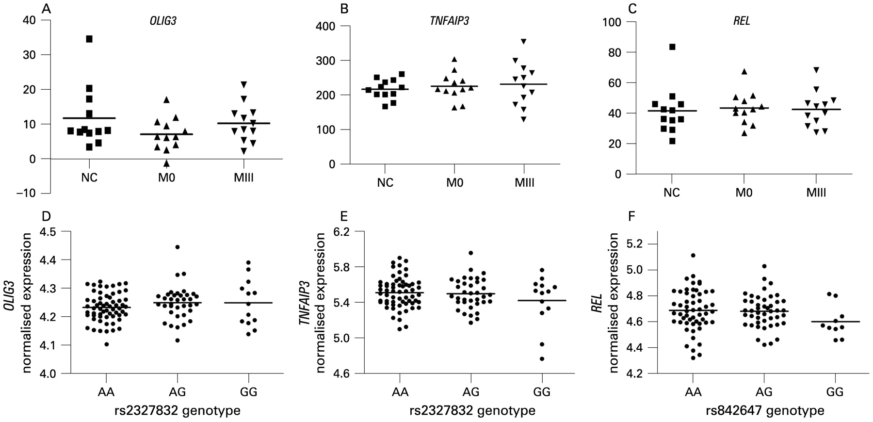
To assess the functional role of the SNPs in two associated regions, we have investigated the available datasets of the genome-wide association studies of gene expression.12 13 No significant effect of the rs2327832 SNP on the OLIG3 or TNFAIP3, nor of rs842647 on REL, was observed in these datasets. We also compared the RNA expression profiles of the OLIG3, TNFAIP3 and REL genes in small-intestine tissue from healthy controls and from treated and untreated coeliac patients. None of the genes showed significant differential expression between the three groups (fig 2A–C).

Expression of candidate genes in small intestine tissue from 12 normal healthy controls (NC), 12 treated coeliac disease patients with a normal histology of the small intestine, Marsh 0 (M0) and 12 untreated coeliac disease patients with villous atrophy, Marsh III (MIII) (A–C). Correlation of the rs2327832 genotype with OLIG3 and TNFAIP3 (D,E) and rs842647 genotype with REL expression (F). Expression levels were determined from the whole blood PAXgene samples from 110 patients with coeliac disease on a gluten-free diet. Bars show group means (A–F).
We correlated cis gene expression in the whole blood samples from 110 patients with coeliac disease for the OLIG3/TNFAIP3 and rs2327832, as well as REL with rs842647 genotypes. No significant effect of genotype on gene expression was observed (fig 2D–F). We also investigated if any other SNP in the 1 Mb window around each of the two associated SNPs affected gene expression. No cis effect was observed after correcting for multiple testing (supplementary data 2).
As one of our aims was to search for shared autoimmune genes, we also combined our results with those from the WTCCC GWAS, in particular the results for RA, T1D and Crohn’s disease compared to the WTCCC blood donor control group. Two of the variants were found to be associated to T1D and RA in the WTCCC GWAS: the OLIG3/TNFAIP3*rs2327832 SNP and the AFF3-LONRF2*rs1160542 SNP. All associations in the WTCCC dataset were observed to the same allele as in our coeliac disease analysis. Combining the analysis of all inflammatory diseases (including coeliac disease, T1D, RA and Crohn’s disease) rs2327832 in OLIG3/TNFAIP3 (p = 3.78×10⁻¹²) showed genome-wide significance to immune-related diseases (table 2). A second SNP, rs1160542 in AFF3-LONRF2, showed modest association (p = 2.49×10−06). The AFF3-LONRF2 locus has previously been nominally replicated in T1D in an independent case/control (p = 0.02) and a family (p = 0.01) datasets,5 further strengthening a role for this locus in immune-related disorders.
DISCUSSION
In this study we performed an extensive replication of moderately associated genetic variants from the GWAS study in coeliac disease, including variants located in immune-related genes, and potentially shared autoimmune SNPs. The strongest association to both coeliac disease and immune-related diseases was seen for rs2327832, located in a 60 kb block of linkage disequilibrium (LD) between the OLIG3 and TNFAIP3 genes. Interestingly, two independent SNPs located in the same LD block have been previously associated to RA;14 15 one of the RA associated SNPs (rs6920220) is a perfect proxy (r2 = 1 in CEU HapMap samples) for rs2327832, the SNP found to be associated in this study. Recently, the same region was found to be associated to systemic lupus erythematosus (SLE), another immune-related disease.16 17 For SLE a second, independent variant in the TNFAIP3 gene was also found to be associated.16 This indicates the TNFAIP3 gene region as a new, shared autoimmune locus. The scheme of association among different autoimmune disorders is presented in fig 3, indicating the complexity of the association pattern within this locus. Both the same and different variants are associated to various autoimmune traits.

Location of the signals in the four autoimmune diseases associated to the OLIG3/TNFAIP3 locus. The figure shows the linkage disequilibrium (LD) block between the OLIG3 and TNFAIP3 genes (NCBIb36, 137 559 184 bp to 138 486 672 bp) associated with multiple diseases. The plot is based on HapMap CEU data; the D’ plot was generated by Haploview. Single nucleotide polymorphims (SNPs) associated per disease are: SLE*a,b,c and SLE** (SNPs reported to be associated with SLE (Graham et al16 and Musone et al,17 respectively); RA* and RA** (SNPs associated with RA in Plenge et al14 and Thomson et al,15 respectively); T1D (SNP associated to T1D in the WTCCC study7); coeliac disease (SNP associated to coeliac disease in the current study). RA, rheumatoid arthritis; SLE, systemic lupus erthymatosus; T1D, type 1 diabetes; WTCCC, Wellcome Trust Case Control Consortium.
TNFAIP3 is an attractive candidate for both inflammatory and autoimmune pathogenesis. The TNFAIP3 gene product A20 is required for termination of the NF-κB signal mediated by innate immune receptors via the de-ubiquitylation of several NF-κB signalling factors.18 Genetic deficiency of A20 in mice leads to persistent activation of NF-κB by toll-like receptors, resulting in multi-organ inflammation, cachexia and neonatal lethality.19 20 It has been suggested that loss of A20 breaks down the tolerance of the innate immune system to the commensal intestinal microflora.21 Although we could not observe an effect of the associated polymorphisms on expression, this does not exclude a role for A20 in coeliac disease; there might well be an effect on the protein level. A20 regulation is rather complex and can be modified by A20-binding proteins such as ABINs or TAX1BP1, as well as by post-translational modifications in A20 protein.22 We do not exclude that subtle changes in the protein structure could lead to modification of A20 activity and, together with other coeliac-associated risk variants, cause the disease. Further extensive studies, including fine mapping with sequencing as well as functional studies (including those on a protein level), are required to identify the true causal variants.
The second new gene associated with coeliac disease – REL – is a component of the NF-κB transcription complex that plays a critical role in promoting immune and inflammatory responses including through the production of pro-inflammatory cytokines. In another study we observed a moderate association of REL polymorphisms to ulcerative colitis (p = 0.001), another intestinal inflammatory disorder,3 suggesting that this gene may not be unique to coeliac disease pathogenesis. Association of coeliac disease to both TNFAIP3 and REL points to a role for innate signalling via NF-κB in the pathology of coeliac disease, this is a novel finding and has not been reported before.
In this study we have extended the replication of our GWAS in coeliac disease and searched for genes shared by coeliac disease and other autoimmune and inflammatory intestinal disorders. We discovered two new loci associated to coeliac disease: REL and OLIG3/TNFAIP3. The OLIG3/TNFAIP3 locus can be considered to be a general immune-related locus as it has now been associated to four autoimmune disorders (fig 3). This supports the recent observation that many disease susceptibility genes contribute to multiple diseases.23 So far, the pathways associated with coeliac disease have pointed to T cell signalling and multiple cytokine involvement. Our observation that the NF-κB signalling pathway is also important adds a new player to the field.
NF-κB is a transcription complex that plays a key role in regulating the cellular immune response to infections, stress, cytokines and other stimuli. Activation of NF-κB in various inflammatory disorders, including asthma, arthritis and inflammatory bowel disease (IBD) has been described.24
Coeliac disease can now be added to the list of complex disorders that show association to the 6q23 region. This strengthens the importance of A20 in controlling inflammation in autoimmune diseases and points to A20 as an attractive candidate for drug targeting, as suggested recently by Coornaert et al.22
How the newly discovered genes interplay with the previously identified coeliac loci can only be speculated. On the one hand, activation of the NF-κB complex leads to overexpression of inflammatory cytokines and, together with previously identified cytokines and cytokine receptor genes such as CCR5, RGS1, IL12A and IL18RAP, this pathway would be important in fine-tuning the immune response. On the other hand, the NF-κB pathway may play an independent role in the innate mechanisms of disease development. Strikingly, genes involved in the innate immune response have recently been associated with various autoimmune diseases, suggesting a role for microbial and viral triggers in disease development. In coeliac disease an increased frequency of rotaviral infections have been observed, suggesting that viral infections may, for example, trigger an innate immune response.25
It is interesting that the knockdown studies of A20 in dendritic cells show a shift in the subset of activated T cells, hyperactivation of cytotoxic T lymphocytes and T helper cells, and suppression of regulatory T cells. This shift results from enhanced expression of co-stimulatory signals and proinflammatory cytokines when inhibiting A2026 and would fit in celiac disease being a Th1 mediated disease.
These two novel loci can be added to the list of eight known, non-HLA, genetic risk factors for coeliac disease that have a smaller risk effect than HLA. Extending the list of common variants that account for coeliac disease will improve the genetic prognosis of patients and may help to predict the likelihood of individuals from the at-risk group developing coeliac disease.
Acknowledgments
We thank J Swift, P Kumar, D P Jewell, S P L Travis, L Dinesen and K Moriarty for collection of UK-GWAS and additional coeliac case samples. We acknowledge use of DNA from the British 1958 Birth Cohort collection, funded by the UK Medical Research Council grant G0000934 and the Wellcome Trust grant 068545/Z/02. We thank H van Someren and F Mulder for clinical database management, E Oostrom, R van ‘t Slot and the genotyping facilities at UMCG and UMC Utrecht (the Netherlands) for technical assistance. We thank C Feighery and J McPartlin for sample collection. Irish control DNA was supplied by the Irish Blood Transfusion Service/Trinity College Dublin Biobank. We thank all coeliac and control individuals for participating in this study. We thank J Senior for critically reading the manuscript.
REFERENCES
Supplementary materials
Web only appendices 58;8:1078
Files in this Data Supplement:
Footnotes
Competing interests: None.
Funding: The study was supported by grants from Coeliac UK (to DAvH); the Coeliac Disease Consortium (an innovative cluster approved by the Netherlands Genomics Initiative and partly funded by the Dutch Government, grant BSIK03009 to CW); the European Union (STREP 036383); the Netherlands Organization for Scientific Research (VICI grant 918.66.620 to CW); the Science Foundation Ireland; the Higher Education Authority PRTLI; The Irish Health Research Board; and the Wellcome Trust (GR068094MA Clinician Scientist Fellowship to DAvH; New Blood Fellowship to RMcM).
Ethics approval: Ethics approval was from Oxfordshire REC B or East London and the City REC 1 (UKGWAS, UK2), the Medical Ethical Committee of the University Medical Centre Utrecht (Dutch), the Institutional Ethics Committee of St James’s Hospital (Irish) and the Ethics Committee of the Fondazione IRCCS Ospedale Maggiore Policlinico, Mangiagalli e Regina Elena (Italian).
▸ Two supplementary files giving additional data for the SNPs in this study are published online only at http://gut.bmj.com/content/vol58/issue8
Linked Articles
- Digest