Article Text

Abstract
Background and aims Patients with hyperplastic polyposis syndrome (HPS) receive endoscopic surveillance to prevent malignant progression of polyps. However, the optimal treatment and surveillance protocol for these patients is unknown. The aim of this study was to describe the clinical and pathological features of a large HPS cohort during multiple years of endoscopic surveillance.
Methods Databases were searched for patients with HPS, who were analysed retrospectively. Endoscopy reports and histopathology reports were collected to evaluate frequency of endoscopic surveillance and to obtain information regarding polyp and the presence of colorectal cancer (CRC).
Results In 77 patients with HPS, 1984 polyps were identified during a mean follow-up period of 5.6 years (range: 0.5–26.6). In 27 (35%) patients CRC was detected of which 22 (28.5%) at initial endoscopy. CRC was detected during surveillance in five patients (cumulative incidence: 6.5%) after a median follow-up time of 1.3 years and a median interval of 11 months. Of these interval CRCs, 4/5 were detected in diminutive serrated polyps (range: 4–16 mm). The cumulative risk of CRC under surveillance was 7% at 5 years. At multivariate logistic regression, an increasing number of hyperplastic polyps (OR 1.05, p=0.013) and serrated adenomas (OR 1.09, p=0.048) was significantly associated with CRC presence.
Conclusions HPS patients undergoing endoscopic surveillance have an increased CRC risk. The number of serrated polyps is positively correlated with the presence of CRC in HPS, thus supporting a ‘serrated pathway’ to CRC. To prevent malignant progression, adequate detection and removal of all polyps seems advisable. If this is not feasible, surgical resection should be considered.
- Hyperplastic polyposis syndrome
- colorectal cancer
- conventional adenoma
- hyperplastic polyp
- serrated adenoma
- serrated pathway
- surveillance
- colonic polyps
- endoscopy
- polyposis
Statistics from Altmetric.com
- Hyperplastic polyposis syndrome
- colorectal cancer
- conventional adenoma
- hyperplastic polyp
- serrated adenoma
- serrated pathway
- surveillance
- colonic polyps
- endoscopy
- polyposis
Introduction
Colorectal cancer (CRC) ranks as the third most common cause of cancer-related death in the Western world.1 A well known mechanism describing CRC development is the adenoma–carcinoma sequence which, in the majority of cases, is initiated by activation of the Wnt signalling pathway.2 3 Much information regarding this pathway has been derived from polyposis syndromes such as familial adenomatous polyposis (FAP) and MYH-associated polyposis (MAP). In addition to this classical adenoma–carcinoma sequence, a proposed ‘serrated neoplasia pathway’, describes the progression of serrated polyps (ie, hyperplastic polyps, sessile serrated adenomas and traditional serrated adenomas) to CRC.4 It is proposed that this possible alternative pathway is also associated with hyperplastic polyposis syndrome (HPS). There are strong indications that mixed pathways also exist in which both conventional adenomas and serrated polyps are involved.5
Clinically, the condition HPS is characterised by the presence of multiple hyperplastic polyps (HPs) spread throughout the colorectum and is associated with an increased risk of CRC. Indeed, numerous patients with CRC and concurrent HPS have been reported.6–12 While previously the indicated management of patients with HPS was unknown, experts presently believe that these patients should undergo regular endoscopic surveillance to prevent malignant progression of polyps.7 13 However, the optimal treatment and surveillance protocol for HPS patients is largely speculative. Therefore it seems possible that a proportion of patients with HPS may be insufficiently treated and consequently be at risk of developing CRC under surveillance (interval CRC).
The aim of this study was to describe the clinical and pathological features of a large HPS cohort (n=77) during multiple years of endoscopic surveillance. Furthermore, we assessed the cumulative incidence and incidence rate of CRC during surveillance and its association with the interval and frequency of surveillance endoscopies. Finally, we analysed possible predictive variables that may be associated with the occurrence of CRC in HPS.
Patients and methods
Study population
Databases of seven medical centres in the Netherlands were searched for patients satisfying the diagnostic criteria of HPS (ie, at least five histologically diagnosed HPs proximal to the sigmoid colon, of which two greater than 10 mm in diameter, or more than 20 HPs distributed throughout the colon) and undergoing endoscopic surveillance.8 11 Owing to the common presence of both HPs and (sessile) serrated adenomas (SAs) in HPS and the difficult histological differentiation between these two groups, both HPs and SAs were used to fulfil the criteria.14–17 Of these patients, clinical data from May 1982 to June 2008 were analysed retrospectively. Adherence to the described criteria was assessed by analysing endoscopy reports with corresponding histopathology reports as well as histopathology reports of colonic surgical resection specimens. Patients with a known germline APC mutation or bi-allelic MYH mutation were excluded from the study.
Clinical characteristics
Demographic data of patients concerning age, sex and history of CRC were ascertained. Endoscopy reports with corresponding histopathology reports during follow-up were collected to evaluate the duration, interval and frequency of endoscopic surveillance and to derive information regarding the, number, size, distribution and histology of polyps. If applicable, histopathology reports of surgical colonic resection specimens were also used to obtain the above mentioned polyp characteristics. Also, if genetic mutation analysis was performed, these data were retrieved.
Polyps were classified as HP, serrated adenoma (SA), mixed polyp (MP) or conventional adenoma. Because the distinction between sessile serrated adenoma and traditional serrated adenoma had not been made throughout the study period by each medical center and because they are both considered to be precursor lesions in the ‘serrated pathway’, the category SAs comprised both types of lesions.16 Regarding the number of polyps detected in this study, all polyps were tallied once, ie, when a detected polyp was not removed during endoscopy this polyp was not re-tallied at subsequent endoscopies.
Information concerning the nature and reason of performed colorectal surgery was obtained if applicable. Detailed information regarding co-existent CRC and CRC incidence during surveillance was examined by evaluating histopathology reports of colectomy resection specimens and/or endoscopy reports. An interval CRC was defined as a CRC detected after HPS diagnosis after at least two previous endoscopies.
Statistical analysis
Statistical analyses were performed by using a statistical software package (SPSS 15.0.1). The cumulative risk of developing CRC during follow-up was analysed by Kaplan–Meier survival analysis. Observation time was measured from date of HPS diagnosis to the incidence of carcinoma or end of the study period. Univariate binary logistic regression was performed for chosen variables that may be associated with the presence of CRC. For multivariate regression analyses, only variables which showed an association (p<0.1) on univariate analysis were used in a final multivariate model.
Results
Patients
Data of 77 patients with HPS from the period 1982–2008 were analysed retrospectively in this study. Clinical characteristics of patients are shown in table 1. The median age at diagnosis of HPS was 56 years (range: 40–74). There were 42 males and 35 females. Fifty-nine of 77 patients (77%) had >5 proximal HPs (of which two were larger than 10 mm) or >20 HPs spread throughout the colon. The other 17 patients had >5 proximal HPs/SAs (of which two were larger than 10 mm) or >20 HPs/SAs spread throughout the colon. In 52/77 (68%) patients, germline APC and MYH-mutation analysis was performed. In all cases mutation analysis was negative except for one patient with a mono-allelic MYH-mutation (Y165C) who had ≥ 15 adenomas. In all patients harbouring ≥15 adenomas (n=5) mutation analysis was performed. The main reasons for initial presentation were colorectal polyps detected elsewhere (n=23), a positive family history for CRC or colorectal polyps (n=16), bloody stools/positive faecal occult blood test/iron-deficiency anaemia (n=15), altered defaecation pattern (n=8), abdominal pain with or without altered bowel habits (n=5), polyps detected at sigmoidoscopy screening programme (n=4), and personal history of CRC (n=3).
Characteristics of patients with hyperplastic polyposis syndrome (HPS) from multiple centres in The Netherlands, divided per centre
Cumulatively, a median number of 15 HPs per patient were found in this cohort. In 47 (61%) patients HPs ≥10 mm were detected. SAs and adenomas were identified in 52% and 69% of patients, respectively. CRC was diagnosed in 27 (35%) patients: 22 (28.5%) at initial endoscopy and 5 (6.5%) during follow-up.
A surgical colonic resection was performed in 33/77 (43%) patients: two total colectomies, 13 subtotal colectomies, eight hemi-colectomies (six left-sided) and 10 (recto)sigmoidal resections. Seven patients underwent a surgical resection because of extensive polyposis or due to difficult advancement of the endoscope during examination resulting in incomplete visualisation of the colon. The other 26 patients underwent a colonic resection due to CRC diagnosis. Consequently, during (a part of) follow-up 44 patients received endoscopic surveillance of the intact colon; 17 patients of the remaining segment proximal to the rectosigmoid colon; 15 patients of the remaining distal colon segment and two patients did not receive endoscopic surveillance after undergoing a proctocolectomy.
The mean follow-up period of patients was 5.6 years (range: 0.5–26.6) and from the point HPS was diagnosed this was 4.0 years (range: 0.4–21.0). During follow-up, the number of surveillance endoscopies varied among patients. In the time period observed, 207 surveillance endoscopies were performed (median 3, range 0–11). One patient was diagnosed with HPS based on the surgical resection specimen and had not yet undergone surveillance endoscopies. The median interval between surveillance endoscopies was 10 months (range: 1–96).
Polyps
Polyp characteristics are outlined in table 1. In this study, 847/1407 (60%) HPs, 197/302 (65%) SSAs and 165/273 (60%) adenomas were detected proximal to the sigmoid colon. The maximum size of HPs and SAs was 30 mm which were located in the ascending and transverse colon, respectively. The largest adenoma detected in this cohort was 75 mm, which was located in the ascending colon. Polyps were detected during endoscopy with standard or high-resolution white-light endoscopy. Narrow-band imaging was used in 22/294 (7%) endoscopies in 22 patients.
Colorectal cancer
Of the 77 HPS patients included in this study, 27 (35%) patients had CRC (median age 56 years; range 36–75). In 14/27 (52%) of these patients, CRC was located proximal to the sigmoid colon. One patient had two separate synchronous CRCs, one proximally and one distally located.
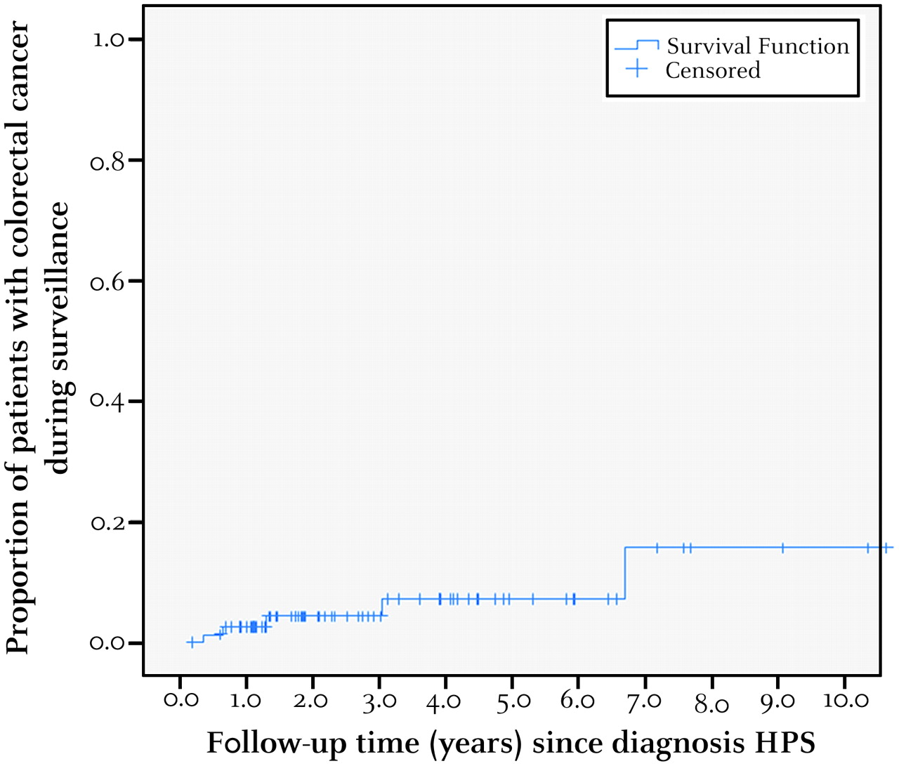
While CRC was diagnosed at initial colonoscopy in the majority (22/27) of HPS patients, in five patients (cumulative incidence: 6.5%) with a median age of 58 years (range 49–68) CRC was detected during surveillance after the diagnosis HPS was made (mean follow-up time 5.6 years) without any prior history of CRC. The median follow-up time in this group was 1.3 years (range: 0.4–6.7) with a median of three endoscopies (range: 2–4). Clinical and histological data of these patients are summarised in table 2. During a total of 294.6 person years of follow-up, this corresponds with a CRC incidence rate during surveillance of 17 per 1000 person-years. In four of the five (80%) patients, CRC was detected during a planned endoscopy and was located within a HP (3/4) or a SA (1/4) without causing clinical symptoms. The median size of these polyps was 10 mm (range: 4–16 mm). One patient (patient 2) presented with weight loss and fatigue after a surveillance interval of more than 3 years. At endoscopy a large CRC was detected. In four of five patients, CRC was located proximally to the sigmoid colon. The median interval between surveillance endoscopies in patients with an interval CRC was 11 months (range: 3–43) compared to 10 months (range: 1–96) in HPS patients without an interval CRC (NS). The median interval between the last surveillance endoscopy and CRC detection in patients with interval carcinomas was also 11 months (range: 4–43). The calculated cumulative risk of CRC in HPS during surveillance was 7% at 5 years (figure 1). When analysing the cumulative risk separately for patients with an intact colon and for patients with a surgical colonic resection, the 5-year cumulative risk was 6% and 4%, respectively.
Characteristics of patients with hyperplastic polyposis syndrome (HPS) in which an interval carcinoma was detected

Cumulative proportion of patients with hyperplastic polyposis syndrome (HPS) and with colorectal cancer during surveillance.
To analyse an association with CRC in HPS, univariate logistic regression was performed for eight independent variables: age, sex, number of HPs, number of SAs, number of adenomas, largest HP, largest SA and largest adenoma (table 3). At univariate logistic regression, the number of HPs and the number of SAs were associated with CRC (p<0.1). At subsequent multivariate logistic regression the number of HPs (p=0.013) and the number of SAs (p=0.048) were significantly associated with CRC with corresponding OR of 1.05 (95% CI: 1.01 to 1.10) and 1.09 (95% CI 1.00 to 1.19) respectively.
Results of univariate and multivariate logistic regression analysis: independent prognostic variables and corresponding odds ratios for the presence of colorectal cancer in hyperplastic polyposis syndrome
Discussion
This multicentre cohort study showed that in a total of 27/77 (35%) HPS patients, CRC was detected. Interestingly, CRC was detected in 5/77 (6.5%) patients during surveillance of which four CRCs within a diminutive serrated polyp (HP or SA) resulting in a cumulative risk of CRC under endoscopic surveillance of 7% in 5 years. This is substantial considering that the lifetime risk of developing CRC in the general population is estimated to be 6%.18 Of these patients with interval CRCs, two CRCs were detected within a year (table 2: 11.4 and 4.3 months) after the diagnosis HPS was made and after two previous endoscopies. The high frequency of endoscopies in a short time period suggest that these patients were probably still in an orientating treatment phase when CRC was detected. If surveillance was defined as endoscopies performed after HPS diagnosis after at least 1-year follow-up, the cumulative risk under surveillance would be 4% at 5 years.
Although different management protocols have recently been advised, thus far no uniform and adequately substantiated management protocol exists for the endoscopic management of HPS patients. Consequently, lack of clarity exists regarding the recommended surveillance interval and which polyps to remove. Recent studies recommend surveillance intervals ranging from 1 to 3 years and concerning the removal of polyps, advice varies from removal of only proximally located polyps to complete removal of all polyps >5 mm.7 13
This absence of a standardised treatment protocol may also be associated with the incidence of interval CRCs in this retrospective multicentre study dating back to 1982. Possible explanations for the incidence of interval CRCs could be that previously an association between HPS and CRC was not made or that only proximal and/or larger lesions were considered clinically significant, resulting in incomplete removal of polyps. Also when considering the relatively short median interval between endoscopies (median interval: 11 months), it is likely that the interval CRCs were also present at prior surveillance endoscopies but were not removed. This was indeed the case for two of five of these patients who underwent incomplete removal of all detected polyps during the last surveillance endoscopy before CRC diagnosis (table 2: patient 2 and 4), underlining the importance of comprehensive polyp removal during surveillance
Conversely, in three of five patients (patients 1, 3 and 5) all detected polyps were biopsied or removed at previous endoscopy. A possible explanation for this contrary finding could be that these CRCs originating in serrated polyps were simply missed. This could possibly be due to the multiplicity of polyps seen in HPS patients resulting in a sub-optimal overview of all colorectal polyps. Alternatively, typical HPs and (sessile) serrated adenomas seldom exceed 10 mm in size, suggesting that most polyps in HPS are diminutive.19–25 It has been shown that the miss-rate of polyps <10 mm can be as high as 23%.26 This could explain why these relatively small CRCs originating in diminutive serrated polyps were not detected at previous endoscopy.
Nevertheless, considering their small size and the unknown progression rate in HPS, it cannot be excluded that these CRCs, originating in serrated polyps (4/5) developed since the last endoscopy. A previous retrospective polyp study of consecutive patients with an average risk for CRC showed that the estimated growth rates of HPs and SAs (both sessile and traditional) compared to conventional adenomas were similar or significantly higher.27 Moreover, a recent case report described the progression of a sessile serrated adenoma to carcinoma within 8 months.28 In this respect, it is conceivable that in HPS a subset of serrated polyps have an increased progression rate leading to CRC. This is an interesting point considering that the risk of high grade dysplasia or even invasive cancer in diminutive lesions (<10 mm) has been shown to be <2%.29–31 The finding of CRC within a small serrated polyp in 4/5 (80%) interval carcinomas suggests that in HPS small polyps have a greater malignant potential than in the general population.
When considering the management of patients with HPS, this study suggests that the absence of a clear treatment protocol plays a role in the presence of CRCs during surveillance. Considering that CRCs detected in this study were as small as 4 mm (detected in a HP: patient 3), removal of all polyps seems advisable but needs to be prospectively assessed. Although these recommendations seem of importance in preventing malignant progression in HPS, practical difficulties may present when trying to comply with these guidelines in a clinical setting. Firstly, besides being small, detection of HPs and SAs is also complicated by their predominantly flat shape, unremarkable colour and mucus coating which possibly increases polyp miss rates.19 32 Secondly, removal of all detected polyps during endoscopic surveillance sessions in HPS patients with a large quantity of polyps can be time-consuming and unfeasible, especially when endoscopic mucosal resection (EMR) is indicated for predominantly flat serrated polyps.
With regard to polyp detection, previous randomised controlled trials demonstrated that chromoendoscopy and narrow-band imaging (NBI) increased the detection of HPs.33–38 Although not formally investigated, these techniques could in this respect also be of value for the detection of serrated polyps in HPS. Concerning polyp removal, the multiplicity of polyps and the use of EMR can indeed lead to increased duration of endoscopic procedures in patients with HPS. In this respect, it is important that these endoscopies are performed by endoscopists experienced in EMR for complete, prompt and safe polyp removal and that allowances are made for sufficient procedure time. Annual surveillance by an experienced endoscopist specialised in HPS, having advanced imaging techniques available such as chromoendoscopy and NBI seems therefore advisable. Alternatively, when complete endoscopic removal of all polyps is not feasible, surgical colonic resection should seriously be considered since these patients have an increased risk of malignant progression of polyps.
In this study, at multivariate logistic regression, an increasing number of HPs and SAs was significantly associated with CRC presence (OR of 1.05 and 1.09 respectively per polyp). In other words, the CRC risk will increase by 5% and 9% respectively with each additional HP or SA. Concordantly, results from previous literature reports strongly suggest that SAs in particular play a role in a ‘serrated pathway’ leading to CRC in HPS.19 39–41 A possible explanation for the significant association between HPs and CRC in this study could be that HPs and SAs (primarily sessile serrated adenomas) are histologically hard to distinguish, leading to incorrect differentiation and misdiagnosis of these serrated polyps. Indeed, it has recently been shown that even at re-evaluation the interobserver agreement for the differentiation of serrated polyps remains only moderate (к=0.55).14 15 42 Nevertheless, the significant association between serrated polyps and CRC in this study supports the hypothesis of a ‘serrated pathway’ leading to CRC in HPS (figure 2).

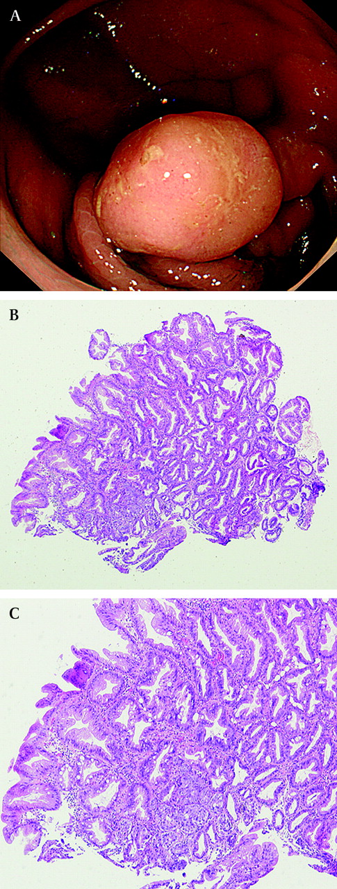
Endoscopic image of a serrated adenoma (10 mm, ascending colon) detected in a patient with hyperplastic polyposis syndrome (A). At microscopy a focus of adenocarcinoma was seen within the serrated adenoma (B,C).
In conclusion, HPS is associated with an increased personal CRC risk, even under endoscopic surveillance. Considering that these advanced lesions were detected in polyps as small as 4 mm (median: 10 mm), which were not recognised as such, all polyps in HPS seem at risk of representing advanced lesions warranting removal of all polyps. However, the miss rate of polyps <10 mm (which represents the majority of polyps in HPS) has been shown to be as high as 23% with standard white-light endoscopy suggesting that a considerable number of polyps in HPS are missed.26 Advanced endoscopic imaging techniques such as chromoendoscopy and NBI may in this respect be of additional value for the detection of polyps in HPS. Alternatively, if endoscopically unfeasible, preventive colonic resection should be considered. An increasing number of serrated polyps are associated with the presence of CRC in HPS, supporting the theory of a ‘serrated pathway’ leading to CRC. Future prospective data from large HPS cohorts, undergoing a standardised treatment protocol are required to further enhance our knowledge with regard to the rate of polyp progression in these patients and to determine the optimal treatment and surveillance protocol for these patients.
References
Footnotes
Linked articles 195032.
Competing interests None.
Ethics approval This study was conducted in accordance with the research code of our institutional medical ethics committee on human experimentation, as well as in agreement with the Helsinki Declaration of 1975, as revised in 1983.
Provenance and peer review Not commissioned; externally peer reviewed.