Article Text

Abstract
Background and Aims Endoscopic resection is safe and effective to remove early neoplasia (ie,high-grade intra-epithelial neoplasia/early cancer) in Barrett's oesophagus. To prevent metachronous lesions during follow-up, the remaining Barrett's oesophagus can be removed by stepwise radical endoscopic resection (SRER). The aim was to evaluate the combined experience in four tertiary referral centres with SRER to eradicate Barrett's oesophagus with early neoplasia.
Methods Design: Retrospective cohort study.
Setting: Four tertiary referral centres.
Participants: 169 patients (151 males, age 64 years (IQR 57–71), Barrett's oesophagus 3 cm (IQR 2–5)) with early neoplasia in Barrett's oesophagus ≤5 cm, without deep submucosal infiltration or lymph node metastases, treated by SRER between January 2000 and September 2006.
Intervention: Endoscopic resection every 4–8 weeks, until complete endoscopic and histological eradication of Barrett's oesophagus and neoplasia.
Results According to intention-to-treat analysis complete eradication of all neoplasia and all intestinal metaplasia by the end of the treatment phase was reached in 97.6% (165/169) and 85.2% (144/169) of patients, respectively. One patient had progression of neoplasia during treatment and died of metastasised adenocarcinoma (0.6%). After median follow-up of 32 months (IQR 19–49), complete eradication of neoplasia and intestinal metaplasia was sustained in 95.3% (161/169) and 80.5% (136/169) of patients, respectively. Acute, severe complications occurred in 1.2% of patients, and 49.7% of patients developed symptomatic stenosis.
Conclusions SRER of Barrett's oesophagus ≤5 cm containing early neoplasia appears to be an effective treatment modality with a low rate of recurrent lesions during follow-up. The procedure, however, is technically demanding and is associated with oesophageal stenosis in half of the patients.
- Barrett's oesophagus
- cancer
- intestinal metaplasia
- intraepithelial neoplasia
- endoscopic resection
- Barrett's carcinoma
- Barrett's metaplasia
- endoscopy
Statistics from Altmetric.com
- Barrett's oesophagus
- cancer
- intestinal metaplasia
- intraepithelial neoplasia
- endoscopic resection
- Barrett's carcinoma
- Barrett's metaplasia
- endoscopy
Significance of this study
What is already known about this subject?
Endoscopic therapy for early Barrett's neoplasia has been proven a safe and effective alternative to surgical resection in selected patients.
Mono-therapy of neoplastic lesions with endoscopic resection is associated with a significant number of recurrences (30%) in the residual Barrett's mucosa during follow-up.
Stepwise radical endoscopic resection (SRER) of the complete Barrett's segment, to decrease the risk of metachronous lesions during follow-up, has been proven feasible in a number of small, single centre studies with limited follow-up.
What are the new findings?
This largest series worldwide shows that in expert hands, SRER is safe and highly effective for complete removal of all neoplasia (97.6%) and all Barrett's mucosa (85.2%).
Neoplasia recurrence after complete eradication of the Barrett's segment was rare during a follow-up of 32 months, which shows that all Barrett's mucosa should be removed for a persistent treatment effect.
Thorough endoscopic work-up of Barrett's patients with early neoplasia accurately identifies the most involved area: all submucosal lesions were resected during the first treatment session; and after removal of the most suspicious area, histological evaluation did not reveal worse tumour characteristics during subsequent treatment sessions.
Almost half of the patients developed an oesophageal stenosis. The rate of oesophageal stenosis was related to the length of the resected Barrett's segment (Mantel–Haenszel test; p=0.002).
How might it impact on clinical practice in the foreseeable future?
Complete eradication of Barrett's oesophagus with early neoplasia appears to decrease the rate of recurrences during follow-up. SRER can be used for initial staging of the most involved area, and for subsequent safe and effective removal of all residual Barrett's mucosa. Given the high rate of oesophageal stenosis; however, future research should be aimed at prevention of oesophageal stricturing or different techniques may be used to eradicate the whole Barrett's segment after diagnostic endoscopic resection.
Introduction
In patients with Barrett's oesophagus containing high-grade intraepithelial neoplasia (HGIN) or early cancer (EC), oesophagectomy used to be the treatment of choice.1–3 However, since lymph node involvement occurs rarely with these early lesions (0% for HGIN and less then 3% for early cancer),4–7 treatment with less invasive endoscopic techniques has emerged and has been shown to be an effective and safe alternative for selected patients.8–11 The cornerstone of endoscopic treatment is endoscopic resection (ER) of focal lesions, which provides a relatively large tissue specimen for accurate histological assessment. Focal ER, however, is associated with recurrent lesions elsewhere in the Barrett's oesophagus in 25–33% of patients during follow-up if the residual Barrett mucosa is left untreated.9–11 To minimise this risk of recurrence, not only the neoplasia but all Barrett mucosa can be removed by stepwise radical endoscopic resection (SRER). In SRER the whole Barrett's oesophagus is resected by subsequent ER-sessions resulting in complete removal of the Barrett's oesophagus with histological correlation of the whole Barrett segment. SRER has been shown to effectively eradicate neoplastic Barrett's oesophagus in relatively small-sized single-centre studies.12–18 The aim of the current multi-centre study was to retrospectively evaluate SRER in four European tertiary referral centres for early Barrett neoplasia, all using a prospective treatment protocol. This study aimed at evaluating the safety and efficacy of SRER in a significantly larger cohort with a longer follow-up period than reported thus far.
Patients and methods
Data collection
All participating centres used a comparable protocol for SRER treatment. In two centres, data were prospectively entered into a dedicated database whereas the other two centres documented their findings in endoscopy and pathology reports. For the purpose of this study, all centres were visited by two researchers experienced in the field of endoscopic treatment of early Barrett neoplasia. Standardised case record forms were used to extract relevant data from the prospective databases and/or endoscopy and pathology reports. The collected data were then entered into a central database for further evaluation. After a first analysis, all centres were revisited by the same researcher to update follow-up data for included patients, collect missing data, and to review charts of all patients that underwent ER at the participating centres again to ensure that no patients were inadvertently excluded.
Selection criteria
For this retrospective multi-centre cohort study, all patients that underwent an ER at the University Hospital Eppendorf (Hamburg), Charité-Campus Virchow (Berlin); Cliniques Universitaires Saint-Luc (Brussels) and Academic Medical Centre (Amsterdam), from January 2000 until September 2006, were reviewed. Patients were included if they met all of the following criteria:
Maximum estimated Barrett's oesophagus length of 5 cm, with intestinal metaplasia (IM) upon biopsy
A histological diagnosis of HGIN or invasive cancer in biopsies or ER-specimens
In the case of a diagnostic ER (always performed during the first ER session), specimens could not show any of the following criteria: invasion >T1sm1, poorly/undifferentiated cancer (G3/G4), lymph–vascular invasion, irradical deep resection margins (Note: T1sm1 cancer with G1/G2 differentiation and no lymph–vascular invasion, was considered a relative indication for endoscopic treatment, if patients had serious contraindications for surgery or refused surgery. Both endoscopic and surgical options were discussed with these patients before treating them with SRER);
No signs of lymph node or distant metastasis on endosonography or CT
Patients were considered eligible for complete eradication of their Barrett's oesophagus by means of SRER.
Patients were excluded if, after focal ER of visible lesions, they were not additionally treated by SRER, but with endoscopic ablation (eg, argon plasma coagulation (APC), photodynamic therapy (PDT) or radiofrequency ablation (RFA)), or if residual Barrett's oesophagus was kept under surveillance.
Treatment protocol
In the case of visible lesions suspicious for submucosal invasion, patients first underwent a diagnostic ER to assess their eligibility for further endoscopic treatment by means of SRER. If there was no suspicion on deep submucosal infiltration on endoscopy or endosonography, 50% of the circumference of the Barrett's oesophagus, including the most suspicious lesion, was removed during the first SRER session. Subsequent SRER sessions were performed with an interval of 4–8 weeks until endoscopic eradication of all Barrett's oesophagus mucosa and neoplasia was considered complete. Histological eradication of IM and neoplasia was confirmed by biopsies from the neosquamous mucosa and immediately distal to the neo-squamocolumnar junction (neo-SCJ) (figure 1).
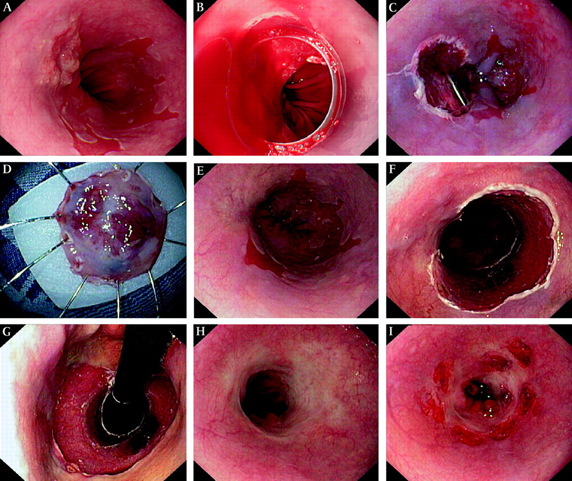
Endoscopic images a Barrett's oesophagus with early cancer treated by stepwise radical endoscopic resection (SRER). (A) C1M4 Barrett's oesophagus with at the 10 o'clock position a 0–IIa–IIc lesion. (B, C) Acute bleeding during diagnostic endoscopic resection (ER) of the lesion treated by placement of a clip. (D) Resection specimen pinned down on paraffin. (E) Prior to the second ER session, 6 weeks after the diagnostic ER a scar is observed at the 9 o'clock position. (F) Resection wound after the SRER session. (G) View on the resection wound with the endoscope in the retrograde position. (H) Complete regeneration of squamous mucosa after SRER and four dilatations sessions for symptomatic stenosis. (I) Extensive biopsies from the neo-squamous mucosa during follow-up.
Patients were prescribed high-dose proton pump inhibitor therapy (esomeprazole/omeprazol 40 mg b.i.d.) as maintenance medication during the entire treatment phase and after completion of the protocol.
Endoscopic resection procedures
SRER procedures were generally performed on an outpatient basis using propofol sedation, or conscious sedation with midazolam and fentanyl. Therapeutic procedures were performed using standard therapeutic video endoscopes (Olympus GIF-1T140/160; Olympus Europe, Hamburg, Germany). ER was performed using either the ER-cap technique, with oblique caps (diameter 12.8/14.8/18 mm, MAJ-296/297 or D206-5; Olympus Europe),19 20 the simple snare technique using a monofilament steel wire (30–50 mm Erlangen-type polypectomy snare; Ch. Grosse, Daldorf, Germany),12 or multi-band mucosectomy (Duette Multi-Band Mucosectomy kit; Cook Endoscopy, Limerick, Ireland).14 20 In all cases, the area to be resected was delineated with coagulation markings, followed by ER of the target area using one of the above-mentioned techniques that have been described in detail elsewhere.12 14 20 All ER specimens were retrieved, pinned on cork or paraffin and fixed in formalin.
Additional ablation
APC was used to ablate small areas of Barrett's oesophagus mucosa that could not be resected (eg, tissue bridges between adjacent resections, areas difficult to reach due to stenosis), or to ablate the neo-SCJ in some patients. A 2.3 mm forward spraying APC probe (Erbe APC 300; Erbe Elektromedizin, Tubingen, Germany) was used (power 80–99 W, argon flow 1.6–2.0 l/min).
Histopathological evaluation
Formalin-fixed biopsies and ER specimens were processed to haematoxylin & eosin stained slides for routine histological evaluation by experienced GI pathologists. Biopsies and ER specimens were evaluated for the presence of (subsquamous) intestinal metaplasia and neoplasia, graded according to the WHO classification.21 Furthermore, tumour infiltration depth, differentiation grade, lymph–vascular involvement and radicality at the deep resection margins were assessed in ER specimens as described elsewhere.22
Follow-up
Follow-up started after complete removal of all endoscopically visible columnar epithelium in the oesophagus, confirmed histologically by biopsies. All patients underwent at least the first follow-up endoscopy in the centre where they were treated and a strict biopsy protocol was applied with random four-quadrant biopsies distal to the neosquamocolumnar junction and for every 1–2 cm of the neosquamous epithelium. Subsequent follow-up endoscopies were performed every 3–6 months during the first year with six monthly or annual endoscopic follow-up thereafter. Follow-up endoscopy consisted of standard video endoscopy combined with chromoendoscopy using Lugol staining or narrow-band imaging to detect recurrent columnar epithelium. Targeted biopsies were obtained from visible columnar-like epithelium in the oesophagus, and randomly from neosquamous mucosa and immediately distal to the neo-SCJ.
Endpoints
Primary endpoints were assessed at the end of the treatment phase and at the end of the follow-up:
Complete eradication of all HGIN/cancer, defined as absence of HGIN/cancer in biopsies (complete response of neoplasia, CR-N)
Complete eradication of all Barrett's oesophagus, defined as absence of IM in biopsies obtained from neosquamous mucosa and immediately distal to the neo-SCJ (complete response of IM, CR-IM).
Secondary endpoints were:
Number of treatment sessions and need for additional treatment during the treatment phase to achieve complete eradication of neoplasia and complete endoscopic removal of all Barrett's oesophagus
Number of patients that required surgery
Histological outcome of subsequent ER procedures
Complications during the treatment phase, defined as ‘acute’ (during procedure), ‘early’ (0–48 h) and ‘late’ (>48 h). Complications were only recorded if they were clinically significant and graded as ‘mild’ (unplanned hospital admission, hospitalisation <3 days, haemoglobin drop <3 g, no transfusion), ‘moderate’ (4–10 days hospitalisation, <4 units blood transfusion, need for repeat endoscopic intervention, radiological intervention), ‘severe’ (hospitalisation >10 days, ICU admission, need for surgery, > 4 units blood transfusion, in the case of stenosis: >5 dilatations, stent placement or incision therapy) or ‘fatal’ (death attributable to procedure <30 days or longer with continuous hospitalisation)
Need for re-treatment during follow-up.
Statistical analysis
For intention-to-treat analysis the primary endpoints at the end of the treatment phase and at the end of follow-up were accounted for in all patients who were included for this study. Patients lost to follow-up were considered a failure for both primary endpoints. For patients who discontinued treatment or follow-up, CR-N and CR-IM were defined at the exit from the study and in case these endpoints were unknown, they were considered a failure. The treatment approach used to achieve and sustain CR-N and CR-IM was a secondary endpoint.
For the per-protocol analysis at the end of the treatment phase, patients who were lost for further treatment or who discontinued treatment for unrelated reasons were censored. For per-protocol analysis at the end of the follow-up phase, patients who were lost to follow-up were censored and for patients who discontinued follow-up CR-N and CR-IM were defined at the exit from the study. Patients undergoing surgery during the treatment or follow-up phase were considered a failure
For descriptive statistics, mean (±SD) was used in case of a normal distribution of variables and median (25%–75%) was used for variables with a skewed distribution. Where appropriate, the Student t test, Mann–Whitney U test, or Mantel–Haenszel test was used.
Results
Patients
From a total of 341 patients undergoing ER in Barrett's oesophagus at the four centres from January 2000 until September 2006, 172 patients were excluded. Exclusion reasons were: Barrett's oesophagus >5 cm (n=91); histological evaluation of diagnostic ER specimens showing contraindications for endoscopic treatment (n=27); no signs of HGIN/EC in biopsies or ER specimens (n=18); ER monotherapy followed by surveillance (n=15), extensive APC (n=2), PDT (n=8) or RFA of residual Barrett's oesophagus (n=11).
A total of 169 patients (151 males, median age 64 years (57–71), median Barrett's oesophagus 3 cm (2–5)) were included. Visible lesions were identified in 127 patients (78%) and the worst histological grade prior to ER was HGIN in 88 patients and EC in 54 patients. Twenty-seven patients had no histological diagnosis based on biopsies prior to ER, but underwent immediate diagnostic ER of focal lesions that were endoscopically suspicious for neoplasia (all showing HGIN/EC in resected specimens).
EUS was performed in 86 patients, of which 12 underwent EUS-FNA (fine-needle aspiration) without any signs of malignancy.
Primary endpoints
Intention-to-treat analysis at the end of the treatment phase
A total of 169 patients were included, there were no deaths during the treatment phase. Five patients discontinued SRER treatment due to the following non-related reasons: withdrawal of consent (n=1), poor mental state (n=1), cardiovascular co-morbidity (n=1), second primary cancer detected during the treatment phase (n=2). Status at time of discontinuation in these five patients was: CR-N (n=5) and CR-IM (n=0). Three patients were lost for further treatment and were considered as a failure for both endpoints.
A total of 161/169 patients (95%) finished the SRER protocol. Complete eradication of neoplasia (CR-N) was reached in 160/161 patients (99%). Complete eradication of all IM
(CR-IM) was reached in 144/161 patients (89%), with small remnants of endoscopically visible Barrett mucosa in ten patients (6%) and a once-only histological finding of IM in seven patients (4%) during the first follow-up in biopsies immediately distal to the neo-SCJ (n=2, 1%) and underneath neosquamous mucosa (n=5, 3% ‘buried Barrett's’).
According to an intention-to-treat analysis the overall primary endpoints after the treatment phase for the whole cohort of 169 patients were: CR-N 165/169 (97.6%) and CR-IM 144/169 (85.2%) (figure 2).

Flowchart illustrating patient flow as well as primary and secondary endpoints in 169 patients with Barrett's oesophagus containing high-grade intraepithelial neoplasia (HGIN)/cancer, treated with stepwise radical endoscopic resection (SRER).
Of the four patients in whom CR-N was not reached, three were lost to follow-up as described above and one patient, initially treated for HGIN, was a failure of the endoscopic treatment protocol. This patient had a C2M3 Barrett's oesophagus with inconspicuous HGIN in biopsies. After four SRER sessions, with HGIN as the worst histology in ER specimens, a rim of Barrett's oesophagus mucosa with HGIN persisted at the gastro-oesophageal junction. The area was treated with APC, since ER was impeded by scarring after prior ERs. However, a visible lesion developed in this area and repeat-ER showed a partially removed, poorly differentiated mucosal carcinoma. Subsequent surgery revealed residual HGIN and a lymph node metastasis at the celiac trunk. Seventeen months after surgery the patient was diagnosed with abdominal metastasis and died 12 months later.
Per-protocol analysis at the end of the treatment phase
For the per-protocol analysis patients who were lost for further treatment (n=3) or who discontinued treatment for unrelated causes (n=5) were censored. Patients undergoing surgery (n=3) were considered a failure for both endpoints. By per-protocol analysis the primary endpoints at the end of the treatment phase were: CR-N 158/161 (98.1%) and CR-IM 141/161 (87.6%).
Intention-to-treat analysis at the end of the follow-up phase
A total of 160 patients entered the follow-up phase after eradication of neoplasia. Median follow-up time from the start of the treatment until the last follow-up endoscopy was 32 (19–49) months, and 27 months (12–42) from the last therapy session to the last follow-up endoscopy. Patients underwent a median of 4 (2–5) follow-up endoscopies. Follow-up was discontinued in 10 patients due to unrelated death (n=7), advanced age (n=2), or a second primary cancer detected during the follow-up phase (n=1). Status at the time of discontinuation of these 10 patients was: CR-N (n=10) and CR-IM (n=7). Four patients were lost to follow-up and were considered failures for both endpoints. By the time of the last data review, 146 patients were alive and under follow-up, of which nine patients underwent re-treatment during the follow-up phase (see below). Complete eradication of neoplasia (CR-N) was sustained in 146/146 patients (100%) whereas sustained eradication of all IM (CR-IM) was accomplished in 129/146 patients (88%): seven patients (5%) had a small island (<5 mm) of non-dysplastic Barrett mucosa and in 10 patients (7%) focal IM was found in biopsies obtained immediately distal to the neo-SCJ at the last follow-up endoscopy. The overall primary endpoints at the end of the follow-up phase were: CR-N 156/160 (97.5%) and CR-IM 136/160 (85.0%).
The overall primary endpoints at the end of treatment and follow-up phase according to intention-to-treat analysis were: CR-N: 161/169 (95.3%) and CR-IM: 136/169 (80.5%) (figure 2).
Per-protocol analysis at the end of the follow-up phase
For per-protocol analysis at the end of the follow-up phase, patients who were lost to follow-up (n=4) were censored and for patients who discontinued follow-up (n=10) CR-N and CR-IM were defined at the exit from the study. Patients undergoing surgery (n=2) were considered a failure for both endpoints. By per-protocol analysis the primary endpoints at the end of the follow-up phase were: CR-N 152/154 (98.7%) and CR-IM 134/154 (87.0%). The overall primary endpoints at the end of treatment and follow-up phase according to per-protocol analysis were: CR-N: 152/157 (96.8%) and CR-IM: 134/157 (85.4%).
Secondary endpoints
Number of treatment sessions and the need for additional treatment during the treatment phase
Patients underwent a median number of two (two to three) ER sessions, with a median number of four (two to six) resections per session. The ER-cap technique was used in 46%, multiband mucosectomy in 23%, simple snare technique in 22% and a combination of these techniques in 8% of procedures (table 1).
Overview of the different endoscopic resection (ER) techniques used in the participating centres
Additional ablation with APC was performed in 103 patients (61%), either during the ER procedure, or at a separate APC session (n=55 patients, median of one additional APC session). In 57 patients APC was used to ablate small islands (<10 mm2), in 20 patients areas between 10–20 mm2 were ablated and in 26 patients APC was performed for areas >20 mm2.
Overall, patients underwent a median of three (two to four) endoscopic treatment sessions, including all ER and additional APC sessions.
Number of patients who required surgery
A total of five patients underwent surgical oesophagectomy (5/169, 3%). Two patients were referred for surgery to remove persisting neoplasia that could not be removed endoscopically; one of these patients, with intra-abdominal tumour recurrence, has been described above; the other patient had a T1m3 G1 cancer in a C0M1 Barrett's oesophagus removed by ER; however, residual HGIN could not be removed by ER due to pre-existing scarring resulting from reflux ulceration. Two patients were treated surgically for a perforation caused by ER (n=1) or by dilatation of a stricture (n=1). Finally, one patient in whom a C2M4 Barrett's oesophagus with a T1m2 G1 cancer was completely removed during two SRER sessions, had recurrence of neoplasia during follow-up (see below). Repeat ER showed a radically removed T1sm1 cancer, for which the patient was referred for subsequent surgery. However, no tumour rest or positive lymph nodes were found in the surgical resection specimen.
Histological outcome
Table 2 displays the worst histological outcome at the subsequent ER sessions. The worst overall diagnosis in ER specimens per patient was a radically removed T1sm1 cancer in seven patients (4%); mucosal cancer in 69 patients (41%); HGIN in 72 patients (43%); LGIN in 10 patients (6%); and no neoplasia in 11 patients (7%). The latter 21 patients with LGIN/no dysplasia in their ER specimens, all had confirmed HGIN/cancer in biopsies obtained during endoscopic work-up.
Worst histological diagnosis in work-up biopsies and endoscopic resection (ER) specimens from subsequent ER sessions
During subsequent ER sessions, histological evaluation did not reveal worse tumour characteristics as diagnosed during the first ER session (ie, no submucosal invasion, no G3/G4 differentiation, no lymph–vascular invasion).
Complications during the treatment phase
Four acute complications occurred during 415 ER procedures, all perforations (1.0% of procedures, 2.4% of patients). Two perforations were considered severe: one was treated surgically; the other by placement of a covered stent (Esophageal Choo Stent; Fujinon Medical Holland, Veenendaal, Netherlands) and ICU admission. Two perforations were graded as moderate: one was closed with clips and the patient was hospitalised for 5 days, the other was hospitalised for 7 days with conservative treatment.
A moderate, early complication occurred in four patients (1.0% of procedures, 2.4% of patients), all delayed bleedings treated by repeat endoscopy and placement of haemoclips (n=3).
Late complications, all symptomatic stenoses, developed in 84 patients (49.7%). The rate of oesophageal stenosis was related to the length of the resected Barrett's oesophagus (Mantel–Haenszel test; p=0.002). Using either Savary bougienage or balloon dilatation, all patients were adequately treated by a median of 3 (IQR 2–6) dilatation sessions, supplemented by placement of a stent (n=2) or incision therapy (n=4). In 56 (67%) patients the stenosis was graded as a moderate complication due to the need for endoscopic dilatation, and in 28 (33%) patients the stenosis was graded as severe since >5 endoscopic dilatations, stent placement or incision therapy were required, or because a perforation occurred during dilatation (n=2, treated conservatively (n=1) and surgically (n=1)). None of the patients had persisting complaints of dysphagia after dilatation therapy.
Need for re-treatment during follow-up
Three patients underwent repeat ER for recurrence of HGIN (n=2) and T1sm1 cancer (n=1); in all cases located immediately distal to the neo-SCJ without a clearly visible Barrett segment in two patients and with a small columnar-lined tongue in one patient. Six patients were additionally treated for visible columnar epithelium in the tubular oesophagus with IM upon biopsy; using APC (n=2), repeat ER (n=1) or a biopsy forceps (n=3). In all cases these were small islands (<5 mm), likely overlooked during the treatment phase.
Discussion
Until recently, oesophagectomy was considered the standard therapy for patients with HGIN/EC in Barrett's oesophagus.1 Although modern series in centres of excellence have reported a mortality of almost 0% for HGIN and <5% for EC,2 3 oesophagectomy remains an invasive procedure and less invasive endoscopic alternatives have therefore been considered. Endoscopic resection is the cornerstone of endoscopic therapy, since it provides a relatively large tissue specimen for histopathological evaluation, enabling proper selection of patients for subsequent endoscopic versus surgical therapy.8–11 22 In the case of submucosal invading lesions (T1sm), the significant risk for lymphatic involvement (15–30%) warrants surgical oesophagectomy with resection of surrounding lymph nodes.6 7 However, in selected patients with HGIN or EC limited to the mucosal layer (T1m), the risk of lymphatic involvement is minimal, and ER in these patients has been reported to have a 5-year disease specific survival of 98%.4–8 In addition, endoscopic treatment is associated with few complications, most patients are treated in an outpatient setting and the functional integrity of the oesophagus is preserved.8–11 After focal ER of HGIN/EC, the residual Barrett's oesophagus stills holds the potential of malignant degeneration, and metachronous lesions occur in 30% of patients.10 11 Additional treatment of the residual Barrett's oesophagus after focal ER is therefore advocated; for example, using stepwise radical endoscopic resection (SRER). In this retrospective multi-centre study we evaluated the results of SRER for Barrett's oesophagus containing HGIN/EC in four tertiary referral centres. The protocol involved stepwise resection of the whole Barrett's oesophagus in multiple endoscopic sessions and was limited to patients with a Barrett's oesophagus ≤5 cm in length.
A total of 169 patients were included in the study and according to intention-to-treat analysis complete eradication of all neoplasia was reached in 97.6% of patients. Three of the four failures were due to patients who were lost to follow-up during the treatment phase and were considered failures according to the intention-to-treat analysis. In one patient (0.6%), however, neoplasia progressed under treatment with eventually a fatal outcome.
The SRER protocol proved to be relatively safe: acute, severe complications occurred in 1.2% of patients and the vast majority of patients were treated with endoscopic procedures only, generally in an outpatient setting, with oesophagectomy being performed in 2.9% of patients.
Complete eradication of all histological IM was reached in 85.2% of patients, with only small remnants of visible Barrett mucosa in ten patients and seven patients without any endoscopic signs of residual Barrett's oesophagus had ‘hidden' IM in biopsies from neosquamous mucosa or immediately distal to the neo-SCJ at the first follow-up endoscopy. Due to our strict criteria, these patients were considered failures for the CR-IM endpoint although we feel that the extent of residual Barrett's oesophagus might be considered negligible.
A prior study from Amsterdam has demonstrated that the neosquamous mucosa that regenerates after SRER is free of oncogenic abnormalities as present in the pre-treatment Barrett's oesophagus.23 This may be reflected in the low recurrence rate during follow-up: three patients (1.8%) developed recurrence of neoplasia that was effectively treated in all cases. After median follow-up of 32 months, intention-to-treat analysis resulted in a sustained complete eradication of neoplasia in 95.3% of patients. The recurrence rate for neoplasia after SRER is thus much lower than the 25–33% that can be expected after mono-therapy with ER.10 11 Also bearing in mind that these series have reported their outcomes per-protocol and not according to the intention-to-treat principle as in our current study. For comparison, per-protocol analysis of our study showed sustained complete eradication of neoplasia in 98.7% of patients.
SRER is, however, technically demanding and a number of issues need to be discussed. First, after SRER it may be difficult to assess if the distal end of the Barrett's oesophagus has been completely resected, since it is almost impossible to endoscopically distinguish residual Barrett's from gastric mucosa at the level of the gastric folds. It is therefore important that the ER extends deep enough into the cardia since additional resections at a later stage may prove difficult due to scarring. Completeness of SRER should ideally be confirmed by biopsies taken immediately distal to the neo-SCJ. Despite absence of endoscopically visible Barrett's oesophagus, focal IM was incidentally found in a small number of patients during follow-up and since all three recurrences of neoplasia occurred in the neo-SCJ area, this may be a relevant finding. Other groups have also reported on the issue of neoplasia developing in the cardia months to years after complete removal of the Barrett's segment.24 25 However, the reason for these undesirable events is still unknown and it is therefore recommendable to focus on the neo-SCJ during follow-up, by detailed endoscopic inspection in the antegrade as well as retrograde position and by obtaining four-quadrant biopsies from this area.
Histological IM underneath neosquamous mucosa (buried Barrett's) was diagnosed in a small number of patients at a single follow-up endoscopy. The clinical relevance of buried Barrett's is still unclear. Despite anecdotal reports of subsquamous cancers that supposedly originated from areas with buried Barrett's after APC and PDT,26 some believe that the risk of malignant degeneration in buried glands is negligible since they are protected from the harmful effects of gastro-esophageal refluxate.27 In this series, the clinical relevance of buried glands may also be questioned since they were only diagnosed early during follow-up, without being reproduced at a later stage.
In 21 patients no HGIN/EC was found in any of the ER specimens during SRER treatment, even though all these patients had HGIN/EC in pre-treatment biopsies, confirmed after revision by a second pathologist. There are three possible explanations for this discrepancy: first, patients did not have HGIN/EC and the diagnosis was misinterpreted by multiple expert pathologists; second, the very small foci of HGIN/EC were effectively removed by biopsies; and third, residual HGIN/EC removed during SRER was overlooked either due to histological misinterpretation or because the area of the ER specimen containing neoplasia was not assessed by the pathologist. However, irrespective of the reason for this discrepancy, the decision to treat the patients with SRER was made according to standard guidelines. In countries were endoscopic management of Barrett's oesophagus neoplasia is less well accepted, these patients would likely have been considered for surgery.
Symptomatic oesophageal stenosis occurred in almost half of the patients and in 33% of these patients the stenosis met our definitions of a severe complication because they required >5 dilatation sessions, additional incision therapy, stent placement or dilatation resulted in a perforation. The stenosis rate was correlated with the length of the resected Barrett's oesophagus and although we anticipate that SRER may also be successful in patients with Barrett's oesophagus >5 cm, we feel that this currently is the upper limit for safe and effective SRER. To decrease the high stenosis rate after SRER, studies on methods to prevent stricturing after ER would be helpful; for example, placement of biodegradable stents, injection of steroids, ‘prophylactic’ dilatation during the healing phase, or application of autologous stem cells.28 29
Recent studies combining focal ER of neoplasia with RFA of residual Barrett's oesophagus have shown high efficacy rates for eradication of Barrett's oesophagus and associated neoplasia, with absent or low rates of stenosis even when used for long-segment Barrett's oesophagus.30 Yet, a downside of all ablation techniques is the lack of histological correlation. This may pose patients at unnecessary risk for lymph node metastases if a lesion with submucosal invasion, poor differentiation or lymph-vascular invasion is not diagnosed as such, and ablated. In this respect, the total histological correlation of the SRER protocol may be considered an important advantage. In the current study, however, the most suspicious area within the Barrett's oesophagus was identified during endoscopic work-up and removed during the first SRER session. In all patients, the histological findings of the first SRER procedure corresponded with the overall worst histopathology of the patient: all T1sm1 cancers were identified as a suspicious lesion and removed during the first procedure and no G3/G4 cancers or lymph–vascular invasion were diagnosed at subsequent ER sessions. This may suggest that after thorough endoscopic work-up and ER of the most involved area with histological correlation, the remaining Barrett's oesophagus can be safely treated with ablation therapy without significant risk of leaving submucosal lesions undiagnosed and under-treated. A randomised comparison of SRER and focal ER supplemented with RFA for residual Barrett's oesophagus is currently under way.31
This study has a number of limitations that need to be addressed. First, the endoscopists at the four study centres had extensive experience in endoscopic detection and work-up for early neoplasia in Barrett's oesophagus, and in performing safe and effective piecemeal resections in Barrett's oesophagus. The safety and efficacy results of SRER reported in this study may, therefore, be different in centres where endoscopic treatment of early Barrett neoplasia is practised less frequently.
Second, this was a retrospective study without a predetermined mutual protocol and without central histopathological assessment. We acknowledge that review by a single pathologist would have increased the homogeneity of our cohort. However, all four centres have a tertiary referral function for early Barrett's neoplasia, which ensures a certain level of histopathological expertise.
Lastly, despite prospective registration of SRER data, the retrospective inclusion of patients may have resulted in exclusion of patients that were initially considered for SRER treatment, but who did not finish treatment because of adverse effects, for example poor healing, stricturing or otherwise. However, according to the intention-to-treat principle these cases should be included in the cohort. After the initial data analysis it was therefore decided to re-visit all centres to review not only patients treated with SRER, but to review all patients undergoing an ER between January 2000 and September 2006. By surveying the course of all ER patients at each centre, we aimed at minimising the number of intention-to-treat patients who were missed during the first data collection.
In summary, focal endoscopic resection for early Barrett's neoplasia has been proven safe and effective with 5 year survival rates up to 98%.8 To minimise the 30% risk of recurrent neoplasia in the residual Barrett's segment during follow-up,9–11 SRER can be used to remove all Barrett mucosa at risk for malignant progression, with histological correlation. The results of this study demonstrate that SRER of Barrett's oesophagus ≤5 cm containing HGIN/EC is an effective treatment modality to remove all neoplasia and all Barrett's mucosa, with a low rate of recurrent lesions during follow-up. However, the high rate of oesophageal stenosis in almost half of the patients is a significant drawback of this approach that needs to be overcome by developments to prevent post-ER stricturing.
References
Footnotes
Details of authors: R.E. Pouw (conception and design, analysis and interpretation of data, drafting the article, final approval to be published); S. Seewald (conception and design, critical revision of the manuscript for important intellectual content, final approval); J.J. Gondrie (conception and design, analysis and interpretation of data, drafting the article, final approval); P.H. Deprez (conception and design, critical revision of the manuscript for important intellectual content, final approval); H. Piessevaux (conception and design, critical revision of the manuscript for important intellectual content, final approval); H. Pohl (conception and design, critical revision of the manuscript for important intellectual content, final approval); T. Rösch (conception and design, critical revision of the manuscript for important intellectual content, final approval); N. Soehendra (conception and design, critical revision of the manuscript for important intellectual content, final approval); J.J. Bergman (conception and design, analysis and interpretation of data, critical revision of the manuscript for important intellectual content, final approval).
Competing interests None.
Provenance and peer review Not commissioned; externally peer reviewed