Article Text

Abstract
Objective Inflammatory bowel diseases (IBDs) feature multiple cellular stress responses, including endoplasmic reticulum (ER) unfolded protein responses (UPRs). UPRs represent autoregulatory pathways that adjust organelle capacity to cellular demand. A similar mechanism, mitochondrial UPR (mtUPR), has been described for mitochondria. ER UPR in intestinal epithelial cells (IECs) contributes to the development of intestinal inflammation, and since mitochondrial alterations and dysfunction are implicated in the pathogenesis of IBDs, the authors characterised mtUPR in the context of intestinal inflammation.
Methods Truncated ornithine transcarbamylase was used to selectively induce mtUPR in a murine IEC line. Dextran sodium sulphate (DSS) was administered to PKR (double-stranded-RNA-activated protein kinase) knockout mice to induce IEC stress in vivo and to test for their susceptibility to DSS-induced colitis. Expression levels of the mitochondrial chaperone chaperonin 60 (CPN60) and PKR were quantified in IECs from patients with IBDs and from murine models of colitis using immunohistochemistry and Western blot analysis.
Results Selective mtUPR induction by truncated ornithine transcarbamylase transfection triggered the phosphorylation of eukaryotic translation initiation factor (eIF) 2α and cJun through the recruitment of PKR. Using pharmacological inhibitors and small inhibitory RNA, the authors identified mtUPR-induced eIF2α phosphorylation and transcription factor activation (cJun/AP1) as being dependent on the activities of the mitochondrial protease ClpP and the cytoplasmic kinase PKR. Pkr−/− mice failed to induce CPN60 in IECs upon DSS treatment at early time points and subsequently showed an almost complete resistance to DSS-induced colitis. Under inflammatory conditions, primary IECs from patients with IBDs and two murine models of colitis exhibited a strong induction of the mtUPR marker protein CPN60 associated with enhanced expression of PKR.
Conclusion PKR integrates mtUPR into the disease-relevant ER UPR via eIF2α phosphorylation and AP1 activation. Induction of mtUPR and PKR was observed in IECs from murine models and patients with IBDs. The authors' results indicate that PKR might link mitochondrial stress to intestinal inflammation.
- Intestinal epithelial cells
- unfolded protein response
- mitochondrial stress
- intestinal inflammation
- dsRNA-activated kinase (PKR)
- IBD basic research
- epithelial cells
- molecular biology
- inflammation
- signal transduction
- stress
- inflammatory bowel disease
- molecular biology
- molecular mechanisms
- IBD—genetics
- probiotics
- genetics
- IBD
- fibrosis
- bacterial interactions
- TGF beta
Statistics from Altmetric.com
- Intestinal epithelial cells
- unfolded protein response
- mitochondrial stress
- intestinal inflammation
- dsRNA-activated kinase (PKR)
- IBD basic research
- epithelial cells
- molecular biology
- inflammation
- signal transduction
- stress
- inflammatory bowel disease
- molecular biology
- molecular mechanisms
- IBD—genetics
- probiotics
- genetics
- IBD
- fibrosis
- bacterial interactions
- TGF beta
Significance of this study
What is already known about this subject?
Endoplasmic reticulum (ER) unfolded protein responses (UPRs) in intestinal epithelial cells (IECs) contribute to the development of intestinal inflammation.
Mitochondrial dysfunction and alterations in energy metabolism, in general, are implicated during the onset and the course of inflammatory bowel diseases (IBDs).
ER and mitochondria are functionally linked, but mitochondrial UPR (mtUPR) has not yet been investigated in the context of intestinal inflammation.
What are the new findings?
The cytoplasmic kinase PKR (double-stranded-RNA-activated protein kinase) mediates mtUPR in IECs.
PKR integrates mtUPR into the disease-relevant ER UPR signalling cascade via phosphorylation of eukaryotic translation initiation factor 2α and activation of the transcription factor AP1.
Pkr−/− mice fail to upregulate the mtUPR surrogate marker chaperonin 60 in IECs in response to short-term dextran sodium sulphate administration and later on are almost protected from colitis induced by dextran sodium sulphate.
Chaperonin 60 and PKR are induced in IECs from two murine models of colitis and patients with IBDs under inflammatory conditions.
How might it impact on clinical practice in the foreseeable future?
These data imply a role for mtUPR in the pathogenesis of IBDs. Signaling of mtUPR, particularly PKR, could be a potential target for disease intervention.
Introduction
Multiple cellular stress responses have been implicated in metabolically driven pathologies such as obesity, diabetes and cardiovascular disease, but also in immunologically mediated disorders such as allergies or inflammatory bowel diseases (IBDs). These chronic diseases, even though phenotypically different, share cellular stress signalling pathways, in particular endoplasmic reticulum (ER) unfolded protein responses (UPRs).1 Ulcerative colitis (UC) and Crohn's disease (CD), the two main idiopathic pathologies of IBDs, are chronic immunologically mediated disorders of the gastrointestinal tract. These multifactorial diseases are characterised by alterations in the innate and adaptive immune system, the microbiota and epithelial functions.2 Accumulating evidence indicates that intestinal epithelial cells (IECs) constituting an interface between the two major factors influencing intestinal inflammation—the gut microbiota and the immune system—are crucial for maintaining intestinal homeostasis.3 Conversely, failure to control inflammatory processes at the IEC level may critically contribute to IBD pathogenesis, a hypothesis strengthened by recent findings suggesting ER stress in the epithelium as both a cause and a consequence of intestinal inflammation.4–6
ER UPR is triggered by accumulation of unfolded proteins within the ER, leading to the activation of proximal effectors (IRE1, ATF6 and PERK) that mediate ER stress signalling. The aim of ER UPR is to restore ER homeostasis by (1) enhancing the degradation of misfolded proteins by ER-associated degradation; (2) translating attenuation through phosphorylation of the α subunit of eukaryotic translation initiation factor (eIF) 2; and (3) expanding the protein folding capacity of the cell through upregulation of ER chaperones such as glucose-regulated protein 78 (GRP78). However, if ER stress is prolonged or excessive, ER UPR can ultimately lead to apoptosis—for example through the proapoptotic transcription factor CHOP (CCAAT/enhancer-binding protein homologous protein).7 Among the initial triggers leading to the accumulation of unfolded proteins in the ER are bacterial infection, oxidative stress and changes in calcium homeostasis.
In addition, ER protein folding is dependent on calcium, metabolite and energy exchange between the ER and mitochondria.8 Consistently, mitochondria have been shown to modulate ER UPR.9–11 Mitochondrial dysfunction and alterations in energy metabolism, in general, have been implicated during the onset and the course of neoplasia, metabolic diseases and inflammation.9 ,11 ,12 Interestingly, it has also been repeatedly suggested that chronic intestinal inflammation represents an energy deficiency disease involving mitochondria and featuring alterations in epithelial cell oxidative metabolism.12 ,13 Recently, a mitochondrial UPR (mtUPR) similar to that of the ER has been described.14–16 The mitochondrial matrix contains its own set of molecular chaperones for folding newly synthesised or imported proteins.15 ,17 Upon accumulation of unfolded protein within the mitochondrial matrix, the transcription of nuclear genes encoding mitochondrial stress proteins is upregulated. Most of the mtUPR-responsive genes are activated through CHOP and include mitochondrial proteases and chaperones such as chaperonin 60 (CPN60), which promotes the refolding and proper assembly of unfolded polypeptides generated under stress conditions in the mitochondria.15 ,18 ER UPR and mtUPR seem to be two distinct signalling pathways, as genes encoding stress proteins of the ER or cytosol are not upregulated during mtUPR15 even though both pathways share the transcription factor CHOP.16 MtUPR has been shown to be crucial for tumour cell survival during cancer treatment.19 Hence, it is likely that mitochondrial stress and ER stress participate in the pathology of chronic diseases including IBDs, but the contribution of mtUPR and its possible inter-relation with ER stress are virtually unknown.
Here, we show that mtUPR is dependent on PKR (double-stranded-RNA-activated protein kinase), which integrates mtUPR into the pathology-relevant ER UPR signalling. Mice lacking functional PKR fail to induce CPN60 upon stress induction via short-term dextran sodium sulphate (DSS) treatment. Subsequently, Pkr−/− showed protection against DSS-induced colitis. Moreover, we report that increased expression of CPN60 is associated with an augmented expression of PKR in two murine models of colitis and in patients with IBDs.
Materials and methods
Ethics statement
Animal use protocols were approved by the institutional animal care and use committee of the University of North Carolina at Chapel Hill or approved by the Bavarian animal care and use committee (AZ 55.2-1-54-2531-164-09). Human studies were approved by the ethics committee of the University of Regensburg and by the institutional review board of Hospital Clinic i Provincial of Barcelona. Written consent was obtained from all patients included in the study. Samples from patients were collected in accordance with the Declaration of Helsinki.
Animals
Adoptive CD4 T cell transfer: CD4 donor T cells were isolated from splenocytes of specific pathogen-free 129SvEv mice (Wt) and IL-10−/−(interleukin (IL)-10-deficient) 129SvEv mice, using the CD4 T cell isolation kit (Miltenyi Biotec) as described in supplementary methods. Lymphocyte-deficient 129SvEv Rag2−/− (recombination activating gene) and 129SvEv Rag2−/−×IL-10−/− mice were reconstituted at 8 weeks of age by an intraperitoneal injection of 3.5×10⁵ CD4 T cells from either Wt or IL-10−/− mice. One week and 4 weeks later, mice were killed by cervical dislocation. Non-reconstituted Rag2−/− and Rag2−/−×IL-10−/− mice served as controls.
Bacterial mono association and dual association: Germ-free 129SvEvTAC (Wt) and germ-free IL-10−/− 129SvEvTAC mice were mono-associated or dual-associated at 12–14 weeks of age with the colitogenic Enterococcus faecalis strain OG1RF and/or Escherichia coli NC101, as previously described.20 The mice were maintained in the National Gnotobiotic Rodent Resource Center at the University of North Carolina at Chapel Hill. Bacterial mono association or dual association and the absence of contamination by other bacterial species were confirmed as previously described.20 Mice were killed by cervical dislocation 16 weeks later. Wt mice mono-associated or dual-associated with En faecalis and/or E coli served as controls.
Pkr−/− mice (129/terSv×BALB/C) harbouring a targeted disruption of the catalytic domain of PKR21 were a generous gift from JC Bell (Ottawa Hospital Research Institute, Ontario, Canada). At 12 weeks of age, male Pkr−/− and CTRL BALB/C mice received 1% DSS for 3 days to induce stress in IECs or two cycles of 1% DSS in drinking water for 7 days, followed by 7 days of water, to induce chronic colitis.22 Mice were kept under conventional conditions, and Disease Activity Index (DAI) was scored daily (for the criteria for scoring, see supplemental table 1). Mice receiving water served as controls. For histological scoring, see supplementary methods.
Patients
Ileal and/or colonic tissue was obtained from patients with active CD (n=8) or UC (n=8) or from patients with colorectal carcinoma (n=7) who underwent surgical resection as previously described4 and as described in supplementary methods. Information on individual patients is presented in supplemental table 2.
Immunohistochemical labelling and quantification
Immunostaining was performed according to the protocol provided by Cell Signalling and as described in supplementary methods. Stained sections were viewed on a Leica confocal microscope using LAS AF V.2.3.0 (Leica Microsystems). Pictures were quantified using Volocity 5.4.1 Software (PerkinElmer). Epithelial cell regions were defined as regions of interest, and the mean intensity of the fluorescence signal per micrometer square was measured.
Isolation of primary mouse IECs
Primary IECs were purified as previously described23 and as described in supplementary methods. For purity, see supplemental figure 1.
Cell culture
The small IEC line Mode-K (passages 10–25) was cultured as previously described.24 Pkr−/− murine embryonic fibroblasts (MEFs) were derived from mice harbouring a targeted disruption of the catalytic domain of PKR21 and genetic background controls. MEFs with a resulting disruption of the Pact gene25 and genetic background controls were a generous gift from GC Sen (Cleveland Clinic Foundation, Cleveland, Ohio, USA). MEFs were cultured as described in supplementary methods.
Cell culture transfection and stimulation
Mode-K cells or MEFs (50% confluent) were transfected using FuGENE (Roche) for ornithine transcarbamylase (OTCΔ) cDNA transfection and using Lipofectamine (Invitrogen) for small inhibitory RNA (siRNA) transfection according to the manufacturers' instructions. The OTCΔ plasmid15 was provided by N Hoogenraad (La Trobe University, Melbourne, Australia). Synthetic Pkr (NM_011163)-specific and control siRNA were purchased from Qiagen (Hilden, Germany). cDNA (2 μg/ml) and siRNA (10 nmol/l) were used for transfection. Whenever indicated, cells were incubated with non-toxic concentrations of PKR inhibitor (1 μmol/l; Calbiochem), PD98059 (20 μmol/l; Calbiochem), TCS-JNK5a (20 μmol/l; Tocris) or Z-LY-CMK (1 μmol/l; Bachem). For additional information on inhibitors, see supplementary methods.
Mitochondrial isolation
Thirty-six hours after OTCΔ transfection, mitochondria were isolated from Mode-K cells by ultracentrifugation as described in supplementary methods.
Western blot analysis
Western blot analysis was performed as previously described24 and as described in supplementary methods.
Co-immunoprecipitation
Co-immunoprecipitation was performed according to the protocol provided by Cell Signalling and as described in supplementary methods.
Chromatin immunoprecipitation
Nuclear extraction and chromatin immunoprecipitation (ChIP) were performed using the ChIP-IT Express Enzymatic kit (Active Motif, Carlsbad, California, USA) according to the manufacturer's instructions and as described in supplementary methods. Primer sequences are given in supplementary methods. PCR products (10 μl) were subjected to electrophoresis on 1% agarose gels.
RNA isolation, reverse transcription and real-time PCR
Total RNA was isolated using the RNeasy Mini Kit (Qiagen) according to the manufacturer's instructions. Reverse transcription was performed using 1 μg of total RNA. Real-time PCR was performed using the Light Cycler® 480 system (Roche Diagnostics, Mannheim, Germany) and the Universal Probe Library system. Primer sequences are given in supplementary methods. Relative induction of mRNA expression was calculated using the Light Cycler® 480 software and 18S expression for normalisation.
Statistical analysis
All statistical computations were performed using SigmaStat software (Systat). Differences between groups were considered significant if p<0.05.
Results
Truncated OTCΔ induces mtUPR in murine IECs
To study mtUPR in IECs, we transfected the murine IEC line Mode-K with a truncated variant of the mitochondrial matrix protein OTCΔ. The deletion prevents the imported protein from folding properly in the mitochondrial matrix and produces mtUPR.15 To show expression and mitochondrial translocation of OTCΔ and induction of mtUPR by OTCΔ in Mode-K cells, we performed Western blot analysis with whole-cell and mitochondrial lysates using antibodies recognising both the truncated and the Wt variant of OTCΔ and CPN60, respectively (figure 1A). The presence of OTCΔ in mitochondria and the associated induction of mitochondrial CPN60 demonstrated induction of mtUPR in the transfected cells. Increased expression of CPN60 in whole-cell lysates was only detectable at late time points (>56 h) (data not shown).
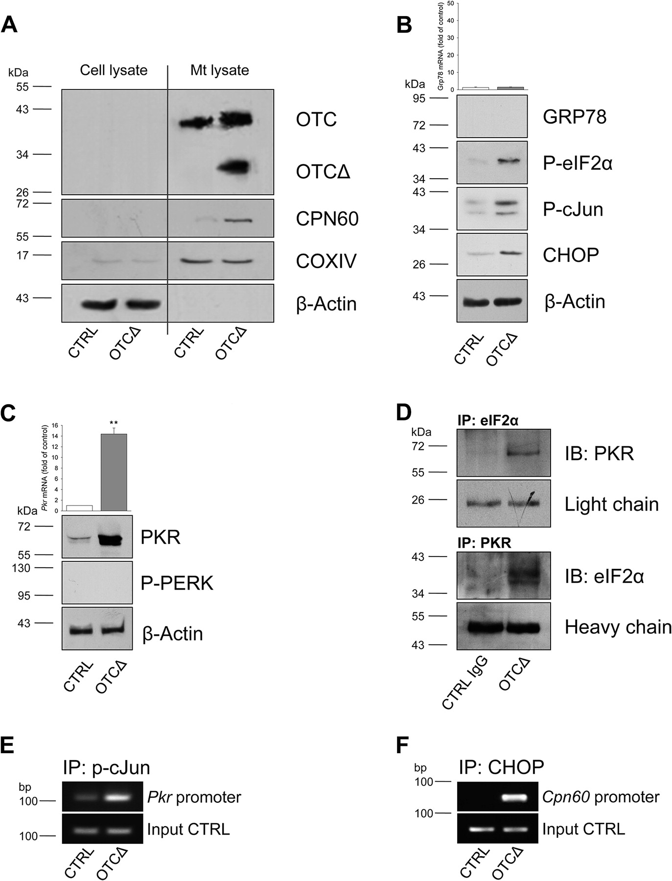
Mitochondrial UPR (mtUPR) induces PKR via AP1. Ornithine transcarbamylase (OTCΔ) induces mtUPR in Mode-K cells. Mode-K cells were transfected with OTCΔ cDNA for 36 h. (A) OTCΔ expression and translocation to the mitochondria and CPN60 recruitment to the mitochondria analysed in mitochondrial and whole-cell protein lysates by Western blot analysis. Cytochrome c oxidase (COX) IV serves as mitochondrial loading control. Bar charts: relative (B) Grp78 gene or (C) Pkr gene expression 36 h after OTCΔ transfection. Data are shown as mean±SD (**p<0.01, t test). (B) Phosphorylation of eukaryotic translation initiation factor (eIF) 2α and cJun 30 h after OTCΔ transfection, and of glucose-regulated protein 78 (GRP78) and CHOP expression 36 h after OTCΔ transfection, as determined by Western blot analysis. (C) PERK phosphorylation 30 h after OTCΔ transfection and PKR expression 36 h after OTCΔ transfection, as determined by Western blot analysis. (D) Cell lysates were prepared 30 h after OTCΔ transfection, followed by immunoprecipitation with anti-eIF2α or anti-PKR antibody and Western blot analysis for PKR or eIF2α. (E) AP1 recruitment to the Pkr promoter and (F) CHOP recruitment to the Cpn60 promoter following OTCΔ transfection analysed by chromatin immunoprecipitation (ChIP) using anti-P-cJun or anti-CHOP antibodies and subsequent PCR analysis.
mtUPR induces PKR via AP1
To confirm the exclusive induction of mtUPR, we determined the mRNA and protein expression of the ER chaperone GRP78 following transfection with OTCΔ. Consistent with published results, OTCΔ transfection did not affect GRP78 expression but induced the phosphorylation of cJun, a component of the transcription factors AP1 and CHOP (figure 1B).7 ,16 CHOP binding to the Cpn60 promoter is required for CPN60 induction under mtUPR14 ,16 and was confirmed in Mode-K cells (figure 1F). Remarkably, mtUPR led to phosphorylation of eIF2α (figure 1B). It is established that this event efficiently inhibits translation under ER stress,7 but this process has not yet been described as a consequence of mtUPR. Under ER UPR, the ER membrane-associated PERK is largely responsible for eIF2α phosphorylation7; however, phosphorylation and, thereby, activation of PERK were not detected following OTCΔ stimulation (figure 1C).
To identify the kinase responsible for eIF2α phosphorylation upon mtUPR, we screened for the recruitment of known mammalian eIF2α kinases. In response to mtUPR, we found PKR to be selectively induced. This was demonstrated at the levels of mRNA expression and total protein (figure 1C). To verify that the phosphorylation of eIF2α was directly mediated by PKR, we used coimmunoprecipitation analysis (figure 1D). In addition to eIF2α phosphorylation, PKR is also known to activate signalling cascades, regulating stress-activated protein kinases such as JNK.26 ,27
Applying the Genomatix Gene2Promotor software, we screened for putative transcription factor binding sites in the Pkr promoter and found a predicted AP1 binding site. Considering the fact that we found the AP1 component cJun to be phosphorylated under mtUPR (figure 1B) and that mtUPR employs AP1 to activate CHOP transcription,14 we performed ChIP analysis to determine AP1 binding to the Pkr promoter. Indeed, phosphorylated cJun and thus AP1 binding to the Pkr promoter were detected after mtUPR induction (figure 1E).
mtUPR signalling is dependent on PKR
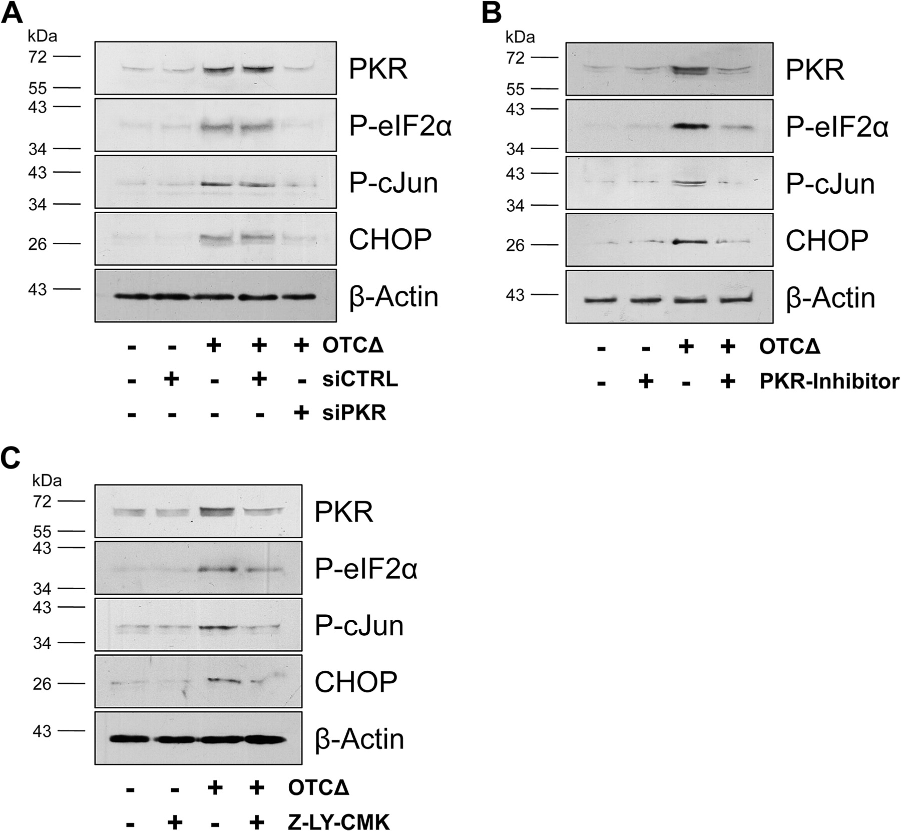
Investigating the dependency of mtUPR signalling on PKR, we used specific siRNA to knock down PKR in Mode-K cells. SiRNA knockdown of PKR prior to OTCΔ transfection completely abrogated eIF2α phosphorylation (figure 2A). Furthermore, by applying a specific inhibitor of PKR, we were able to demonstrate that mtUPR-mediated induction of PKR itself was dependent on PKR activity (figure 2B). Autoregulation of PKR expression upon its activation has been described before,27 and previous reports indicated that mitochondrial-to-nuclear signalling uses a JNK pathway, including the mitogen-activated protein kinase kinase MEK and JNK2,16 to activate AP1 under mtUPR. Applying specific inhibitors for MEK and JNK2/3 mimicked the effect of PKR knockdown on Mode-K cells, validating the AP1 dependency of PKR induction under mtUPR (supplemental figure 2A,B). In Caenorhabditis elegans, it has been shown that mtUPR signalling requires peptides generated by the mitochondrial protease ClpP.28 ,29 In line with these data, the ClpP inhibitor Z-LY-CMK was able to diminish mtUPR signalling also in mammalian (murine) IECs (figure 2C).

Mitochondrial unfolded protein response (mtUPR) signalling is dependent on PKR and ClpP in Mode-K cells. (A) Mode-K cells were transfected with small inhibitory RNA (siRNA) specific for Pkr or control siRNA (10 nmol/l) for 12 h, (B) or pretreated with PKR inhibitor (1 μmol/l) (C) or the ClpP inhibitor Z-LY-CMK (1 μmol/l) for 10 h. Subsequently, cells were transfected with ornithine transcarbamylase (OTCΔ) cDNA for 30 h or 36 h to determine protein phosphorylation (eukaryotic translation initiation factor (eIF) 2 and cJun) and expression (PKR and CHOP), respectively.
To further confirm the importance of PKR for mtUPR signalling, we transfected PKR-deficient MEFs with OTCΔ and showed that eIF2α phosphorylation was only detectable in Pkr+/+ MEFs (supplemental figure 3A). In the absence of infection, PKR can be activated by the protein activator PACT.27 Yet, OTCΔ-induced mtUPR was not impaired in Pact−/− MEFs (supplemental figure 3B).
Pkr−/− mice fail to induce CPN60 in response to DSS-induced stress and show reduced sensitivity to DSS-induced colitis
To evaluate the disease relevance of PKR-mediated mitochondrial stress mechanisms, we subjected Pkr−/− mice that have a deletion in the catalytic subunit of the Pkr gene21 to a short-term DSS feeding protocol to induce stress in IECs. These mice do not have an obvious phenotype, showing no impairment in tumour suppression, anti-viral response, apoptosis induced by tumour necrosis factor or eIF2α phosphorylation.21 After administration of 1% DSS in drinking water for 3 days, IECs were isolated from the colon. During treatment, control and Pkr−/− mice did not show any sign of disease in terms of the DAI. However, PKR expression and CPN60 expression were induced in control mice receiving DSS, whereas CPN60 expression was not enhanced in DSS-fed Pkr−/− mice (figure 3A,B). After two cycles of 1% DSS for 7 days, control mice showed severe weight loss compared to Pkr−/− mice, consistent with an exaggerated colitic response and IEC loss (table 1, supplemental figure 4). The persistent induction of PKR and CPN60 in IECs from CTRL mice and the absence of CPN60 induction in IECs from Pkr−/− mice were confirmed by immunohistochemistry (IHC) and fluorescence intensity measurements at this time point (supplemental figure 5) These results suggest a PKR-independent regulation of CPN60 under normal conditions; however, under inflammatory conditions, PKR-mediated mitochondrial stress signalling seems to be essential for CPN60 induction and appears to accelerate disease progression.

Pkr−/− mice fail to induce chaperonin 60 (CPN60) in intestinal epithelial cells (IECs) in response to stress induced by dextran sodium sulphate (DSS). Pkr−/− and CTRL mice received 1% DSS for 3 days, and colonic IECs were isolated (n=5). (A) IECs were analysed for expression of CPN60 and PKR by Western blot analysis. (B) Cpn60 and Pkr gene expression in IECs analysed by quantitative RT PCR. Bars represent fold induction±SD compared to Pkr−/− or CTRL mice receiving water. (a, b) Different from CTRL mice receiving water (two-way ANOVA followed by Tukey test; p=0.005 and p=0.019, respectively).
Pkr−/− mice show reduced sensitivity to DSS-induced colitis
CPN60 and PKR are induced in primary IECs under inflammatory conditions
Further investigating the in vivo relevance of PKR-mediated signalling, we determined the protein expression of the mtUPR hallmark protein CPN60 and PKR in primary IECs. Rag2−/− mice, as well as Rag2−/− mice backcrossed to IL-10−/− mice, were reconstituted with CD4 T cells from either Wt or IL-10−/− donor mice. The CD4 T cell population consists of colitogenic CD25− T cells and regulatory CD25+ T cells that mediate their protective function primarily by IL-10.30 Histological analysis confirmed the presence of inflammatory changes in both recipient mouse strains, which gradually develop mild to severe colitis over 4 weeks (figure 4A). Supporting previous results,31 we induced CPN60 in IECs under inflammatory conditions but preceding histological changes and, accordingly, PKR induction accompanied histological changes (figure 4A). IHC and fluorescence intensity measurements in colonic tissue sections further verified the induction of CPN60 and PKR in the intestinal epithelium after T cell transfer (figure 4B,C).

Chaperonin 60 (CPN60) and PKR are induced in primary intestinal epithelial cells (IECs) in experimental colitis. Rag2−/− and Rag2−/−×IL-10−/− recipients were adoptively transferred with CD4 T cells from Wt or IL-10−/− mice (n=5). Mice were killed 1 week and 4 weeks later. (A) Bar charts: mean histopathologic score±SD: (b) different from (a), (f) different from (e), (h) different from (g) and (i) (ANOVA on Ranks followed by Holm–Sidak test, p<0.01); (d) different from (c) (ANOVA on Ranks followed by Dunn's test, p<0.05). Isolated large IECs for expression of CPN60 and PKR were determined by Western blot analysis. (B) Immunohistochemical staining of CPN60 and PKR in colonic tissue sections 4 weeks after T cell transfer (CPN60; PKR (green), DAPI (blue), 1800×). (C) Bar charts show the mean intensity/μm2±SD of the fluorescence signal of each group (n=5 per group; five IEC regions per mouse): (b) different from (a) and (c), (e) different from (d) and (f) (one-way ANOVA followed by Holm–Sidak test, p<0.001). Immunohistochemical double staining of CPN60 with PKR or E-cadherin, respectively, in colonic tissue sections of Rag2−/− (D) and Rag2−/−×IL-10−/− (E) recipients reconstituted with CD4 T cells from IL-10−/− mice for 4 weeks (PKR, E-cadherin (red), CPN60 (green), DAPI (blue), 1800×). (F) Germ-free Wt and IL-10−/− mice were mono-associated or dual-associated with En faecalis and/or E coli. Mice were killed 6 weeks later. Bar charts: mean histopathologic score±SD: (b) different from (c) and (d), (d) different from (a) and (c) (ANOVA on Ranks followed by Holm–Sidak test, p<0.01). Isolated large IECs for expression of CPN60 and PKR were determined by Western blot analysis.
In addition, a bacteria-driven model of colitis—germ-free IL-10−/− mice mono-associated or dual-associated with non-pathogenic En faecalis and/or E coli strains—was investigated in terms of mtUPR marker proteins. We have previously shown that the different bacterial strains induce distinct disease phenotypes at different time courses and only in the genetic susceptible host, IL-10−/− mice.20 Also in this model, the induction of CPN60 and PKR reflecting inflammatory changes in the intestinal epithelium could be recovered (figure 4D). Taken together, these results strongly suggest that the induction of PKR was not due to viral infections, as the animals were maintained under a specific pathogen-free environment or under germ-free conditions, ensuring mono association and dual association, respectively.
Most importantly, the induction of CPN60 associated with elevated PKR protein levels was also observed in IECs from human patients with IBDs, as determined by Western blot analysis (figure 5A) and IHC (figure 5B,C) in different patients. Double staining of CPN60 with PKR or E-cadherin, respectively, confirmed the presence of the mtUPR hallmark protein CPN60 and of the mtUPR signalling-associated PKR in IECs under inflammatory conditions (figure 5D,E). This was true for the IECs of patients with UC (WB: n=4, IHC: n=4) and also for the IECs of patients with CD (WB: n=3, IHC: n=5; controls: WB: n=2, IHC: n=5). Fluorescence intensity measurements of individual patients and information on disease status and medication can be found in supplemental table 6 and supplemental table 2, respectively.

Chaperonin 60 (CPN60) and PKR are induced in primary intestinal epithelial cells (IECs) from patients with inflammatory bowel diseases. (A) Primary IECs were isolated from the surgical specimens of patients with colorectal cancer (CC; non-inflammatory control), active Crohn's disease (CD) and ulcerative colitis (UC). UC patient 6: IECs of non-inflamed (N) and inflamed (I) tissue regions. Expression of CPN60 and PKR was determined by Western blot analysis. Patients 1–5 and 7–9 were analysed on the same Western blot. (B) Immunohistochemical staining of CPN60 and PKR in the surgical specimens of patients with colorectal cancer (control), active CD or UC (not the same patients as in (A)). (C) Bar charts show the mean intensity/μm2±SD of the fluorescence signal of each group (n=5 for CTRL and CD, n=3 for UC; 10 IEC regions per patient). *Different from CTRL (ANOVA on Ranks followed by Dunn's test, p<0.05). The fluorescence intensity measurements of individual patients can be found in supplemental table 2. (D) Immunohistochemical double staining of CPN60 with PKR or E-cadherin, respectively, in the surgical specimens of patients with CD and UC (PKR and E-cadherin (red), CPN60 (green), DAPI (blue), 1800×).
Discussion
Although ER stress has been identified to participate during the onset and the course of neoplasia, metabolic diseases and inflammation,32–34 and although functional alterations in mitochondria and energy metabolism, in general, have been implicated in these diseases,9 ,11 ,12 little is known regarding the cooperation of ER and mitochondria in the development of these pathologies. Mitochondria and ER interact physically and functionally35; consistently, ER stress impacts mitochondrial gene expression36 ,37 and, vice versa, mitochondria have been shown to modulate ER UPR.9–11
Interestingly, we found that the highly selective mtUPR pathway employs PKR to recruit signalling molecules associated with ER-UPR, namely eIF2α and cJun (AP1) (figure 6). It has been reported that PKR participates in thapsigargin-induced ER stress and apoptosis.38 Moreover, PKR-mediated eIF2α phosphorylation has been shown to be responsible, at least in part, for the translational inhibition of cytoprotective inducible heat shock proteins in colonic IECs under inflammatory conditions.31 Significantly, Nakamura et al39 suggested that PKR-coordinated signalling may represent a central mechanism for the integration of innate immunity into metabolic pathways that are critical in metabolic diseases. These diseases comprise obesity, insulin resistance and type II diabetes, and are characterised by low-grade local inflammation. PKR can be activated by various triggers, including TLRs, growth receptor signalling, cytokines and palmitic acid.27 ,39 In turn, PKR is able to modulate signalling induced by tumour necrosis factor26 and IkB kinase26 ,27 activity, and can induce insulin receptor substrate phosphorylation at serine 307, thereby blocking insulin action.39 These broad functions of PKR are reflected by the observation that Pkr−/− mice, in response to a high-fat diet, exhibit significantly reduced levels of several inflammatory cytokines.39

Schematic illustration of the integration of mitochondrial unfolded protein response (mtUPR) and endoplasmic reticulum unfolded protein response (ER UPR). PKR is activated by ClpP-dependent mtUPR signalling and in turn induces its own transcription via MEK, JNK2 and AP1. Enhanced PKR signalling then amplifies eukaryotic translation initiation factor (eIF) 2α and cJun phosphorylation, resulting in transcriptional activation of stress-related genes. Signaling of ER UPR and mtUPR converges at the levels of eIF2α phosphorylation and AP1 activation. As a consequence of ER UPR and mtUPR, nuclear-encoded compartment-specific chaperones such as glucose-regulated protein 78 (GRP78) and chaperonin 60 (CPN60) are induced.
Regarding the diverse properties of PKR, one cannot exclude the possibility that the induction of PKR seen in the animal models of colitis and in human patients is not (or not solely) due to mitochondrial signalling. However, our finding that PKR is selectively induced during mtUPR and acts in concert with ER-UPR-derived signals implicates that PKR is a central player in mitochondrial–nuclear communication.
Alongside, it cannot be ruled that the reduced sensitivity to DSS-induced colitis seen in our experimental setup is due to other functions of PKR, yet the early induction of PKR and CPN60 in control mice while the CPN60 level remained unaltered in Pkr−/− mice provides strong evidence for a role of PKR in stress-induced CPN60 expression. We focused on mtUPR in IECs; however, this pathway is also most likely important to other cell types involved in inflammatory processes. Future experiments need to address the contribution of haematopoietic versus nonhematopoietic cells to the protective effect seen in Pkr−/− mice. Our results suggest that (1) PKR is only relevant to CPN60 induction under stress conditions and (2) the lack of an additional stress signal amplifying ER UPR signalling might be a crucial factor for favouring adaptive over apoptotic responses in the epithelium, thereby preventing tissue damage.
We found evidence that in mammalian cells, like in C elegans, the efflux of peptides resulting from the activity of proteases induced by unfolded proteins in the mitochondrial matrix may provide the initial signal for mtUPR.29 In C elegans, ClpP degrades unfolded proteins to peptides that are subsequently transported into the cytosol by HAF-1 to activate the transcription factor ZC376.7, leading to the transcription of mitochondrial chaperone genes.28 Several possibilities have been suggested as to how ClpP-generated peptides might activate downstream signalling.28 ,40 A peptide-specific receptor and the rate of peptide efflux have been implicated. Alternatively, ClpP-mediated proteolysis might release a non-peptide ligand that is subsequently transported by HAF-1.28 Of note, the related mammalian ABC transporter ABCB10 has been implicated in heme transport across the mitochondrial inner membrane.41 Alongside, it has been supposed that a mechanism analogous to ER UPR, where sensing of stress appears via binding of GRP78 to unfolded proteins,7 may exist in mitochondria through the association of CPN60 with mutant proteins.17 In this context, it is noteworthy that OTCΔ has already been shown to coimmunoprecipitate with CPN60 and ClpP.15
It has been suggested that Ca2+ release from the ER activates PKR via Ca2+/calmodulin-dependent protein kinase II and that PACT is involved in this activation.42 Nevertheless, we neither found evidence for a contribution of Ca2+, Ca2+/calmodulin-dependent protein kinase II or PACT to our experimental setup. Further investigations are needed to specify the cytosolic signal leading to PKR activation under mtUPR.
Accumulating data have placed mitochondria at the center of diverse cellular functions and suggest mitochondria as integrators of various signalling pathways. Mitochondria participate in cellular calcium homeostasis and constitute a major source of cellular reactive oxygen species (ROS),17 thereby affecting processes such as autophagy and inflammatory signalling.43 Moreover, recent work has confirmed the role of mitochondria in immune responses by linking mitochondrial ROS production and autophagy to the activation of NLRP3 inflammasome,44 a multiprotein complex involved in proteolytic maturation and release of IL-1β and IL-18.45 Polymorphisms in NLRP3 have been associated with CD in a candidate gene study,46 and a single-nucleotide polymorphism within the IL-18 receptor accessory protein gene (IL18RAP) has been identified as a risk factor for both CD and UC.47 Consistently, expression of IL-1β and IL-18 is enhanced in IBDs, particularly in the epithelium.48 ,49
In addition, polymorphisms in the genes encoding IRGM and UCP2 (proteins that directly impact mitochondrial function) have been identified as disease susceptibility factors in CD (and, in the case of UCP2, also in UC).50 IRGM, by affecting mitochondrial fission, has been shown to induce autophagy of intracellular mycobacteria and also to influence mitochondrial membrane polarisation.51 Furthermore, several pathogens specifically target mitochondria to disrupt their function,52 and proinflammatory cytokine-evoked ROS generation is associated with a drop in mitochondrial membrane potential.53 Contrarily, inducing mitochondrial stress in IECs with the oxidative phosphorylation uncoupler dinitrophenol caused decreased transepithelial electrical resistance and increased translocation of E coli.54 Enterocytes of patients with IBDs have been reported to display swollen mitochondria with irregular cristae indicative of impaired function, confirming the relevance of these data.55 In accordance, reduced ATP levels have been found in the colon of some patients with CD.56 It has been suggested that chronic intestinal inflammation might represent an energy deficiency disease involving mitochondria and featuring alterations in epithelial cell oxidative metabolism.12 ,13 In particular, β oxidation is implicated in CD pathogenesis, and a polymorphism in SLC22A557 encoding the carnitine transporter OCTN2 has been described as a risk factor in IBDs. Carnitine is essential to the energy metabolism of IECs by transporting long-chain fatty acids into mitochondria for β oxidation.58 Consequently, genetic ablation of OCTN2 results in experimental colitis.59 Sustaining energy supply might therefore be particularly important in IECs metabolically challenged by alterations in the microbiota and/or in the context of energy-consuming inflammatory processes.60
Multifactorial diseases such as IBDs require both genetic susceptibility and environmental triggers in their etiologies. Furthermore, recent data highlight how a specific microbe can determine the phenotype of a host carrying the autophagy-related ATG16L1 risk allele for inflammatory disease.61 These complex interactions underscore the necessity to identify cellular check points at which different signals converge and which may thus be promising targets for therapeutical interventions.
MtUPR and ER UPR might represent those check points that integrate disease-relevant functions such as energy supply, ROS generation and cytokine production. Studies with chemical chaperones, phenyl butyric acid and tauro-ursodeoxycholic acid confirmed that ER can be chemically targeted to enhance its functional capacity.62 In murine models of obesity and diabetes, administration of these chaperones increased insulin sensitivity, reduced fatty liver disease and suppressed inflammatory signalling.63 The use of mitochondria-specific anti-oxidants such as acetyl-l-carnitine and r-alphalipoic acid could complement such strategies by additionally targeting mitochondrial stress. Whether it is possible to adapt those strategies for IBD treatment is currently unknown, but they provide promising evidence for new therapeutic approaches.
Acknowledgments
We thank Professor Dr Martin Klingenspor and Dr Tobias Fromme for instructions and help with mitochondrial isolation; Dr Roger Vogelmann, Dr Sylvia Steininger and Viktoria Doll for help with confocal microscopy; Nadine Waldschmitt for help with MEF culture; and Professor Dr Gerhard Rogler for providing IEC samples from patients with IBDs.
References
Supplementary materials
Supplementary Data
This web only file has been produced by the BMJ Publishing Group from an electronic file supplied by the author(s) and has not been edited for content.
Files in this Data Supplement:
- Download Supplementary Data (PDF) - Manuscript file of format pdf
- Download Supplementary Data (PDF) - Manuscript file of format pdf
Footnotes
ER and EB contributed equally to the manuscript.
Funding This work was supported by Die Deutsche Forschungsgemeinschaft grants GRK 1482 and HA 3148/2-1, the German Academic Exchange Service, NIH DK RO1 DK 53347, P40 RR018603 and the Crohn's and Colitis Foundation of America.
Competing interests None.
Ethics approval This study was approved by the ethics committee of the University of Regensburg and by the institutional review board of Hospital Clinic i Provincial of Barcelona.
Provenance and peer review Not commissioned; externally peer reviewed.