Article Text

Statistics from Altmetric.com
Visceral hypersensitivity is currently the holy grail....it is widely regarded as the reason for the development of functional gastrointestinal diseases, including functional dyspepsia and irritable bowel syndrome.1 Although the field has advanced considerably in the past decade, it is necessary to take stock and realistically appraise our current understanding. Identifying issues of controversy will help address the directions or priorities to advance this field in the future. Thus our objectives are to review the current understanding of the neuroanatomy and physiology of gut sensation, briefly review examples of the relationship between symptoms and sensorimotor dysfunction, and discuss controversies and speculations requiring more thorough study. In pursuing this discussion, we have drawn on experience and data from several regions of the gut as work in a single region does not provide the comprehensive or broad perspectives necessary.
Neuroanatomy and physiology of sensation
Enteroendocrine cells in the lining of the gut serve as chemical and mechanical transducers for local reflexes (for example, peristalsis) or initiation of afferent projections to the central nervous system.1 ,2 As with somatic sensation, gut afferent signals reach conscious perception through a three neurone chain.2 The first order neurone, whose cell body is in the dorsal root ganglion, terminates in the dorsal column laminae of the spinal cord (fig 1). En passant fibres project to noradrenergic neurones in prevertebral ganglia, and this reflex centre results in modulation of viscus functions, including motility. Somatic and visceral afferents converge on dorsal horn neurones and result in viscerosomatic projection or referred pain. Descending modulatory fibres (serotonergic, adrenergic, and possibly others) from brain stem centres such as the periaqueductal grey alter the sensitivity of the dorsal horn neurones and thus serve to centrally control the intensity of perception during visceral stimulation (fig1).1

Three order neurone chain resulting in visceral perception; note the descending pathways converging on the dorsal horn neurone modulating the projections from this relay station to the surface and to the brain.
The second order neurone projects from the dorsal horn of the spinal cord to the thalamus and reticular formation in the brain stem (fig 1). The ascending pathways are located in the spinoreticular and spinothalamic tracts. Recently, a nociceptive spinal pathway was identified in the dorsal column in primates; these project nociception from viscera such as the colorectum, pancreas, and duodenum.3 ,4
These second neurones synapse with autonomic and satiety centres and with the third order neurone that leads to emotional responses (limbic system) and conscious perception (sensory cortex). These projections lead to changes in pulse rate, blood pressure, appetite, and emotions in response to visceral pain. The loci of projection in the sensory cortex are still not fully understood; there is evidence that the anterior cingulate cortex, insula, and cerebellum are activated during distensions of the oesophagus, stomach, and rectum.
Currently, we have a limited understanding of the cerebral processing of visceral stimuli, the pathways and mediators of visceral afferents, the role of end organ modulation of sensation, and association of symptoms with some sensorimotor dysfunctions. These will be reviewed below but it is essential to acknowledge the many gaps in our understanding. These gaps are particularly evident in comparison with the thorough characterisation of transmitters/mediators5-8 involved in the reflex responses at the prevertebral ganglia. The candidate transmitters in visceral perception include serotonin (5-HT), calcitonin gene related peptide, substance P, norepinephrine, and opiates (peripherally at kappa receptors, centrally at mu receptors).1 ,9 ,10There are receptor subtypes for all of these transmitters and advances in therapy will require full characterisation of the roles of transmitters and receptors along the sensory neural axis.
Definitions of visceral sensation parameters
Prior to discussing the pros and cons of measurements and the relationship between physiological parameters and symptoms, a few definitions are essential.
- (1)
- Accommodation is relaxation of the stomach in the early postprandial period. Using the barostat, wall tone has been assessed by the relative change (that is, not an absolute measurement) of stomach volume under constant pressure. However, the volume of accommodation can also be measured with novel imaging methods (for example, magnetic resonance imaging or single photon emission computed tomography, discussed below).
- (2)
- Compliance is the volume response (y axis) to an imposed pressure (x axis) and it has a sigmoid relationship with initial reflex relaxation (change in wall tension without volume change), followed by a linear section that reflects partly the elasticity of the viscus wall, and a final plateau phase (fig 2). Compliance is inversely related to elastance. Wall compliance is measured by means of isobaric stepwise (ramp) distension (for example, with a barostat). The nature of the balloon (latex v polyethylene) and the method of inflation influence the results.11-13
- (3)
- Wall tension has been calculated using the law of Laplace (T≈P.r), derived with a slight difference in the formula for the actual shape (for example, cylinder, sphere) of the balloon in the viscus segment tested. This measurement assumes several factors which are unknown in most human experiments (discussed below under “Stretch versus tension modulation of afferent discharges”). It was claimed that measurement of tension would be more relevant to understand sensory changes14 but correlation of sensation in response to tension based stimuli may not be significantly better than with pressure based stimuli.
- (4)
- Hypersensitivity, hyperalgesia, allodynia. Hypersensitivity refers to increased sensation of stimuli. In practice, this is appraised by measurement of threshold volumes or pressures for first sensation or pain. Alternatively, it refers to the increased scores of symptoms (including pain) in response to standard stimuli. Hyperalgesia refers to increased pain sensation in response to a certain stimulus. Allodynia refers to the appreciation that a stimulus, which was previously not perceived as being painful, becomes painful. These terms, which were originally used in reference to somatic sensation, have been adapted to studies of visceral sensation.
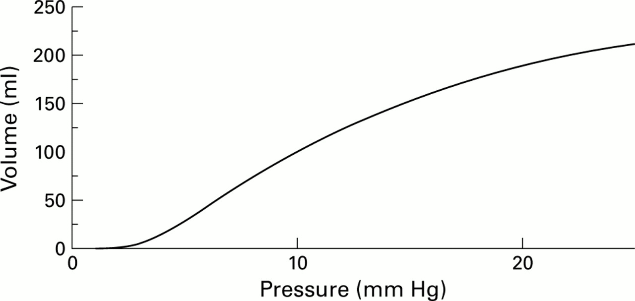
Example of a compliance curve (volume response to imposed pressure) in a segment of human colon. The curve is obtained by stepwise increments of intraballoon pressure and simultaneous measurement of intraballoon pressure. Note the initial “cushion” in which an increase in pressure does not result in any change in volume. The second portion of the compliance curve is more linear and partly reflects the elasticity of the viscus.
Stretch versus tension modulation of afferent discharges
Although the reasons for hypersensitivity to distension are unclear, mechanoreceptors must be activated to initiate, convey, or perceive the distending stimulus. It has been proposed that mechanoreceptors are either in series or in parallel with muscle fibres. In animals, in parallel mechanoreceptors respond to stimuli that elongate the stomach wall; in series mechanoreceptors respond to stimuli that increase the tension within the stomach wall.15 ,16 Figure 3 illustrates the responses of tension mechanoreceptors (in series) and elongation mechanoreceptors (in parallel) to different stimuli (distension, relaxation, and contraction of smooth muscle). In series receptors are activated during distension and contraction against a resistance; conversely, in series receptors are inactivated during relaxation. In parallel receptors are activated during distension and relaxation and are inactivated during contraction.

Schematic illustration of responses of tension mechanoreceptors (in series) and elongation mechanoreceptors (in parallel) to distension, relaxation, and contraction of gastric smooth muscle. Mechanoreceptors are modelled as coils and positioned in parallel (right) or in series (left) to the muscle. In series, receptors are activated during distension and contraction against a resistance; they become inactivated during relaxation. In parallel, receptors are activated during distension and relaxation, and become inactivated during contraction.
In human studies, gastric distension results in elongation as well as increased wall tension of the stomach; the stomach also reflexly responds to the stimulus applied by contracting. Hence it is unclear which of these two types of mechanoreceptors mediates sensitivity to distension of the proximal stomach in vivo. Observations made in recent studies provide arguments for involvement of both types of receptors. For example, during postprandial gastric accommodation, an increase in proximal gastric volume (and hence elongation) occurs but this does not cause enhanced perception17 ,18 until a large volume of food stimulates a sense of satiety and fullness. The latter is probably mediated by a change in wall tension from the pressures imposed by food and reflex contractions. In humans, one way to assess the roles of volume versus tension in gastric sensation is to evaluate the effects of high volume clamping by fixed volume inflation of a balloon while pressure is allowed to vary. This allows measurement of muscle tension while changes in muscle length are “neutralised” by the high volume clamp. In this experimental setting, phasic contractions which are associated with phasic increases in wall tension cause enhanced perception, probably through in series receptors which are under increased “load” with viscus contraction.19
Thumshirn and colleagues20 demonstrated that wall tension, estimated by Laplace's law, was significantly correlated with sensation scores (r=0.4, p<0.05) during a pharmacological study of gastric sensitivity, suggesting that >80% of the sensation variance (1−r 2) is attributable to other factors, such as visceral afferent functions or central control (spinal or supratentorial). To assess the relative contribution of both receptor types in mediating sensation in vivo, novel approaches and more sensitive methodology are needed. Distrutti and colleagues14 suggested that a “tensostat” may facilitate this task but the relationships (r values) between symptoms and tension or pressure in their work differed minimally. An alternative method, which has been used to measure rectal wall tension, is impedance planimetry; increased wall tension was associated with greater sensation of the need to defecate. However, similar increases in sensation were observed with increases in distension pressure.21
It has been suggested that the simplified law of Laplace can be used to estimate wall tension during barostat studies of hollow organs, and that this level of wall tension determines the level of perception during distension studies.14 However, this formula makes a number of assumptions that are not necessarily fulfilled by the extant experimental conditions. Those assumptions include: the wall of the viscus is infinitely thin; the intraluminal balloon and viscus have a perfectly defined (for example, spherical) shape that can be defined mathematically; the pressure external to the viscus is known and is evenly distributed. Most importantly, the Laplace formulae do not take into account the modulatory effects of changes in the contractile state of the viscus which may occur reflexly or in response to neurohumoral or pharmacological modulation and which are superimposed on the compliance of the hollow organ. The limitations in current assessments of tension with tensostat or barostat were recently discussed by Gregersen22 who emphasised the error in the assumption that tension is equal throughout the proximal stomach (that is, isotropic), particularly in view of the complex and variable geometry of the stomach within and between individuals. These differences in geometry render invalid a theoretical attribution of shape (for example, spherical v ellipsoidv more complex).
A strict application of Laplace's law to isobaric measurements also appears to lead to conclusions that conflict with daily experiences. For example, relaxation (or elongation of the circumference) of the proximal stomach under isobaric conditions would predict increased wall tension as T∼r when P is constant. Thus if tension was the determining factor in sensation, Laplace's law would predict increased sensation at times when the viscus is relaxed, for example during the 3–5-fold increase in gastric volume that occurs within five minutes of starting a meal. Conversely, under conditions of adequate accommodation and of pharmacological relaxation, the experimental evidence suggests that symptoms elicited by mechanical stimuli are usually reduced in healthy subjects,17 ,20unless massive relaxation is induced.
Measurement of visceral sensations
In general, the literature in humans shows two types of visceral sensation measurements in vivo (fig 4). The first addresses the sensation of mechanical, electrical, or other stimuli applied within the gut, and uses standardised symptom based questionnaires (visual analogue scale or adjectival scale) to determine thresholds or severity of symptoms induced. The second method measures changes in cerebral blood flow using positron emission tomography, functional magnetic resonance imaging, or single photon emission computed tomography during visceral stimulation.
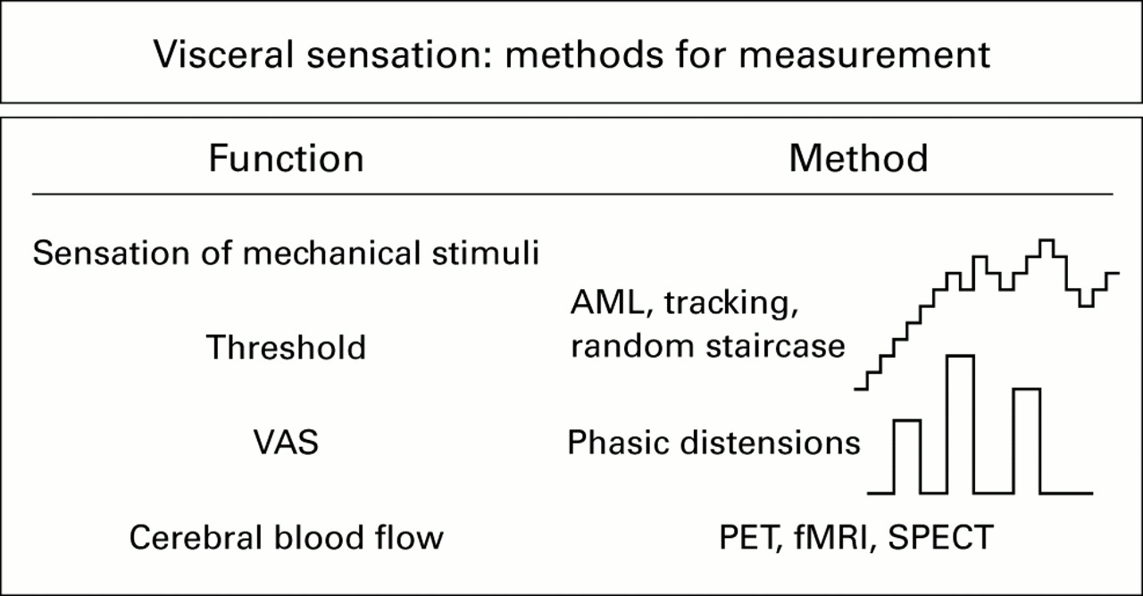
Schema demonstrating distension paradigms used in visceral sensation studies. PET, positron emission tomography; fMRI, functional magnetic resonance imaging; SPECT, single photon emission computed tomography; VAS, visual analogue scale; AML, ascending method of limits.
METHODS OF MEASURING SENSORY THRESHOLDS
Methods of measuring the threshold for initial perception or discomfort/pain have generally used ascending method of limits, tracking, or a random staircase design.12-14 In these methods, sequentially increased pressure or volume distensions are delivered until the subject perceives either first sensation or the symptom of discomfort/pain. After the threshold is identified, a computer method randomly delivers a pressure or volume stimulus which is either above or below the previously identified threshold. This serves to fine tune the level of the threshold for either volume or pressure distensions. In the random staircase method, the stimulus paradigm does not necessarily increase continually as in ascending method of limits but randomly applies the stimulus either of greater or lower intensity to try to avoid response bias.23 While these methods have received a great deal of application in the literature, their sensitivity and potential for response bias have not been adequately assessed. For example, when ascending method of limits and tracking are used to assess first perception and pain, it is possible that the subject will be interrogated 40 or 50 times while assessing the thresholds. This clearly could introduce an element of response bias which is probably worse with increasing number of distensions.
DISTENSIONS USING PRESSURE BASED MECHANICAL STIMULI
To avoid the potential inaccuracies introduced by response bias, we have used a restricted number (three to five) of distensions using pressure based mechanical stimuli which are applied in a randomised order.20 ,24-26 During distensions, the individual is asked to complete a visual analogue scale pertaining to the symptoms that are of interest (for example, pain and gas in the colon; pain and urgency in the rectum; or bloating, nausea, and pain in the stomach).
CEREBRAL BLOOD FLOW MEASUREMENTS
Cerebral blood flow measurements are intended to identify the projections in the brain of visceral stimuli applied in the gut. These will be discussed in greater depth below. However, it is important to realise that changes in cerebral blood flow detected by these methods range from 2% to 5%. Sensitivity to detect increases in cerebral blood flow over background activity, with variations unrelated to the specific stimulus, is somewhat vulnerable because of the relatively low absolute changes in blood flow that can be expected.27-29Thus there is a low signal to noise ratio which renders interpretation difficult.
Symptoms and disturbed sensorimotor functions: any role as a biological marker?
Irritable bowel syndrome (IBS) is associated with rectosigmoid hypersensitivity30 ,31 which has been postulated to represent a biological marker for IBS.32 Several lines of evidence lead us to question this suggestion. Firstly, across studies, the prevalence of rectal hypersensitivity in IBS is 20–80% and may only be demonstrable in response to repetitive stimuli rather than single stimuli. Secondly, there was only a weak correlation between rectal sensory thresholds and current pain, and no significant correlation with pain severity in the prior two weeks.33Thirdly, changes in rectal sensory thresholds did not predict response to therapy in one study reported to date.34 Fourthly, demonstration of rectal hypersensitivity has not yet contributed to the diagnosis or alteration of pharmacotherapy in IBS. Fifthly, the increased sensation has been shown to coincide, in some studies, with the timing of reflex rectal contractions, suggesting that there is also a motor component to the augmented sensitivity.31 Hence it should to be emphasised that application of rectal sensation tests in the diagnosis or management of IBS is premature. The methodology for sensory testing is not yet standardised (for example, ascending method of limits, tracking, random staircase) or completely validated (for example, coefficient of variation, test-retest reliability, sensitivity, specificity).
Several studies have confirmed that, as a group, patients with functional dyspepsia are hypersensitive to isobaric and isovolumetric balloon distension of the proximal stomach,17 ,35 that is, the thresholds for first perception and discomfort are lower in dyspeptics than in controls. Hypersensitivity to gastric distension is a feature of functional and not organic dyspepsia.36 It is not known if gastric sensory thresholds are correlated with current or recent symptom severity or if hypersensitivity is associated with specific symptoms in functional dyspepsia patients. A preliminary study reported that almost half of functional dyspepsia patients have hypersensitivity to gastric distension, and that postprandial pain is significantly more prevalent in these patients.37 However, one can question the relevance of fasting perception thresholds as a biological marker for functional dyspepsia, which is by definition a symptom complex that occurs in the postprandial period. Patients who report pain during fasting gastric distensions are potentially more prone to report postprandial pain; pressure induced increased sensation during the postprandial period may be a better marker of functional dyspepsia.38 ,39
Several authors have shown that functional or non-ulcer dyspepsia patients have reduced postcibal gastric accommodation compared with controls.18 ,38 Among 40 consecutive non-ulcer dyspeptics who underwent measurement of gastric accommodation, Tack and colleagues18 demonstrated that early satiety and weight loss were significantly more frequent in patients with impaired versus normal accommodation. As fasting gastric compliance is normal, these data suggest heightened sensitivity of the sensory apparatus from gut to cortex, with presumed modulation of hypothalamic or other satiety centres.
Controversial interpretation of visceral sensation data
In the next section we will address a number of controversial issues pertaining to visceral sensation. Such controversial statements and interpretations include the following:
- (1)
- If a drug alters perception without a change in compliance, the drug must affect visceral afferent function.
- (2)
- Wall tension determines the level of sensation in a viscus.
- (3)
- Relaxation results in reduced perception.
COMPLIANCE AND PERCEPTION
The interpretation of perception data in response to pharmacological perturbations has incorporated measurements of organ compliance. Thus when compliance is unchanged but sensation is, it has been implied that afferent nerve function has been altered. This is illustrated by the example of octreotide, which alters sensation without changing compliance,40-43 suggesting that its effect must be on afferent nerve function. However, octreotide also reduces postprandial colonic tone, suggesting that it may blunt reflex contraction to physiological stimuli44; another study suggested octreotide did not alter either rectal sensation or compliance.45
Compliance data are far more complex than would be suggested from the linear interpretation of data that have typically been applied to the compliance curves. Thus in the gut, compliance curves have an initial “cushion” in which an increase in pressure does not result in any change in volume (see fig 2). This accommodation to a pressure stimulus can be altered by a pharmacological perturbation, such as with an α2 adrenergic agonist. This portion of the compliance curve can be assessed mathematically by estimating the β component on a power exponential analysis of the compliance curve26 or the inflection point in the curve.32 The physiological contribution of tone and tension to this part of the compliance curve has not been fully evaluated.
The second portion of the compliance curve is more linear and partly reflects the elasticity of the viscus. This may explain why compliance is rarely altered by diseases, except those associated with replacement fibrosis (scleroderma, radiation) or by drugs. Clonidine significantly alters the linear portion of the colonic compliance curve,26 suggesting that neuromuscular function may partly alter this aspect of compliance.
TENSION
The relationship between tension in the wall and sensation14 is also more complex and, as indicated in the previous discussion, requires more thorough study. When a compliance curve is shifted to the left or right with no change in volume but a change in pressure, a simple application of Laplace's law would suggest that there has been a change in wall tension. Such a change may not be identified if only the slope of a linear model is applied to the compliance curve.
ISOVOLUMETRIC STIMULATION: RELAXATION VERSUS ANTINOCICEPTION
Another pitfall in the interpretation of data in the literature pertains to observations with isovolumetric stimuli. These volume based mechanical stimuli are vulnerable to misinterpretation; thus changes in wall tension or relaxation will result in greater volumes required to achieve the threshold of sensation. At first glance, this would suggest a true sensory effect of the perturbation or drug. However, as is shown from a recent example in the literature,46 volume based mechanical stimuli are vulnerable because changes in compliance or relaxation are associated with greater volumes to achieve threshold, but no such change in the pressure thresholds. In a study of IBS patients, Delvaux and colleagues46 demonstrated that alosetron 0.25 mg twice daily or 4 mg twice daily was associated with increases in the perception threshold and pain threshold to volume distensions. However, there was no significant difference in pressure thresholds, suggesting absence of an antinociceptive action of alosetron. Studies of colonic compliance performed in the same evaluation provide an explanation for the observed differences in volume thresholds as alosetron significantly altered the compliance of the colon. Other data suggest that relaxation alone is unlikely to result in reduced sensation. For example, in a study by Tack and colleagues47 performed in 18 healthy volunteers who were pretreated for five days with cisapride 10 mg four times daily or placebo, the accommodation response of the stomach was significantly greater with cisapride. However, cisapride did not alter the pressure or volume of first perception and it actually decreased pain thresholds to both volume and pressure stimuli relative to placebo in the fasting state.
Surveying the literature, there are several other examples (table 1) in which drugs have been demonstrated unequivocally to reduce sensation to volume distension but effects on pressure induced discomfort have either not been evaluated or have not been as clearly demonstrated as effects on volume distension. Examples of altered volume thresholds include effects of ondansetron and granisetron, 5-HT3antagonists,48 alosetron (as described above46) octreotide,43sumatriptan,49 and cisapride.47 In contrast, there is good evidence that the α2 adrenergic agonist clonidine increases compliance of the colon, reduces colonic tone, and also markedly reduces pain sensation during mechanical distension of the descending colon.26 Similarly, the kappa opioid agonist fedotozine50 reduces sensation to both volume and pressure stimuli, suggesting it has antinociceptive activity. The role of cytokines, purines, neurokinins, and other mediators on sensation and compliance requires further study in animals and humans.10
Pharmacological modulation of visceral tone: documented and potential sensory effects
Differences between relaxant and antinociceptive actions were demonstrated convincingly in comparisons between the effects of nitroglycerin and clonidine in gastric physiology in humans.20 Both these classes of drugs result in relaxation of the stomach during fasting and postprandially. However, whereas clonidine reduced the sensation of pain in a dose dependent manner no such effect of nitroglycerin was observed. In fact, healthy volunteers exposed to nitroglyercin tended to have a greater sensation of nausea. The selective antinociceptive activities of clonidine were also demonstrated in experiments performed in the human descending colon. Thus whereas saline (placebo) was associated with a stepwise, intensity related perception of gas and pain, clonidine markedly diminished the median sensation score for pain, and the α2 antagonist yohimbine increased pain sensation.26 Intriguingly, neither clonidine nor yohimbine significantly altered the sensation of gas relative to saline placebo. These findings suggest that clonidine has a selective effect on nociceptive pathways26 rather than on all afferents.
CEREBRAL BLOOD FLOW MEASUREMENTS
Changes in cerebral blood flow during viscus stimulation require further study. Positron emission tomography and functional magnetic resonance imaging are the most widely used techniques. After intravenous injection of a radioactive compound, positron emission tomography assesses blood flow or regional cerebral metabolism in brain areas. Functional magnetic resonance imaging detects increases in oxygen concentration in areas of heightened neuronal activity without administration of radioactive compounds. For a more extensive review of these techniques, their advantages and limitations, the reader is referred to Aziz and Thompson.56 It has been demonstrated that changes in cerebral blood flow or evoked potentials are associated with viscus sensations or contractions.57 ,58It is also assumed that changes in cerebral blood flow reflect a change in cerebral control of sensory function23 but little is known of the transmitters involved or the sensitivity of changes in blood flow relative to perception. One of the most intriguing observations has been activation of a focus in the left dorsolateral prefrontal cortex in patients with IBS. Thus in a landmark study by Silvermanet al in 1996, six patients with IBS were found to have activation of the left dorsal lateral prefrontral cortex in anticipation of a painful rectal stimulus.27 Cerebral blood flow in these patients was different from that of healthy controls in two respects. Firstly, there was no activation of the anterior cingulate cortex and secondly, there was no focal change in blood flow during the actual painful rectal stimulus in patients with IBS. Selective activation of the left dorsolateral prefrontal cortex in anticipation of the painful stimulus suggests a change in attention or vigilance as this part of the brain is involved in focus, attention, anticipation, vigilance, and memory recall.27
The initial observations have also been confirmed in another study59 that demonstrated activation of the same focus of the frontal cortex in patients with IBS or fibromyalgia. Interestingly, the rectal distension stimulated this region in IBS patients but not in healthy controls or in those with fibromyalgia. In contrast, the left dorsolateral prefrontal cortex was activated in fibromyalgia patients following somatic stimulation, but not following rectal stimulation. Taken together, these two studies suggest that attention and vigilance are activated in anticipation and in response to specific stimuli that may be related to patient recall as the location of the stimulus is relevant to the patient's symptoms.
While these changes in cerebral blood flow are extremely interesting, it is important to note that several factors may influence such changes. Indeed, during somatic painful stimuli, it has been demonstrated that attention or vigilance,60 the unpleasantness61 experience of the stimulus, gender,62 and even facial expression63 may influence the intensity of changes of cerebral blood flow, as well as the locus of that change. For example, Bushnell and colleagues60 have shown that, during imposed attention by enhancing a somatic stimulus with a thermal task, there was significant activation of the somatosensory cortex. In contrast, during application of the painful somatic stimulus on its own or during an auditory task, there was no such somatosensory cortex activation. This partly explains the previous literature in which human brain imaging studies did not consistently reveal pain related activation of the somatosensory cortex. It also suggests that somatosensory cortex activation is highly modulated by cognitive factors that alter pain perception, including attention and previous experience.
The perceived unpleasantness61 of the stimulus also modulates cerebral blood flow changes. By applying a somatic stimulus and changing the level of unpleasantness by concomitant hypnosis, Rainville et al demonstrated that there was greater activation in the anterior cingulate cortex but not in the somatosensory cortex. Gender differences in cerebral blood flow62 in response to stimulation with noxious heat stimuli applied to the left volar forearm have been reported by Paulsonet al. Thus females had greater activation of the thalamus, anterior insula, and prefrontal cortex compared with males. Finally, facial expression can also change brain activation. Blair et al have shown that sadness resulted in activation of the left amygdala and temporal pole whereas anger was associated with activation of the orbital frontal and anterior cingulate cortex.63
Diagnostic tests to evaluate visceral sensation
Before one can realistically apply the information bank on visceral sensation to clinical practice it is necessary to develop valid, preferably non-invasive diagnostic tests. Currently, most of the tests involve intubation of the viscus of interest and application of mechanical stimuli such as balloon distension with monitoring either perception scores on a visual analogue scale, threshold perceptions, or changes in cerebral blood flow, as described above. These approaches do not lend themselves easily to widespread application in the clinic. In recent years, several groups have attempted to develop a liquid nutrient or non-nutrient load test18 ,64 to identify patients with hypersensitivity due to non-ulcer or functional dyspepsia. However, across studies, only about 50% of functional dyspeptics65 can tolerate a lower volume prior to development of satiety. Moreover, measurement of the volume ingested does not differentiate hypersensitivity of the afferent apparatus from changes in compliance or tone. Thus a non-invasive approach to measurement of gastric accommodation would be of considerable interest.
Recently, two approaches have been proposed to measure gastric accommodation. Several studies from Kunz and colleagues66have demonstrated the use of magnetic resonance imaging for evaluation of gastric emptying and gastric contractility. This method depends on intraluminal contrast material. It is conceivable that imaging of the gastric wall rather than the content of the stomach would allow measurements of the stomach itself. Indeed, this was the rationale for the gastric accommodation test developed at the Mayo Clinic67; intravenous injection of 99mTc pertechnetate resulted in uptake of the isotope by the gastric parietal and other mucosal cells; after single photon emission computed tomography, slices of the stomach can be reconstructed using a three dimensional analysis program such as Analyze to measure gastric volume. Following ingestion of a standardised meal, it is then possible to evaluate accommodation of the entire proximal and distal stomach.
These methods clearly need further validation, as well as assessment of the effects of medications that are known to alter the volume of the stomach. However, the combination of volume measurements with a simple drink test and measurement of symptoms such as satiety, pain, nausea, fullness, and bloating 30 minutes after ingestion of the maximum volume of nutrient or non-nutrient liquid64 might provide a clinically applicable means to assess both accommodation and sensation responses. Symptoms associated with impairment of accommodation may be amenable to gastric relaxation therapy, whereas a normal accommodation response with increased symptoms would suggest that an antinociceptive or antinausea medication would be more appropriate.
Gaps in current knowledge and conclusion
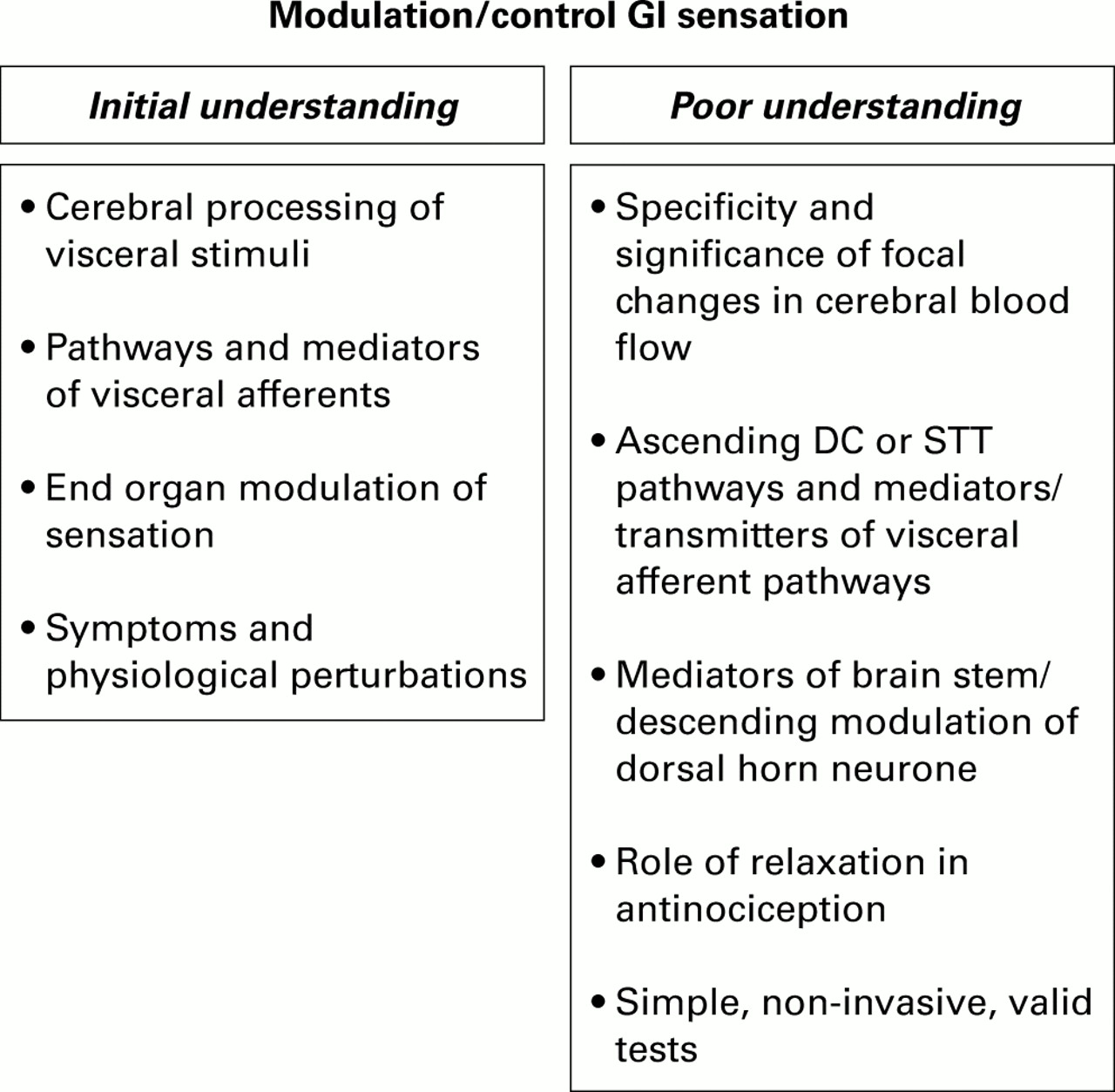
Figure 5 summarises the areas where we have an initial understanding of visceral sensation and those concepts or functions that require further study. These areas deserve high priority for research in the near term.

Gaps in current knowledge of visceral sensation. DC, dorsal column; STT, spinothalamic tract.
The last decade has seen a tremendous surge of interest in the study of the augmented visceral sensitivity of the gut in several disease states. For further advance of this discipline, novel diagnostic tests and treatments are necessary but these await a clearer understanding of the mechanisms and pathophysiology of visceral sensation, with particular emphasis on contrasting the effects of medications on relaxation versus antinociception. The validity of the concept of rectal hypersensitivity as a biological marker in IBS is still unproved. It has been claimed that the holy grail of functional gastrointestinal disease is indeed visceral hypersensitivity; however, the proof of the pudding will lie in its eating. Ultimately, the relevance of the concept will depend on proof that symptoms are attributable to altered sensitivities and that therapies aimed at correction of hypersensitivity result in clinical benefit. A prerequisite for such correction is a more thorough understanding of the transmitters or mediators involved in visceral hypersensitivity and the development of novel, selective approaches to target those transmitters.
Abbreviations used in this paper
- IBS
- irritable bowel syndrome