Article Text

Statistics from Altmetric.com
Endomysial antibodies are a hallmark of coeliac disease. The existence of autoantibodies whose titres fluctuate with ingestion of gliadin is enigmatic. Gliadin seems to drive this antibody secretion as endomysial antibodies are produced in biopsy samples cultured with a peptic/tryptic digest of gliadin.1 The phenomenon of endomysial antibodies has been explained by molecular mimicry between gliadin and the endomysial antigen, or unmasking of cryptic epitopes in the endomysial antigen upon exposure to gliadin.1 ,2Recently, Dieterich and colleagues identified tissue transglutaminase (tTG) as the antigen for endomysial antibodies.3 Their data indicate that tTG forms complexes with gliadin, and they hypothesise that neoepitopes in the complex between gliadin and tTG initiate an immune response that is finally directed against gliadin and tTG.3 ,4 Based on the observation that tTG and gliadin form complexes, we would like to propose an alternative mechanism for the production of antibodies to tTG.
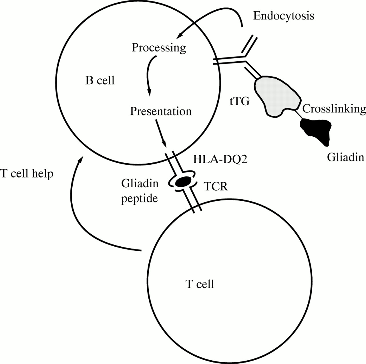
The prevailing view of B cell tolerance states that tolerance to soluble self-antigens, in contrast to tolerance to multivalent self-antigens, is regulated at the level of helper T cells (that is, indirectly by T cell tolerance).5 In the normal state, B cells specific for soluble self-antigens exist, but they secrete antibodies only when given help by T cells. The appropriate help can be provided by linking the self-antigen with a T cell epitope of a foreign antigen (that is, a carrier protein).5 Complex formation between tTG and gliadin will create this situation (fig 1). Gliadin specific CD4+ helper T cells exist in the small intestinal mucosa of patients with coeliac disease.6 ,7 These gliadin reactive helper T cells could, when the gliadin and the tTG antigens are linked, provide help for tTG specific B cells. When gliadin is withdrawn from the diet, T cell help for anti-tTG specific B cells will cease and the titres of antibodies to tTG will decline. If, however, help for tTG specific B cells is provided by tTG specific T cells, the production of antibodies to tTG would continue as long as the tTG antigen is present. Given that tTG antigen is expressed in many organs and released upon tissue damage, one would assume that the immune response to tTG should be chronic and not regulated by gliadin.

: Gliadin specific T cells can aid antibody production by tTG specific B cells. B cells with antibodies specific for tTG are likely to be present in all individuals, but are dependent on T cell help to start secreting immunoglobulin and to initiate isotype switching. Complex formation between gliadin and tTG makes gliadin a carrier protein for tTG.
This concept can also provide an explanation why antibodies to tTG are more disease specific than those to gliadin. The production of antibodies to tTG will require a localised T cell response to gliadin in an environment where gliadin can be linked with tTG—that is, the small intestinal mucosa. Pathology could be the outcome of this localised mucosal immune reaction. Many individuals have antibodies to gliadin but no signs of gut pathology.8 ,9 The formation of these anti-gliadin antibodies can result from T cell and B cell interactions in regional lymph nodes and hence is not always indicative of gut immunopathology.
The restriction of endomysial, but not gliadin, antibodies to individuals who carry HLA-DQ28-10 is further evidence for the underlying role of mucosal gliadin specific T cells in the production of endomysial antibodies. We have observed that gut mucosa derived gliadin specific T cells have a strong preference for using HLA-DQ2 as the restricting element.6 ,10 Gliadin specific peripheral blood T cells do not show a similar striking bias in HLA restriction.11
tTG catalyses protein crosslinking, resulting in the formation of an ε-(γ-glutamyl)-lysine bond. Gliadin can act as one of a limited number of donor substrates, whereas numerous proteins can be acceptor substrates.12 It is therefore conceivable that gliadin can be crosslinked with a variety of self-proteins. There is evidence for the existence of autoantibodies in coeliac disease with several distinct specificities.10 According to our model, the T cell immune response to gliadin would potentially drive antibody responses towards proteins that are crosslinked with gliadin, thus explaining the existence of such antibodies.
Dieterich et al 3 suggest a novel approach for treatment of coeliac disease disease by introducing tTG into the diet to induce oral tolerance. Oral tolerance operates at the level of T cells and the rationale behind the ingestion of tTG is that tTG specific T cells would be “silenced”.13 From the arguments presented here, the existence of tTG specific T cells is not proved by the mere existence of antibodies to tTG. Oral feeding of tTG may therefore not be a rational strategy for treating coeliac disease. Our concept rests heavily on the observation that tTG and gliadin form complexes. The first step to verify this concept is to reproduce the finding that tTG and gliadin can be linked.
Acknowledgments
We thank Markku Mäki for commenting on our manuscript.