Article Text

Statistics from Altmetric.com
- INF-γ, γ-interferon
- mTNF, transmembrane TNF-α
- PET, positron emission tomography
- SPECT, single photon emission computed tomography
- TNF, tumour-necrosis factor
It has long been known that apoptosis of T cells is an important mechanism for terminating inflammatory reactions.1 It was proposed over 10 years ago that defective apoptosis could play a role in the pathogenesis of inflammatory bowel disease.2 There is now substantial experimental and clinical evidence supporting this hypothesis.3
In Crohn’s disease there is an expansion in CD4 T-cell populations mediated by tumour necrosis factor (TNF) and γ-interferon (IFN-γ), which activate macrophages to release interleukin-6, interleukin-12 and TNF.4,5 These cytokines act to perpetuate the inflammatory reaction by reducing the susceptibility of T cells to die by apoptosis.3 In the uninflammed state, lamina propria T cells have a higher susceptibility to apoptosis than unstimulated T cells from the peripheral blood because of high expression of the apoptosis-inducing receptor Fas.6 In contrast to this, lamina propria T cells from patients with Crohn’s disease are resistant to apoptotic stimuli.7 These observations suggest that apoptosis limits the number of CD4 T cells in healthy individuals whereas in Crohn’s disease expansion of T-cell populations can occur without the restriction of apoptosis.
This resistance to induction of apoptosis is mediated by interleukin-12, the interleukin-6 receptor and TNF. Interleukin-12 is one of the most important cytokines in Crohn’s disease promoting Th1 T-cell differentiation. It also renders T cells resistant to Fas-induced apoptosis, possibly through inhibition of caspase 3 and 9, thereby prolonging T-cell survival.8 Early clinical studies have shown that antibodies that block the action of interleukin-12 reduce the severity of Crohn’s disease.9 Such antibodies also increase apoptosis in lamina propria T cells and reduce the severity of trinitrobenzene sulphonic acid experimental colitis.5
Interleukin-6 secreted by lamina propria macrophages and T cells also promotes the survival of T cells by inhibiting apoptosis. Complexes of interleukin-6/interleukin-6 receptor activate lamina propria T cells expressing the cytokine receptor gp130 on their surface. This activates a signal transduction pathway involving the phosphorylation, by JAK kinases, of the transcription factor STAT3. STAT3 increases the expression of the anti-apoptotic protein Bcl-xL thereby increasing the resistance of T cells to apoptosis.4
Perhaps the most compelling evidence for the importance of apoptosis in Crohn’s disease has come from analysis of the mechanism of action of anti-TNF therapy. Though complex, a full understanding of the biology of TNF is essential for an appreciation of its role in the treatment of Crohn’s disease. TNF is a cytokine that has many proinflammatory effects. A precursor form called transmembrane TNF-α (mTNF) is expressed on the surface of activated lymphocytes and macrophages. The extracellular 157 amino acids can be cleaved off mTNF and secreted. Both the secreted and transmembrane forms can induce apoptosis. Secreted TNF can bind either of the two TNF receptors, TNF-R1 (p55) or TNF-RII (p75), and activate the extrinsic apoptosis pathway through caspase 8.10 The transmembrane form can also activate the extrinsic apoptosis pathway by binding to TNF-RII.11
However, a third mechanism of inducing apoptosis may be the most relevant for the treatment of Crohn’s disease with anti-TNF therapy. mTNF can deliver an apoptosis signal into the cell upon which it is expressed by a process called reverse signalling. When an activating antibody binds to mTNF the JNK pathway is activated, which phosphorylates several transcription factors including p53. This upregulates the pro-apoptotic proteins bax and bak, which trigger apoptosis by the intrinsic mitochondrial pathway.10 Cell-cycle arrest is also induced by upregulation of p21. Of additional relevance to Crohn’s disease, secretion of the anti-inflammatory cytokine interleukin-10 is stimulated.12 Together these are powerful mechanisms for reducing T-cell numbers in Crohn’s inflammatory bowel disease.
This detailed analysis of the biology of TNF gives an explanation to one of the most puzzling features of anti-TNF therapy. Infliximab, a chimaeric anti-TNF-α monoclonal antibody, and adalimumab, a fully humanised immunoglobulin-1 anti-TNF-α antibody, can induce remission in Crohn’s disease13,14 whereas etanercept, a TNF-α RII receptor immunoglobulin fusion protein, has no therapeutic effect in Crohn’s disease.15 This is surprising as etanercept is highly effective in rheumatoid arthritis, ankylosing spondylitis and psoriasis.16 The explanation for this paradox is that infliximab can induce apoptosis by the mTNF signalling pathway described above whereas etanercept cannot activate this pathway.12,17 This suggests that in Crohn’s disease, reverse signalling from mTNF is of greater importance than blocking soluble TNF-α, whereas in rheumatoid arthritis blockade of soluble TNF-α is the dominant therapeutic action.
The potential importance of induction of apoptosis in the treatment of Crohn’s disease is further underlined by observations that azathioprine, corticosteroids and sulphasalazine all induce apoptosis in T cells.18–,20
However, although provocative, these studies still do not prove that apoptosis of T cells and macrophages is the key therapeutic action of infliximab. Apoptosis could be a bystander effect with other unrelated effects mediating infliximab’s therapeutic action. More direct evidence is required from measuring apoptosis of lamina propria T cells in patients before and after anti-TNF treatment; however, this is not straightforward. Histological examination of tissue biopsies has been the mainstay of measurement of apoptosis in human samples. However, biopsies do not measure the total number of apoptotic cells in an organ and are severely prone to sampling error. Fluorescence-activated cell sorting can detect apoptosis of leukocytes but has very limited use for cells outside peripheral blood. Recently, a blood test has been developed to measure apoptosis of hepatocytes.21 This is an ELISA for the apoptosis neoepitope of the C-terminal domain of cytokeratin-18. It is works well in conditions where there are high rates of coordinated epithelial apoptosis such occurs as in hepatitis. However, cytokeratin 18 is not present in T cells so this test cannot be applied in this situation.
There has been great interest for several years for imaging apoptosis in vivo. Most methods rely on the use of labelled reagents such as annexin V which bind apoptotic cells.22 This technique takes advantage of the fact that apoptotic cells display phosphatidyl serine on their surface, which can bind annexin V. It is possible to label annexin V with technetium,[99] inject it into patients and image apoptosis by single photon emission computed tomography (SPECT) or positron emission tomography (PET). PET may have some advantages over SPECT as it has greater sensitivity and temporal resolution. Recently, annexin V labelled with I124 for PET has shown promising results.23,24
In this issue, the study by Van der Brande et al (see page 509) adds further evidence for the importance of apoptosis of lamina propria T cells in the action of infliximab.25 It uses SPECT for the non-invasive localisation and quantification of apoptosis of lamina propria T cells in patients with Crohn’s disease. The authors show in two mouse models of colitis, trinitrobenzene sulphonic acid colitis and chronic CD45RBhigh colitis, that apoptosis in the intestine is increased compared with uninflammed intestine. More significantly, they demonstrate, for the first time, that infliximab induces more apoptosis in patients with Crohn’s disease who respond than those who do not respond. This important observation is further refined by the demonstration that the region of apoptosis corresponds to the inflamed intestinal segment. Fluorescence-activated cell sorting of mucosal biopsies identifies lamina propria T cells as the cell type undergoing apoptosis. Together, these data give direct experimental evidence for the importance of apoptosis of lamina propria T cells, and probably macrophages, in the termination of the Crohn’s disease inflammatory reaction. They also give further experimental support for the hypothesis that infliximab induces remission by inducing apoptosis of lamina propria T cells. Apoptosis of intestinal epithelial cells of patients with Crohn’s disease has been reported recently.26 However, this observation was not confirmed in this study.
This highly provocative study raises several important questions. Does the efficiency of induction of apoptosis determine the clinical efficacy of infliximab therapy? The observation that the induction of apoptosis was much greater in patients who responded to infliximab than non-responders certainly supports this hypothesis, though further supporting evidence is required before we can be certain. Is it possible to predict the clinical response to infliximab therapy by determining the susceptibility of lamina propria T cells to undergo apoptosis in response to infliximab? If this was true and a practical laboratory test could be devised, patients likely to respond to infliximab could be selected for therapy, giving safety and economic benefits. These observations also highlight our lack of understanding of the mechanisms of apoptosis resistance of lamina propria T cells and macrophages. Why do some Crohn’s patients have T cells that are responsive to apoptotic stimuli and others do not? Further investigation is required into the molecular lesions in the activation of the extrinsic and intrinsic apoptosis signalling pathways in Crohn’s disease.
REFERENCES
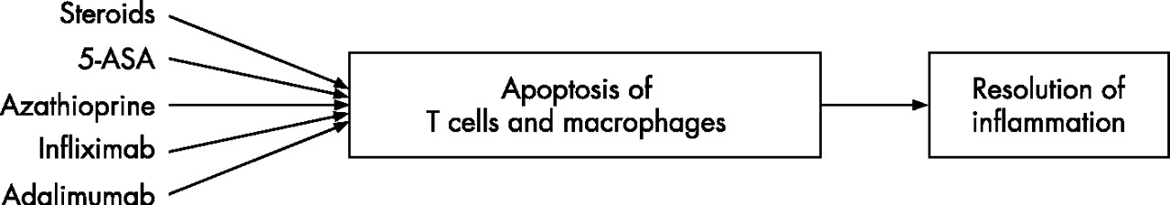
Steroids, 5-aminosalicyclic acid compounds, azathioprine, infliximab and adalimumab all cause apoptosis of T cells in the lamina propria, which contributes to the induction of remission of inflammation in Crohn’s disease.
Footnotes
Competing Interest: None.