Article Text

Statistics from Altmetric.com
We read with interest the paper by Jalanka et al,1 who examined the influence of bowel preparation on intestinal microbiota by using phylogenetic microarray and quantitative PCR analyses of frozen samples. Conventionally, faecal samples are frozen on dry ice or in a deep-freezer (at −80°C) immediately after collection, as done by Jalanka et al, because bacterial taxa can change appreciably within 15 min at room temperature (RT).2 However, immediate deep-freezing is often inconvenient in routine clinical practice, and we wondered whether simple storage of faecal samples at RT in test tubes containing 4 M guanidine thiocyanate solution would be equally effective. Guanidine thiocyanate is a general protein denaturant3 and inhibits bacterial growth.3–5 We collected faecal samples before and after colonoscopy, and divided each into two parts: one was stored frozen and the other at RT. Taxonomic compositions were determined by 16S ribosomal RNA sequence analysis, and the results in the two groups were compared. We also examined the stability of faecal microbiome composition, since Jalanka et al found that the intestinal microbiota is changed by whole-bowel irrigation, but recovers within 14 days.1
First faecal samples were collected immediately at defecation and frozen on dry ice (sample D0_F) or stored at RT in a test tube (D0_R) at home 1 day before colonoscopy (n=8) (figure 1). The test tubes (TechnoSuruga Laboratory, Shizuoka, Japan) at RT contained 100 mM Tris-HCl (pH 9), 40 mM EDTA, 4 M guanidine thiocyanate, and 0.001% bromothymol.4 Second faecal samples were collected on the morning of the day of the test immediately at defecation and similarly frozen on dry ice (D1–1_F) or stored at RT (D1–1_R) at home. On the day of the test, other faecal samples were collected immediately at first defecation during oral administration of bowel-cleansing agent at the hospital and again frozen on dry ice (D1–2_F) or stored at RT (D1–2_R). Intestinal fluid was also sampled during colonoscopy and frozen on dry ice (D1–3_F). Last faecal samples were collected 60 days after colonoscopy, immediately at defecation and frozen on dry ice (D60_F).
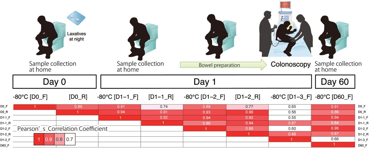
Pairwise Pearson correlation coefficients for microbial composition between eight different sampling and storing conditions (D0_F, D0_R, D1–1_F, D1–1_R, D1–2_F, D1–2_R, D1–3_F, and D60_F; for details, see text). Values are medians over eight subjects.
To compare taxonomic compositions among different sampling conditions, we computed pairwise Pearson's correlation coefficients for taxonomic profiles with median values for the eight individuals (figure 1, see online supplementary figure S1). Frozen samples at different time points showed high (ρ≥0.88, p<0.01) correlations with each other. Remarkably, samples D60_F showed high correlations with the samples collected before colonoscopy (see online supplementary figure S2). Intestinal fluid (D1–3_F) had much lower correlations with faecal samples. Samples collected at the same time points but stored under different conditions showed high (ρ≥0.88, p<0.01) correlations with each other.
Supplemental material
To examine the influence of storage temperature on each taxon, we computed fold changes in taxonomic abundances of 20 dominant genera between frozen samples and RT-stored samples (figure 2, middle). No significant difference (false discovery rate (FDR)-corrected p≤0.1 in Wilcoxon signed-rank test) was found. Our findings indicate that faecal sample storage in test tubes filled with 4 M guanidine thiocyanate solution at RT could be a practical alternative to fresh-frozen storage for taxonomic examination.

Left, fold changes in taxonomic abundance of 20 dominant genera. Middle, comparisons between frozen and room temperature-stored samples from one day before colonoscopy (blue), the test day morning (red) and during bowel cleansing (yellow). Right, comparisons between baseline samples (D0_F) and samples from the test day morning (blue), during bowel cleansing (red), and 2 months after colonoscopy (yellow).
We next investigated the effects of sampling time point (before/after colonoscopy) on taxonomic abundance. Figure 2 (right) compares the fold change in taxonomic abundance in D1–1_F vs D0_F (blue), D1–2_F versus D0_F (red), and D60_F vs D0_F (yellow). No significant difference (FDR-corrected p≤0.1 in Wilcoxon signed-rank test) was found. These findings indicate that the gut microbiota is robust during colonoscopy, in accordance with Jalanka et al's findings1 using different methodology.
Footnotes
YN and SM contributed equally.
Acknowledgements We are grateful to Ms Chika Shima, Ms Keiko Igarashi, Ms Risa Usui, Ms Arisa Kaya, Ms Shoko Ohashi, Ms Tomoko Urushidate, Ms Yuko Shimizu and Ms Naoko Okada (National Cancer Center Research Institute) for expert technical assistance.
Contributors TN, TS, SY and TY contributed to the study concept and design. TN, YS and SY collected the clinical samples. FH and SY performed the experiments. YN, SM, HW and TY performed bioinformatics analyses. YN, SM, SY and TY wrote the manuscript. TS supervised the study.
Funding National Cancer Center Research and Development Fund (25-A-4 and 28-A-4); Grants-in Aid for Scientific Research from the Ministry of Education, Culture, Sports, Science and Technology of Japan (25710016 to TY); PRESTO (Precursory Research for Embryonic Science and Technology) from Japan Science and Technology Agency (TY); the Takeda Science Foundation (SY); the Suzuken Memorial Foundation (SY).
Competing interests None declared.
Patient consent Obtained.
Ethics approval The experimental protocols were approved by the Institutional Review Board at the National Cancer Center (#2013-244). Written informed consent was obtained from all subjects.
Provenance and peer review Not commissioned; externally peer reviewed.