Article Text

Abstract
Introduction The autonomic nervous system (ANS) modulates numerous processes, including metabolic control and visceral pain processing. Accumulating evidence purports that ANS function is disturbed in both inflammatory bowel disease and irritable bowel syndrome (IBS). Whilst the brain is a central hub for regulating autonomic function, the association between resting autonomic activity and subcortical morphology has not been comprehensively studied and thus we sought to address this knowledge gap.
Method In twenty-seven healthy subjects (14 male and 13 female; mean age 30 years (range 22–53 years)), we quantified resting cardiac sympathetic index (CSI) and parasympathetic cardiac vagal tone (CVT) using a Neuroscope. Autonomic parameters were recorded as per international recommendations. In addition, high resolution structural magnetic resonance imaging scans were acquired using a GE Signa HDxt3.0 Tesla scanner. Subcortical shape differences, i.e. “deformation”, contingent on resting ANS activity were studied using FSL-FIRST, by means of positive and negative linear contrasts.
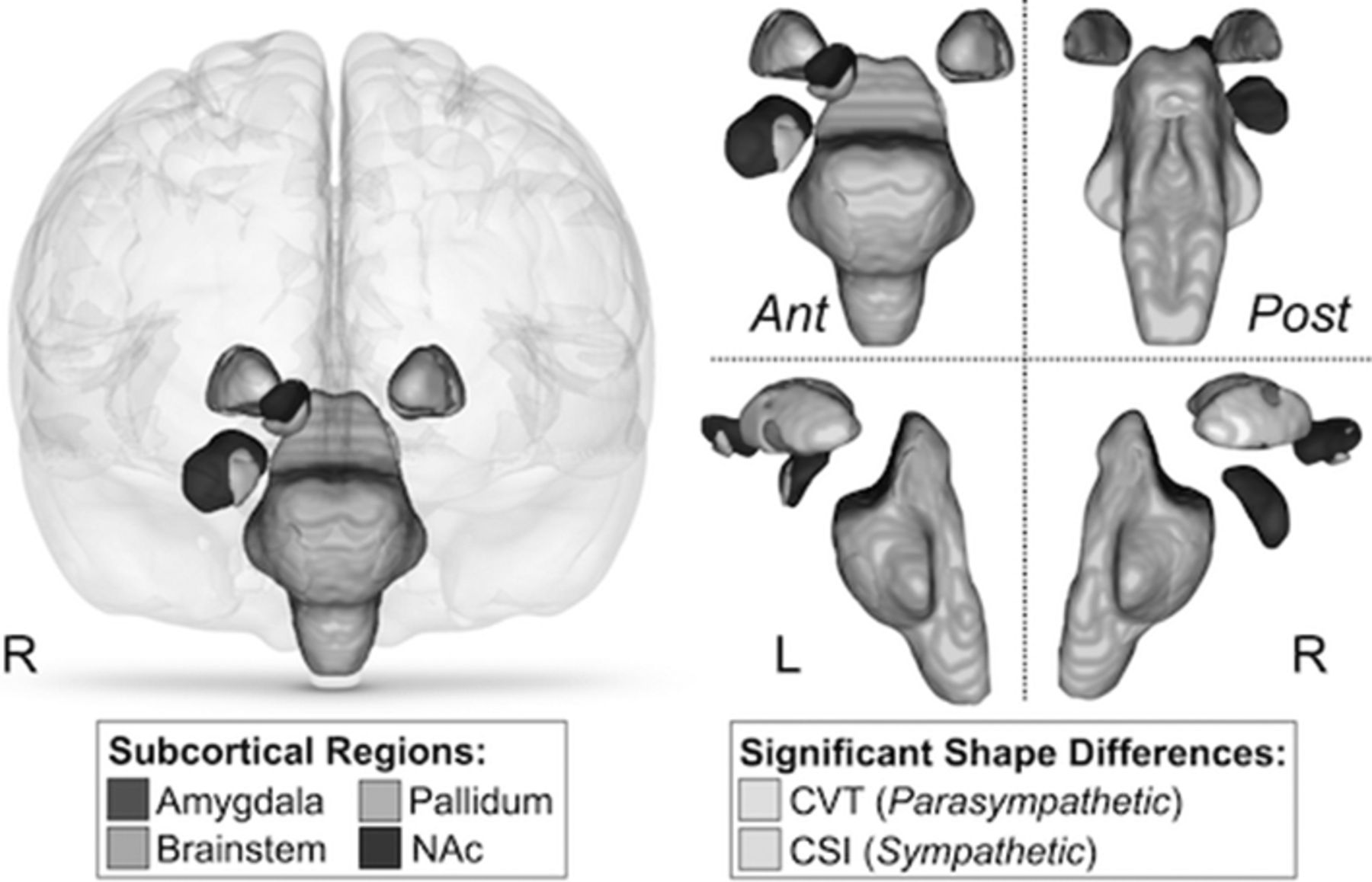
Results CSI was significantly associated with outward deformation of the brainstem, right nucleus accumbens, right amygdala and bilateral pallidum (family-wise error (FWE) corrected p<0.05) (Figure 1, turquoise coloured areas of nuclei represent outward deformation associated with CSI). In contrast, CVT was significantly associated with inward deformation of the right amygdala and pallidum (FWE corrected p<0.05) (Figure 1, yellow coloured areas of nuclei represent inward deformation associated with CVT). Furthermore, bilateral putamen volume positively correlated with resting CVT (p<0.0003).

Conclusion Our study provides novel evidence that resting autonomic state is associated with differences in the shape and/or volume of subcortical nuclei. The Rome working group have highlighted that inter-individual differences are a key limiting factor in the neuroimaging of visceral pain, these data suggest that resting autonomic function reflects an additional important variable. Given that perturbations in autonomic function and brain morphological differences are well described in IBS, our findings suggest that future studies should control for resting autonomic tone as covariates.
Disclosure of Interest J. Ruffle: None Declared, S Coen: None Declared, V Giampietro: None Declared, S Williams: None Declared, A Farmer Conflict with: Medical Research Council Grant, Q Aziz Conflict with: Medical Research Council Grant
- Autonomic Nervous System
- Brain-Gut Axis
- Cardiac Sympathetic Index
- Cardiac Vagal Tone
- Parasympathetic Nervous System
- Sympathetic Nervous System